Corresponding author: Ji Ae Park
Department of Medical Laboratory Science, Jinju Health College, 51 Uibyeong-ro, Jinju 52655, Korea
E-mail: [email protected]
ORCID: https://orcid.org/0000-0002-2957-1865 ORIGINAL ARTICLE
Detection of bla
KPC
and bla
NDM
Genes from
Gram-Negative Rod Bacteria Isolated from a
General Hospital in Gyeongnam
Byoung Seon Yang, Ji Ae Park
Department of Medical Laboratory Science, Jinju Health College, Jinju, Korea
경남지역 종합병원에서 분리된 그람음성막대균으로부터
bla
KPC
및
bla
NDM
유전자 검출
양병선, 박지애
진주보건대학교 임상병리과ARTICLE INFO ABSTRACT
Received January 28, 2021 Revised 1st February 18, 2021 Revised 2nd February 23, 2021 Revised 3rd February 26, 2021 Accepted February 28, 2021
This study investigated the use of real-time PCR melting curves for the diagnosis of blaKPC and blaNDM
genes among the most frequently detected carbapenemase-producing Enterobacteriaceae in Korea. As a means of addressing the shortcomings of phenotype tests and conventional PCR. The modified Hodge test confirmed positivity in 25 of 35 strains, and carbapenemase inhibition testing confirmed positivity in 14 strains by meropenem+PBA or meropenem+EDTA. PCR analysis showed amplification products in 25 strains of Klebsiella pneumoniae carbapenemases (KPC), 10 of K. pneumoniae, 5 of E. coli, 5 of A. baumannii, 4 of P. aeruginosa, and 1 of P. putida. New Delhi metallo β-lactamase (NDM) identified amplification products in 8 strains, that is, 2 K. pneumoniae, 3 E. coli, 1 P. aeruginosa, 1 E. cloacae, and 1 P. retgeri strains. Real-time PCR melting curve analysis confirmed amplification in 25 strains of KPC and 8 strains of NDM, and these results were 100% consistent with PCR results. In conclusion, our findings suggest early diagnosis of carbapenem resistant Enterobacteriaceae by real-time PCR offers a potential means of antibacterial management that can prevent and control nosocomial infection spread.
Copyright Ⓒ 2021 The Korean Society for Clinical Laboratory Science. All rights reserved.
Key words blaKPC blaNDM MHT CIT Real-time PCR 서 론 지난 10여년 동안 전 세계의 입원 환자들 사이에서 내성이 강 한 그람 음성균주 감염에 의한 유병률이 급격히 증가했다[1]. 이 러한 병원 환경에서 다제내성 세균으로 인한 심각한 감염을 치 료하기 위해 사용되는 carbapenem은 강력하고 광범위한 β-lactam계 항생제이나, 광범위한 사용으로 carbapenem
resistant Enterobacteriaceae (CRE)의 보급이 빠르게 증가 하여 공중보건에 심각한 위협이 되고 있다.
그람 음성 세균에서 carbapenem 저항성은 β-lactamase 생산, 유출 펌프의 발현, porins 손실 및 penicillin binding proteins (PBPs)의 변경의 결과일 수 있다[2]. 현재 β- lactamase는 구조적 유사성(A, B, C, D 등급)을 기준으로 4개 로 분류되거나 가수분해 및 억제제 양상(1∼4)에 따라 4개의 그 룹으로 분류된다[3]. 그리고 저항성은 carbapenemases의 유 전자를 암호화하는 이동식 유전 요소인 plasmid, transposons 의 유출로 다른 유전자 간에도 저항 유전자의 성공적인 수평 확 산의 가능성을 제공하였고[4], 이러한 발견 이후 carbapenemase Korean Society for
는 세계적인 문제가 되었다. 한국에서는 2010년 12월에 처음으로 CRE에 감염된 사람이 보고되었고[5], 2014년에는 전국적으로 Klebsiella pneumoniae carbapenemases (KPC) 생성 균주에 감염된 환자가 급증하 였다. 초기에는 병원 기반 발병이 발생하였고, 그 결과 병원 간 또는 지역 간 확산이 발생하여 2015년에는 KPC 생성 균주가 풍 토화되었다[6, 7]. 2020년 Park 등[8]의 연구에 따르면 서울 소재 병원의 Enterobacteriaceae 1,468개 임상검체 중 carba-penemase 유전자의 분자 특성화를 통해 CRE를 1년간 관찰한 결과 가장 흔한 분리된 균주는 Klebsiella pneumoniae 56.5% 과 Escherichia. coli 17.0%이었다. 그리고 일반적으로 최소 1 개의 carbapenemase producing Enterobacteriaceae (CPE) 에 대해 58.1%가 양성반응을 보였으며, KPC-2가 46%로 가장 일반적인 CPE형으로 나타났으며, New Delhi metallo β- lactamase (NDM)-1이 5.9%로 뒤를 이었다. KPC-2에서 K. pneumoniae 73.1%, E. coli는 15.8%로 나타났으며 NDM-1은 Enterobacter cloacae 복합체에서 23.3% 분리되었다. 이러한 CRE 외에도 공중보건에 대한 심각하거나 긴급한 위 협으로 분류되는 항생제 내성 그람 음성 병원체에 해당하는 것 으로 제3세대 cephalosporin-resistant Enterobacteriaceae (3GC-R), CRE, multidrug resistant Pseudomonas aeruginosa (MDRP), multidrug resistant Acinetobacter species (MDRA) 등이 있다[9, 10]. 뿐만 아니라 extended spectrum β-lactamase (ESBL)를 생성하는 Enterobacteriaceae 와 multidrug resistance (MDR) Enterobacteriaceae 또한 치료가 어렵고, 따라서 더 많은 수의 유병균에 의한 감염 위험에 처하게 되었다[1]. 이처럼 그람 음성 세균의 항균제 저항성은 공공의 건강을 계 속 위협하고 있으며 의료의 사회적 비용을 증가시키고 있다. 각 종 항생제에 내성을 가지는 그람 음성 세균에 의한 감염 위험 인 자는 대체로 중복되며, 이전 carbapenem의 사용은 저항력이 높은 여러 그람 음성 세균으로 인해 감염의 위험성을 증가시키 는 것으로 알려졌다[9].
그러나 현재 CRE 진단에 활용되고 있는 clinical laboratory standards institute (CLSI) 가이드 라인[11]을 따른 검출법 및 선별검사법으로 이용되는 modified Hodge test (MHT)의 경 우 진단시간이 많이 소요되며, 특정 효소의 검출에는 낮은 민감 도를 보인다는 단점을 가진다[12]. 그리고 특정 억제제를 이용 하여 유전자형을 확인하는 carbapenemase inhibition test (CIT) 검사법 또한 정확한 유전자형을 알기 위해서는 추가적인 분자진단을 필요로 한다.
따라서 CRE의 적시에 신속하고 정확한 검출은 감염의 임상 적 예방과 치료에 매우 중요하다 할 수 있다. 일반적으로 유전자 형의 검사에는 polymerase chain reaction (PCR)을 이용한 분자 방법을 이용하고 있으나, 사후 분석에 있어 종종 시간과 비 용이 많이 소요된다. 그래서 근래에는 실시간 중합효소 연쇄반 응(real-time PCR) 분석방법을 통해 보다 신속한 유전자형의 진단을 실시하며, 이러한 real-time PCR의 분석방법으로는 유 전자의 정량적 분석과 유전자의 변이 형태 등의 분석에 활용되 는 융해 곡선 분석법 등이 많이 활용되고 있다. Real-time PCR 분석방법 중 융해 곡선 분석은 1997년 처음 소개되었다. 융해는 두 가닥 DNA가 가온되면 한 가닥 DNA로 분리되는 현상을 말하며, 두 가닥 DNA에 비특이적으로 결합하 는 형광 색소(fluorescent dye)인 SYBR Green을 사용하여 융 해과정이 실시간으로 측정된다. 이는 반응 산물의 염기서열, 크 기 및 GC 비율에 따라 특징적인 융해 곡선을 보여 전기영동 없 이도 융해 곡선 분석만으로 반응 산물을 확인할 수 있다. 그리고 SYBR green법은 generic non sequence specific double stranded DNA binding dye 기반으로 double strand DNA 모두 검출하기 때문에 유전자별로 별도의 probe를 준비할 필요 가 없어 저렴한 비용으로 반응을 구축하는 장점을 가진다. 그러 나 광범위하게 활용되는 SYBR Green은 고농도에서 중합효소 연쇄반응을 저하시키고, 융해과정에서 형광색소의 재분포가 일 어나므로 유전자형 결정(genotyping)까지 가능한 고해상 융해 곡선(high-resolution melting curve)분석에는 적합하지 않 다고 알려져 있다[13, 14].
그리고 2013년 Zheng 등[15]은 Enterobacteriaceae에서 NDM과 KPC 유전자의 동시 식별을 위한 이중 Taqman real-time PCR 분석을 도입하였고, multiplex real-time PCR의 결과는 기존 PCR의 결과와 100% 일치한다는 것을 보여 주었다. 이러한 Taqman probe법의 경우 sequence specific probe을 기반으로 하여 비슷한 서열까지 구별해서 검출 특이성 이 높으나 비용적인 부분이 많이 소요되는 단점을 가지고 있다. 최근 수십 년 동안 CRE는 전 세계의 다양한 의료기관에 널리 퍼졌고, 시간이 많이 소요되는 진단 방법과 제한된 치료제 때문 에 환자들 사이의 사망률이 높다. 항생제에 내성이 있는 그람 음 성 세균은 전 세계적으로 심각한 위협을 제기하고 있으므로, 다 른 환자에 대한 교차 전염을 방지하기 위해 적절한 항균 요법의 신속한 구현과 격리 절차 및 공중보건 측면에서의 감염관리를 위해 신속한 검출이 무엇보다 중요하다. 연구에 따르면 감염의 시작과 전달 사이의 48∼72시간 이내에 항생제의 적절한 처리 는 대단히 중요하다[16, 17]. 그러므로 신속한 진단 테스트 결과
가 중요한 시간 동안 항생제 선택에 도움이 되며, 적절한 항생제 치료의 가능성을 높일 수 있다. 따라서 CRE, 특히 CPE의 시기 적절하고 정확한 검출은 임상 치료와 감염 예방에 필수적이라 할 수 있다. 그러나 항생제 내성균의 진단에 이용되는 CLSI 가이 드라인에 따른 약제 감수성 검사, MHT 검사 및 CIT 검사는 일 종의 표현형 검사법으로 많은 시간이 소요되고, 정확한 유전자 의 검출을 위해서는 추가적인 분자 진단이 필요하다. 그리고 기 존의 PCR을 이용한 분자 방법은 사후 분석에 대한 필요성 때문 에 종종 시간과 비용이 많이 소요된다. 이에 본 연구는 감염관리를 위한 CPE의 시기적절하고 정확 한 검출을 위하여 경남지역 종합병원에서 2018년 10월부터 2019년 10월까지 수집된 carbapenem 약제에 내성이 확인된 그람 음성 막대균주를 이용하여 국내에서 가장 빈번히 검출되는 CPE의 유전자형 중 blaKPC 및 blaNDM 유전자의 진단으로 기존 의 표현형적 검사 및 PCR 방법보다 시간 단축 및 사후 분석 등의 단점이 보완된 real-time PCR의 융해 곡선을 이용한 분석방법 에 대해 알아보았다. 재료 및 방법 1. 대상 균주 분석에 사용한 검체는 경남지역 종합병원에서 2018년 10월 부터 2019년 10월까지 수집된 imipenem, meropenem 및 ertapenem에 약제 내성이 확인된 그람 음성 막대균 35균주를 대상으로 분석에 이용하였다. 2. 세균의 동정 분석에 이용한 분리 배양된 35균주는 Gram nagative (GN) kit를 사용하여 VITEK 2 automated instrument ID system (BioMérieux, Marcyl’Etoile, France)으로 세균 동정을 하였 다. 먼저 각각의 test tube에 0.25% NaCl 3 mL를 넣고 탁도계 에서 영점을 맞춘다. 그 후 탁도계를 이용하여 trypcase soy agar (TSA)에서 증식된 단일 집락을 취한 후 부유액의 탁도가 McFarland No. 0.5이 되도록 한다. 균액의 튜브와 GN kit를 카셋트에 꽂아주고, 뾰족한 선 부분이 균액에 들어가도록 한다. 다음으로 VITEK 프로그램을 실행한 후 장치 왼쪽 위의 filler 문 을 열고 카셋트를 넣어 필링을 실시하고 완료되면 카셋트를 빼 낸다. VITEK 장치 오른쪽 아래에 있는 loader에 문이 열리면 카셋트를 넣는다. 그 후 시간이 지나면 card만 잘려 들어가게 되 고, 하루가 지나고 VITEK 프로그램의 ‘setup test post entry’ 에서 ‘accession #’에 샘플번호를 입력하고 저장 버튼을 누른
다. 그 후 동정된 균주 및 정확도를 확인한다.
3. Carbapenemase 선별 시험법
분석에 이용한 35균주는 carbapenemase 선별시험인 디스 크 확산법을 시행하였다. 시험에 이용한 균주는 균주 부유액 탁 도를 McFarland No. 0.5로 맞춘 후 Mueller Hinton 배지에 면봉으로 고루 접종하였다. 접종한 균주가 마른 뒤 배지의 중앙 에 imipenem, meropenem 및 ertapenem (10 μg, BBL, Cockeysville, MI, USA)를 놓은 후 37°C 배양기에서 18∼24 시간 배양한다. 항균제별 최소발육억제농도(minimum inhibitory concentration, MIC)의 해석은 CLSI의 기준을 따라 양성으로 판단하였다[11]. 그 후 carbapenemase 표현형 검출은 CLSI 에서 설명한 modified Hodge test (MHT)를 이용하였다. MHT를 시행하기 위해 carbapenem 감수성 균주인 E. coli ATCC 25922를 McFarland No. 0.5 탁도로 균주 부유액을 만 들었다. 탁도를 맞춘 균주 부유액 0.5 mL를 식염수 4.5 mL에 넣어 1:10으로 희석하였고, 그 후 MacConkey 배지에 고르게 도말하였다. 3∼5분 건조 후 각각 10 μg imipenem, meropenem 및 ertapenem 감수성 검사용 디스크를 배양접 시 가운데에 올려준 후, 검사하고자 하는 균주를 백금이에 묻혀 디스크에서 배양접시 가장자리까지 그어준다. 접종된 배양접시 는 배양기에서 18∼24시간 배양한 후 다음날 획선된 균주 주위 로 E. coli ATCC 25922 균주가 클로버 잎과 같이 움푹 들어간 상태(clover leaf-like indentation)로 자라면 양성으로 판정 하였다[11, 12]. 선별검사에서 유전자형 검출에 이용되는 car-bapenemase inhibition test (CIT)는 특정 억제제를 추가하 여 carbapenemase의 활성을 억제하는 원리를 이용한 검사이 다. Class A carbapenemase (KPC)의 검출을 위해서는 phenylboronic acid (PBA)를, class B carbapenemase (metallo-β-lactamase, MBL)의 검출을 위해서는 ethylen-ediaminetetraacetic acid (EDTA)를 억제제로 이용한다. 이 러한 CIT의 검사방법 및 결과해석은 2015년 질병관리본부과 대한 임상미생물학회에서 제시한 CPE 진단법 지침서를 따랐다 [18]. 검사대상 균주는 McFarland No. 0.5 탁도로 균주 부유 액을 만들고, Muller Hinton 배지에 도말한 후 meropenem 디스크 3개를 적절한 간격으로 놓는다. 3개의 meropenem 디 스크 중 한 개에는 PBA 시약을, 다른 한 개에는 EDTA 시약을 각 각 10 μL씩 떨어트린다. 37°C 배양기에서 16∼20시간 배양한 후 각 디스크의 억제대를 측정한다. 시약을 떨어뜨리지 않은 meropenem의 억제대를 기준으로 시약을 떨어트린 억제대의 직경과의 차이가 ≤3 mm는 negative, 4 mm는 weak
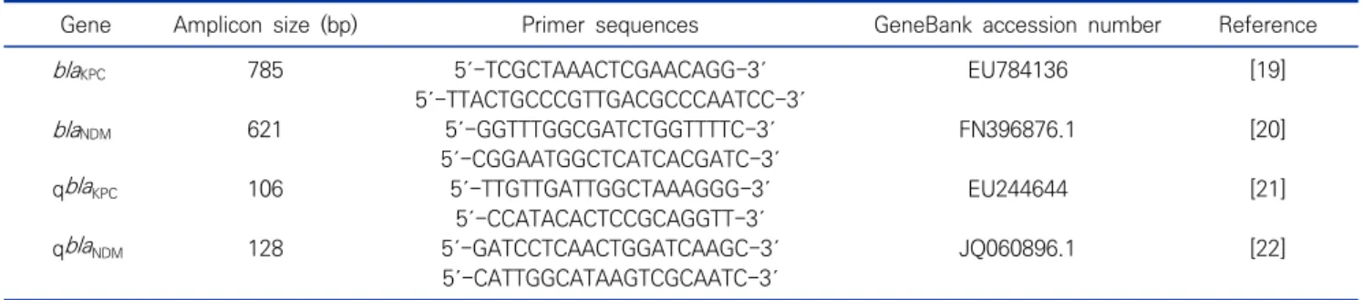
Table 1. Primers for the detection of carbapenemase-producing bacteria
Gene Amplicon size (bp) Primer sequences GeneBank accession number Reference
blaKPC 785 5´-TCGCTAAACTCGAACAGG-3´ EU784136 [19]
5´-TTACTGCCCGTTGACGCCCAATCC-3´
blaNDM 621 5´-GGTTTGGCGATCTGGTTTTC-3´ FN396876.1 [20]
5´-CGGAATGGCTCATCACGATC-3´
qblaKPC 106 5´-TTGTTGATTGGCTAAAGGG-3´ EU244644 [21]
5´-CCATACACTCCGCAGGTT-3´
qblaNDM 128 5´-GATCCTCAACTGGATCAAGC-3´ JQ060896.1 [22]
5´-CATTGGCATAAGTCGCAATC-3´ positive, ≥5 mm은 positive로 판독하였다. 그리고 CRE의
선별검사로 이용되는 MHT와 CIT의 결과를 바탕으로 통합 해 석을 하였다[18].
4. PCR을 통한 blaKPC 및 blaNDM 유전자 검출
Carbapenemase 선별시험인 디스크 확산법에서 KPC 및 NDM 생성균주로 의심되는 균주에서 DNA을 추출하여 PCR을 실시하였다. 먼저 의심 균주는 Wizard genomic DNA purification kit (Promega, Wisconsin, USA)의 Gram negative (GN)방법을 이용하여 DNA 추출을 수행하였다. 그 후 carbapenem 분해효소 중 2종(KPC, NDM) 에 대한 유전자 검사를 시행하였다(Table 1). AccuPower PCR PreMix (Bioneer, Daejeon, Korea) 안에 각각 5 pmol의 primer 1 μL, DNA 2 μL, 증류수를 혼합하여 총 부피 20 μL의 반응용액을 만 들었다. Dual block PCR C-1000 Thermal Cycler (Bio-Rad Laboratories, Inc., California, USA)를 이용하여 95°C에서 5분간 반응 후, 95°C에서 45초, 60°C에서 45초, 72°C에서 1분씩 35회 증폭 반응을 시키고, 72°C에서 5분간 연 장반응을 시켰다. 증폭된 PCR 반응 산물은 ethidium bromide (EtBr)를 포함한 2% agarose gel에서 100 volt 전압 으로 40분간 전기영동하여 밴드를 확인하였다. 확인된 PCR 산 물은 QIA quick gel extraction kit (Qiagen, Hilden, Germany)를 이용하여 정제하였고, 그 후 BigDye Terminator V3.1 sequencing kit (Applied Biosystems, Massachusetts, USA)와 ABI3730XL (Applied Biosystems, Massachusetts, USA)을 이용하여 염기서열 분석을 실시하였다. 결정된 염기서 열의 비교분석은 NCBI에서 제공하는 blast 프로그램을 이용하 였다.
5. Real time PCR의 융해 곡선 분석을 이용한 blaKPC 및 blaNDM 유전자 진단
Real-time PCR의 융해 곡선(melting curve) 분석을 통한
유전자형 진단을 위하여 iQ SYBR Green Supermix (Bio- Rad Laboratories, Inc., California, USA)를 사용하여 실시 간 중합효소 연쇄반응을 실시하였다. 반응용액은 10 µL의 iQ SYBR Green Supermix, 1 μL의 primer 혼합액, 1 µL DNA 및 8 μL의 증류수를 넣어 반응액 20 µL를 제조하였다. 실시간 중합효소 연쇄반응은 CFX96 Touch Real-Time PCR Detection System (Bio-Rad Laboratories, Inc., California, USA)를 이용하였다. 95°C에서 5분 반응 후 95°C에서 10초, 60°C에서 30초, 72°C에서 10초의 반응을 40회 반복하였다. 마지막 PCR 반응이 끝난 후 65°C에서 95°C까지 초당 0.5°C의 속도로 온도 를 증가시키면서 융해 곡선을 모니터링 하였다. 실시간 중합효소 연쇄반응(Real-time PCR)을 이용한 균주 의 분석은 모두 2 반복 실험으로 진행하였다. 그리고 표준곡선 을 이용하여 최소검출농도(limit of detection, LOD)를 확인 하였고 SYBR green에서의 비특이적 증폭 산물의 생성 여부를 확인을 위하여 no-template control (NTC)을 함께 실시하였 다. NTC는 주형을 제외한 나머지 반응액으로 분석에 이용한 반 응조건과 동일하게 진행하였다. 결 과 1. 검체별 특성 본 연구의 분석에 사용한 검체는 경남 지역 종합병원에서 수 집된 35균주를 대상으로 하였고, 균주의 식별은 VITEK 2 automated instrument ID system (BioMérieux, Marcyl’Etoile, France)에 의하여 확인하였다. 분석결과 Klebsiella pneumoniae 12균주, Escherichia coli 7균주, Acinetobacter baumannii 6균 주, Pseudomonas aeruginosa 6균주, Enterobacter cloacae 2균주, Providencia rettgerii 1균주, Pseudomonas putida 1균주를 확인하였다. 분리된 검체의 빈도는 sputum 15개 (42.8%), urine 9개(25.7%), blood 6개(17.1%), pus 2개 (5.7%), ascitic fliud 1개(2.9%), tracheal aspirates 1개
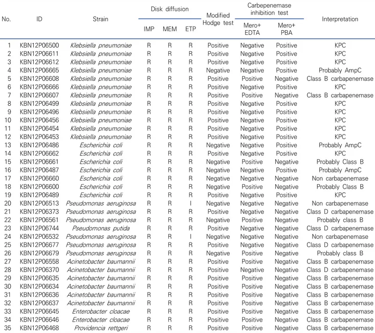
Table 2. Carbapenem resistance gram negative rod bacteria analyzed in screening test No. ID Strain Disk diffusion Modified Hodge test Carbepenemase inhibition test Interpretation
IMP MEM ETP Mero+ EDTA Mero+PBA
1 KBN12P06500 Klebsiella pneumoniae R R R Positive Negative Positive KPC
2 KBN12P06611 Klebsiella pneumoniae R R R Positive Negative Positive KPC
3 KBN12P06612 Klebsiella pneumoniae R R R Positive Negative Positive KPC
4 KBN12P06665 Klebsiella pneumoniae R R R Negative Negative Positive Probably AmpC 5 KBN12P06608 Klebsiella pneumoniae R R R Positive Positive Negative Class B carbapenemase
6 KBN12P06666 Klebsiella pneumoniae R R R Positive Negative Positive KPC
7 KBN12P06607 Klebsiella pneumoniae R R R Positive Positive Negative Class B carbapenemase
8 KBN12P06499 Klebsiella pneumoniae R R R Positive Negative Positive KPC
9 KBN12P06496 Klebsiella pneumoniae R R R Positive Negative Positive KPC
10 KBN12P06456 Klebsiella pneumoniae R R R Positive Negative Positive KPC
11 KBN12P06454 Klebsiella pneumoniae R R R Positive Negative Positive KPC
12 KBN12P06453 Klebsiella pneumoniae R R R Positive Negative Positive KPC
13 KBN12P06486 Escherichia coli R R R Negative Negative Positive Probably AmpC
14 KBN12P06662 Escherichia coli R R R Positive Negative Positive KPC
15 KBN12P06661 Escherichia coli R R R Negative Positive Negative Probably Class B
16 KBN12P06487 Escherichia coli R R R Negative Negative Positive Probably AmpC
17 KBN12P06660 Escherichia coli R R R Negative Negative Negative Non carbapenemase
18 KBN12P06600 Escherichia coli R R R Negative Positive Negative Probably Class B
19 KBN12P06489 Escherichia coli R R R Positive Negative Positive KPC
20 KBN12P06513 Pseudomonas aeruginosa R R I Negative Negative Negative Non carbapenemase 21 KBN12P06373 Pseudomonas aeruginosa R R R Positive Negative Negative Class D carbapenemase 22 KBN12P06561 Pseudomonas aeruginosa R R R Negative Positive Negative Probably class B 23 KBN12P06744 Pseudomonas putida R R R Positive Negative Negative Class D carbapenemase 24 KBN12P06532 Pseudomonas aeruginosa R R I Negative Negative Negative Non carbapenemase 25 KBN12P06677 Pseudomonas aeruginosa R R R Positive Negative Negative Class D carbapenemase 26 KBN12P06679 Pseudomonas aeruginosa R R R Negative Positive Negative Probably class B 27 KBN12P06558 Acinetobacter baumannii R R R Positive Positive Negative Class B carbapenemase 28 KBN12P06370 Acinetobacter baumannii R R R Positive Negative Negative Class D carbapenemase 29 KBN12P06635 Acinetobacter baumannii R R R Positive Positive Negative Class B carbapenemase 30 KBN12P06634 Acinetobacter baumannii R R R Positive Positive Negative Class B carbapenemase 31 KBN12P06636 Acinetobacter baumannii R R R Positive Positive Negative Class B carbapenemase 32 KBN12P06637 Acinetobacter baumannii R R R Positive Positive Negative Class B carbapenemase 33 KBN12P06645 Enterobacter cloacae R R R Positive Positive Negative Class B carbapenemase 34 KBN12P06646 Enterobacter cloacae R R R Positive Positive Negative Class B carbapenemase 35 KBN12P06468 Providencia rettgeri R R R Positive Positive Negative Class B carbapenemase Abbreviations: R, resistant; I, intermedius; IMP, imipenem; MERO, meropenem; ETP, ertapenem; KPC; Klebsiella pneumoniae carbapenemases.
Figure 1. Frequency of detection in specimens.
(2.9%), other 1개(2.9%) 순으로 나타났다(Figure 1).
2. 표현형적 carbapenemase 선별 시험법
임상검체 중 식별된 모든 균주에 대해서는 디스크 확산법을 시행하였다. Imipenem, meropenem 및 ertapenem 약제를 이용하였으며, 항생제 감수성 시험은 CLSI 가이드라인의 항생 제에 대한 내성판정 기준으로 하였고[11, 12] 결과는 Table 2와 같다. 그 후 선별시험인 MHT를 실시하여 결과를 판독하였다 (Figure 2-1). Ertapenem을 이용한 MHT 결과 35균주 중 25 균주에서 양성 결과를 확인하였다. 분석결과 K. pneumoniae
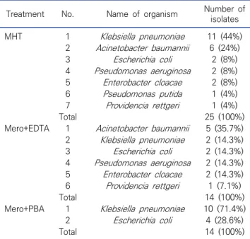
Table 3. Gram negative rod bacteria showing positive results in modified Hodge test and carbepenemase inhibition test (MHT; N=25, Mero+EDTA; N=14, Mero+PBA; N=14)
Treatment No. Name of organism Number of isolates MHT 1 Klebsiella pneumoniae 11 (44%) 2 Acinetobacter baumannii 6 (24%) 3 Escherichia coli 2 (8%) 4 Pseudomonas aeruginosa 2 (8%) 5 Enterobacter cloacae 2 (8%) 6 Pseudomonas putida 1 (4%) 7 Providencia rettgeri 1 (4%) Total 25 (100%)
Mero+EDTA 1 Acinetobacter baumannii 5 (35.7%) 2 Klebsiella pneumoniae 2 (14.3%) 3 Escherichia coli 2 (14.3%) 4 Pseudomonas aeruginosa 2 (14.3%) 5 Enterobacter cloacae 2 (14.3%) 6 Providencia rettgeri 1 (7.1%) Total 14 (100%)
Mero+PBA 1 Klebsiella pneumoniae 10 (71.4%) 2 Escherichia coli 4 (28.6%)
Total 14 (100%)
Abbreviations: MHT, modified Hodge test; Mero, Meropenem; EDTA, ethylenediaminetetraacetic acid; PBA, phenylboric acid.
Figure 3. PCR amplification profile of the blaKPC and blaNDM gene from
the gram negative rod bacteria isolates. 1) blaKPC PCR product size
of 785 bp and 2) blaNDM PCR product size of 621 bp. M, 100 bp DNA
ladder marker.
Figure 2. Result of modified Hodge test (MHT) and carbepenemase inhibition test (CIT). 1) The MHT performed on a MacConkey plate.
Pseudomonas aeruginosa and Acinetobacter baumanni, positive result; Pseudomonas aeruginosa negative result. 2) CIT reading result. (A) negative, (B) Mero+EDTA positive, (C) Mero+PBA positive. 11균주, A. baumannii 6균주, E. coli 2균주, P. aeruginosa 2균주, E. cloacae 2균주, P. putida 1균주, P. rettgeri 1균주 로 나타났다(Table 3).
유전자형 검출을 위하여 carbapenemase inhibition test
(CIT) 검사를 실시하였다. 3개의 meropenem 디스크 중 한 개 에는 class A carbapenemase (KPC)의 검출을 위해 PBA 시 약을, 다른 한 개에는 class B carbapenemase (MBL)의 검출 을 위해 EDTA 시약을 각각 10 μL씩 떨어트렸다. 37°C 배양기 에서 16∼20시간 배양한 후 각 디스크의 억제대를 측정 후 판독 하였다(Figure 2-2). 분석결과 meropenem+PBA에서 35균주 중 14균주에서 양 성 결과를 확인하였고, K. pneumoniae 10균주, E. coli 4균주 로 나타났다. Meropenem+EDTA의 분석결과 35균주 중 14균 주에서 양성을 확인하였고, 균주는 A. baumannii 5균주, K. pneumoniae 2균주, E. coli 2균주, P. aeruginosa 2균주, E. cloacae 2균주, P. rettgeri 1균주로 나타났다(Table 3).
그리고 MHT와 CIT의 결과를 바탕으로 통합 해석을 하였고 [18], 분석결과 KPC 11균주, class B 및 추정 class B cabapenemase 10균주, class D cabapenemase 4균주, AmpC 추정 3균주, non cabapenemase 3균주를 확인하였다 (Table 2). 3. 분자학적 방법을 이용한 blaKPC 및 blaNDM 검출 분석에 이용된 모든 균주에 대해 carbapenem 분해효소 중 2종(blaKPC, blaNDM)에 대한 유전자검사를 시행하였다. 분석결 과 35균주 중 blaKPC 유전자는 25개의 양성 균주를 blaNDM 유전 자는 8개의 양성 균주를 확인하였다(Figure 3). PCR 검사 결과 음성으로 확인된 blaKPC 유전자는 35균주 중 10개의 균주에서 음성을 확인하였고, K. pneumoniae 2균주, E. coli 2균주, P. aeruginosa 2균주, E. cloacae 2균주, A. baumannii 1균주, P. rettgeri 1균주로 나타났다. blaNDM 유전자는 35균주 중 27 개의 음성균주가 확인되었으며, K. pneumoniae 10균주, E.
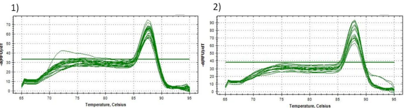
Figure 4. Melting curve analysis of blaKPC and blaNDM. 1) melting curve analysis of blaKPC amplicon was generated that showed the fragment
Tm 87.5°C and 2) melting curve analysis of blaNDM amplicon was generated that showed the fragment Tm 88°C.
coli 4균주, A. baumannii 6균주, P. aeruginosa 5균주, P. putida 1균주, E. cloacae 1균주로 나타났다.
증폭된 blaKPC 25균주의 분석결과, K. pneumoniae 10균 주, E. coli 5균주, A. baumannii 5균주, P. aeruginosa 4균 주, P. putida 1균주로 나타났다. 증폭된 blaNDM 8균주의 분석 결과, K. pneumoniae 2균주, E. coli 3균주, P. aeruginosa 1균주, E. cloacae 1균주, P. rettgeri 1균주로 나타났다. 그 후 염기서열 분석을 통해 유전자형을 확인하였으며, NCBI에서 제 공하는 blast 프로그램을 이용하여 염기서열의 비교분석을 하 였다. blaKPC 유전자의 경우 Genebank No. CP054782.1에 서 상동성(homology)은 평균 99%이며, 원래 서열보다 평균 3 개 염기의 누락(Gap 1%)을 확인하였다. blaNDM 유전자의 경우 Genebank No. LC536682.1에서 상동성(homology)은 평균 99%이며, 원래 서열보다 평균 1.5개 염기의 누락(Gap 1%)을 확인하였다. 4. 융해 곡선 분석을 이용한 blaKPC 및 blaNDM 검출 융해 곡선의 분석에 앞서 NTC 분석결과 검체 이외에는 증폭 된 산물이 없음을 확인하였고, 이후 분석에 이용한 균주는 모두 2 반복 실험으로 진행하였다. 표본이 30 사이클의 교차점 (crossing point, Cp) 이전의 임계값을 초과할 경우 양성으로, Cp가 30보다 클 경우 음성으로 간주하였다. 실시간 중합효소 연쇄반응(real-time PCR)을 이용한 분석결과 blaKPC 유전자 는 25개, blaNDM 유전자는 8개의 균주에서 증폭곡선을 확인하 였다. blaKPC 25균주에는 K. pneumoniae 10균주, E. coli 5 균주, A. baumannii 5균주, P. aeruginosa 4균주, P. putida 1균주로 나타났다. blaNDM 8균주는 K. pneumoniae 2균주, E. coli 3균주, P. aeruginosa 1균주, E. cloacae 1균주, P. rettgeri 1균주로 나타났으며, 이는 PCR 결과와 100% 일치하 였다. 증폭된 유전자의 평균 cycle threshold (Ct) 값은 blaKPC
의 경우 17.01±1.03, blaNDM은 23.04±1.73으로 나타났으 며, 음성으로 나타난 균주에서는 증폭 산물이 없었다. 그리고 증 폭 산물의 융해 곡선 분석 결과 blaKPC는 melting temperature (Tm) 87.5°C, blaNDM은 Tm 88°C를 확인하였다(Figure 4). 양 성으로 확인된 검체를 이용하여 표준곡선분석을 실시였고 100 희석한 검체에서 최소검출농도(limit of detection, LOD)를 확인한 결과 blaKPC 유전자는 22 ng/mL, blaNDM 유전자는 27.96 ng/mL으로 확인되었다. 고 찰 2010년 국내에서 최초로 CRE 보고 이후, 국내의 표본감시에 따르면 CPE의 발생은 해마다 증가하고 있다[5, 23]. 2016년 기 준으로 우리나라에서 발생하는 carbapenem 분해효소의 종류 로는 KPC (70.7%), NDM (13.5%), oxacillinase-48 (OXA-48, 9.6%), Guiana extended spectrum β-lactamase (GES, 3.1%), Verona integron-encoded metallo-β-lactamase (VIM, 2.0%), imipenemase (IMP, 1.1%) 순으로 보고되었다 [24]. 이러한 CRE는 지역사회 수준과 의료 시설 모두에서 중요 한 관심사이며, 세계적으로 CRE 감염의 증가는 높은 이환률과 사망률을 가진 고 독성 세균과의 감염의 연관성으로 인해 매우 우려되는 상황이다. 이에 본 연구는 경남지역 종합병원에서 분리된 35균주를 이 용하여 국내에서 가장 빈번히 검출되는 CPE의 유전자형 중 blaKPC 및 blaNDM 유전자의 real-time PCR의 융해곡선을 이용 한 분석 방법으로 보다 신속한 유전자 진단법에 대해 알아보고 자 하였다.
먼저 분리된 35균주를 동정한 후 식별된 모든 균주에 대해서 는 carbapenem 분해효소 선별시험인 디스크 확산법을 시행 하였고, 항생제 감수성 시험은 imipenem, meropenem 및
ertapenem를 이용하여 CLSI 가이드라인에 따라 시행 및 판독 하였다. 그 후 선별시험인 MHT를 실시하여 결과를 판독하였고 35균주 중 25균주에서 양성을 확인하였다.
기존의 연구[21]에서 MHT의 민감도와 특이성은 90%를 초 과하는 것으로 나타났지만, 일부 연구에서는 특히 세포벽에 porin 결핍을 가진 ESBL 또는 AmpC β-lactamse를 생성하는 CRE에서 높은 수의 위양성 결과가 관찰되었다[25-27]. 또한 A. baumanii, P. aeruginasa와 같은 일부 균주에 대해서는 해석 이 어려울 수 있기 때문에 KPC에 대한 이상적인 형태적 확인 시 험이 아닐 수 있다[21]. 이처럼 MHT는 CLSI가 carbapenemase 생산 균주의 표현형 검출을 위해 권장하는 테스트이지만 MHT 의 해석에 문제가 있고 일부 분리된 균주에서 비교적 높은 위양 성 및 위음성 결과 비율이 있으므로 carbapenemase 생산의 최종 확인에는 부적합하다. 또다른 표현형을 이용한 분석 방법 으로 여러 억제제 기반 테스트가 있으며[2, 28], 본 연구에서는 유전자형 검출을 위하여 억제제 기반 테스트인 CIT 검사를 실시 하였다. CIT에서 KPC 생성 균주의 표현형 시험은 KPC가 boronate 유도체에 의해 억제되는 것을 바탕으로 검사하며, 유도체 중에 서 PBA가 더 유용한 것으로 알려져 있다. MBL 생성 균주의 표 현형 시험은 MBL이 chelating agent에 의해 억제되는 것을 바 탕으로 검사하며 EDTA가 전통적으로 사용되고 있다. EDTA와 같은 화합물은 Zn 이온을 제거함으로써 MBL이 항균제를 가수 분해할 수 없게 불활성화시킨다. 표현형적 검사인 MHT와 CIT 검사결과를 바탕으로 한 통합 해석을 실시하였고[18], 분석결과 KPC 생성균주는 11균주, NDM 생성 추정균주는 14균주를 확 인하였다. 그 후 정확한 유전자 진단을 위해 분석에 이용된 모든 균주에 대해 blaKPC, blaNDM에 대한 유전자 검사를 시행하였으며, 분석 결과 blaKPC 유전자는 25균주, blaNDM 유전자는 8균주에서 양 성을 확인하였다(Figure 3). 양성으로 나온 균주는 염기서열 분 석을 통해 유전자형을 확인하였으며, 그 결과 blaKPC 유전자는 평균 99%, blaNDM 유전자는 평균 99%의 상동성(homology) 을 확인하였다. 그리고 유전자형을 확인할 수 있다고 알려진 CIT 검사와 MHT 결과를 바탕으로 한 통합 해석 결과와 PCR 결과를 비교 분석하였다. 분석결과 통합해석에서 KPC 생성균주는 11균주, PCR 검사는 25균주가 양성으로, 표현형적 검사법에서 14균주 (44%)의 위음성을 확인하였다. 그리고 통합해석 결과에서 NDM 생성 추정 균주는 14균주, PCR 검사는 8균주가 양성으 로, 표현형적 검사법에서 6균주(43%)의 위양성을 확인하였다. 일반적으로 boronate 기반 검사는 KPC 생산 균주의 검출에 높은 민감도를 보이나, 높은 수준의 AmpC형 β-lactamse 발현 및 porin 결핍때문에 carbapenem에 대한 감수성이 감소된 분리균주에서 특이성 문제가 발생할 수 있다[29]. 그리고 β- lactam-chelator 조합에 기초한 MBL 검출 방법은 K. pneumoniae와 E. coli에 대해 우수한 성능을 발휘하지만, 다 른 장내세균 종에 대해서는 체계적으로 검사되지 않았다. 또한, MBL 검출 방법의 특이성은 킬레이트 작용제의 영향으로 손상 되어 비특이적으로 작용하고 다른 프로세스에 영향을 미칠 수 있다는 점을 반드시 감안하여야 한다[30-32]. 그리고 억제제를 이용하여 유전자형을 알아볼 수 있는 CIT 검사에서 MBL 억제 제로 흔히 사용되는 EDTA는 세균 외부 세포막의 투과성을 증가 시켜, 특히 Acinetobacter에 대해 비특정 위양성 결과를 발생 시킬 수 있다[33, 34]. CIT 검사와 PCR 검사의 비교 분석한 결 과, A. baumannii 1균주에서 CIT 검사는 양성이나 PCR 검사 결과에서는 음성으로 위양성 결과를 확인하였고, 이는 EDTA는 세균 외부 세포막의 투과성을 증가시켜, 특히 Acinetobacter 에 대해 비특정 위양성 결과를 발생한 것으로 보인다. 이처럼 MHT 및 CIT 검사의 경우 일종의 표현형 검사방법으 로 많은 검사시간의 소요뿐만 아니라 여러 판독적 오류 및 효소 종류에 따른 낮은 검출 효율로 인해 정확한 항생제 내성 유전자 형을 확인하기 위해서는 분자학적 방법이 필수적이라 할 수 있 다. 그러나 기존의 PCR을 이용한 분자 방법은 사후 PCR 분석에 대한 필요성 때문에 종종 시간과 비용이 많이 소요되므로, carbapenemase 등과 같은 저항성 유전자를 대상으로 한 real-time PCR 방식이 최근 빈번하게 적용됐다[35]. 본 연구에 서는 저렴하고 사용이 간편한 등 많은 장점을 가져 광범위하게 활용되는 SYBR Green을 이용한 융해 곡선 분석을 실시하였으 며, 비특이적 반응 등의 SYBR Green의 단점을 보완하고자 검 증된 primer를 활용하여 분석을 실시하였다. 분석에 이용된 qblaKPC primer는 이전의 연구[21]에서 항균 제 감수성 검사를 통해 β-lactam 항생제의 여러 등급에 내성을 갖는 159 그람음성균주로부터 SYBR Green I를 기반으로 한 real-time PCR 분석에서 blaKPC 유전자의 106 base pair (bp) 산물을 증폭시키도록 설계되었다. SYBR Green I를 이용 한 증폭산물의 융해곡선 분석을 통해 blaKPC 유전자를 확인하였 으며, Tm 값은 약 89°C에서 검출되었다. 그리고 연구결과 표현 형적 검사인 MHT 및 염기서열 분석과 비교하여 실시간 PCR 분 석의 특이성과 민감도는 100%로 평가되었으며, 실시간 PCR 검사는 높은 분석 민감도와 특수성을 가진 KPC-harbing 균주 를 빠르고 정확하게 검출할 수 있었다[21]. 또한 융해곡선 분석
에 이용된 qblaNDM primer는 알려진 모든 blaNDM (blaNDM- 1∼7) 대립유전자와 동일함을 확인하였다[22]. 본 연구에서는 SYBR green을 이용한 융해곡선 분석으로 blaKPC와 blaNDM 유전자의 검출 유무를 확인하였으며, 분석 결 과 기존의 연구와 마찬가지로 real-time PCR의 결과가 100% 일치한다는 것을 확인하였다. CRE, 3GC-R 또는 MDRP로 인한 감염 환자는 제한적인 치 료 옵션을 가지고 있으며, 배양 및 감염성 보고 전에 이러한 감염 을 인식하지 못하는 경우가 많기 때문에 적절한 치료를 받기에 는 매우 취약한 실정이다[36]. 현재의 CLSI 가이드라인에 따른 표현형적 검사법은 carbapenemase 생성 의심균주의 분리 후 첫 결과까지 보통 24∼48시간이 소요되고, carbapenemase 유형을 구분할 수 없어 검사가 시간이 많이 걸린다는 단점을 가 진다. 그러나 검사 표본에서 분석 특이성과 민감도가 높은 균주를 검출하기 위해 개발된 실시간 PCR 검사는 turn around time 단축의 이점을 제공하였으며[37], 임상에서 분리된 저항성 유전 자를 가지는 세균의 검출을 위해 융해 곡선 분석을 이용한 real-time PCR 검사는 다른 진단검사 방법보다 신속하고 정확 하게 저항성 유전자를 가지는 균주를 검출하는 것을 확인하였 다. 그 중 SYBR green법은 유전자별로 별도의 probe를 준비 할 필요가 없어 Taqman probe법보다 저렴한 비용으로 반응을 구축하는 장점을 가지며, 일반적인 PCR 방법보다는 시간과 오 염에 대한 문제점이 감소된다. Carbapenem 항생제의 광범위한 사용과 함께, CPE는 공중 보건에 대한 주요 관심사로 나타났다. CPE는 병원 환경에서 높 은 사망률을 가진 병원내 감염을 일으키는 원인으로 빠르게 퍼질 수 있는 잠재력을 가지고 있으며, 다른 균주로의 carbapenemase 유전자의 수평적 전달은 이용 가능한 모든 항생제에 내성을 갖 는 다제 내성을 초래하므로 정확하고 신속한 진단이 요구된다.
결론적으로 CPE의 조기진단에 있어 real time PCR을 이용 한 검사법은 기존의 표현형적 검사법이나 일반적인 PCR 검사 법과 비교하여 보다 신속하고 특이성이 높게 검출이 가능하여, 신속한 진단으로 항생제 내성의 경향은 부상하는 병원균에 대한 중요한 통찰력을 제공할 뿐만 아니라 공중보건 및 감염 중에 항 균 관리접근법을 통한 병원 내 감염확산 방지 및 통제가 가능할 것으로 사료된다. 요 약 본 연구는 국내에서 가장 빈번히 검출되는 CPE의 유전자형 중 blaKPC 및 blaNDM 유전자의 진단으로 기존의 표현형 검사 및 일반 PCR 검사보다 시간 단축 및 사후 분석의 단점이 보완된 real-time PCR의 융해 곡선을 이용한 분석법에 대해 알아보았 다. 표현형적 검사결과 MHT는 35균주 중 25균주에서 양성을 확인하고, CIT는 meropenem+PBA 및 meropenem+EDTA 에서 각각 14균주의 양성을 확인하였다. PCR 검사결과 KPC 25균주에서 증폭 산물을 확인하였고, K. pneumoniae 10균 주, E. coli 5균주, A. baumannii 5균주, P. aeruginosa 4균 주, P. putida 1균주로 나타났다. NDM은 8균주에서 증폭 산물 을 확인하였고, K. pneumoniae 2균주, E. coli 3균주, P. aeruginosa 1균주, E. cloacae 1균주, P. rettgeri 1균주로 나 타났다. 실시간 중합효소연쇄반응(Real-time PCR)을 이용한 융 해 곡선 분석결과 KPC 25균주, NDM은 8균주에서 증폭을 확 인하였고, PCR 결과와 100% 일치함을 확인하였다. 결론적으 로 real-time PCR을 이용한 신속하고 특이성이 높은 CRE의 조기진단은 공중보건 및 감염 중에 항균 관리접근법을 통한 병원 내 감염확산 방지 및 통제가 가능할 것으로 사료된다.
Acknowledgements: This paper was supported by the Jinju Health College in 2020. The pathogen resources for this study were provided by Gyeongsang National University Hospital Branch of the National Culture Collection for Pathogens (GNUH-NCCP).
Conflict of interest: None
Author’s information (Position): Yang BS, Professor; Park JA, Adjunct professor.
REFERENCES
1. Weiner LM, Webb AK, Limbago B, Dudeck MA, Patel J, Kallen AJ, et al. Antimicrobial-resistant pathogens associated with health-care-associated infections: summary of data reported to the na-tional healthcare safety network at the centers for disease control and prevention, 2011-2014. Infect Control Hosp Epidemiol. 2016;37:1288-1301. https://doi.org/10.1017/ice.2016.174 2. Bedenić B, Plečko V, Sardelić S, Uzunović S, Godič Torkar K.
Carbapenemases in gram-negative bacteria: laboratory de-tection and clinical significance. BioMed Research International. 2014;841951. https://doi.org/10.1155/2014/841951
3. Papp-Wallace KM, Endimiani A, Taracila MA, Bonomo RA. Carbapenems: past, present, and future. Antimicrobial Agents and Chemotherapy. 2011;55:4943-4960. https://doi.org//AAC. 00296-11
4. Diene SM, Rolain JM. Carbapenemase genes and genetic plat-forms in gram-negative bacilli: Enterobacteriaceae, Pseudomonas, and Acinetobacter species. Clinical Microbiology and Infection.
2014;20:831-838. https://doi.org/10.1111/1469-0691.12655 5. Rhee JY, Park YK, Shin JY, Choi JY, Lee MY, Peck KR, et al. KPC
producing extreme drug-resistant Klebsiella pneumoniae isolate from a patient with diabetes mellitus and chronic renal failure on hemodialysis in South Korea. Antimicrob Agents Chemother. 2010;54:2278-2279. https://doi.org/10.1128/AAC.00011-10 6. Kim JO, Song SA, Yoon EJ, Shin JH, Lee H, Jeong SH, et al.
Outbreak of KPC-2-producing Enterobacteriaceae caused by clonal dissemination of Klebsiella pneumoniae ST307 carrying an IncX3-type plasmid harboring a truncated Tn4401a. Diagn. ZMicrobiol. Infect. Dis. 2017;87:343-348. https://doi.org/j.diagmicrobio. 2016.12.012
7. Jeong SH, Kim HS, Kim JS, Shin DH, Kim HS, Park MJ, et al. Prevalence and molecular characteristics of carbapenemase pro-ducing Enterobacteriaceae from five hospitals in Korea. Ann. Lab. Med. 2016;36:529-535. https://doi.org/10.3343/alm.2016. 36.6.529
8. Park SH, Kim JS, Kim HS, Yu YK, Han SH, Kang MJ, et. al. Prevalence of carbapenem-resistant Enterobacteriaceae in Seoul. J Bacteriol Virol. 2020;50:107-116. https://doi.org/10.4167/jbv. 2020.50.2.107
9. Tacconelli E, Carrara E, Savoldi A, Harbarth S, Mendelson M, Monnet DL, et al. Discovery, research, and development of new antibiotics: the WHO priority list of antibiotic-resistant bacteria and tuberculosis. Lancet Infect Dis. 2018;18:318-327. https:// doi.org/10.1016/S1473-3099(17)30753-3
10. Lodise TP, Zhao Q, Fahrbach K, Gillard PJ, Martin A. A systematic review of the association between delayed appropriate therapy and mortality among patients hospitalized with infections due to
Klebsiella pneumoniae or Escherichia coli: how long is too long? BMC Infect Dis. 2018;18:625. https://doi.org/10.1186/s12879- 018-3524-8
11. Clinical Laboratory Standards Institute. Performance standards for antimicrobial susceptibility testing; twenty-first informational supplement M100-S27. Wayne PA: Clinical Laboratory Standards Institute; 2017.
12. Bora A, Sanjana R, Jha BK, Mahaseth SN, Pokharel K. Incidence of metallo-beta-lactamase producing clinical isolates of
Escherichia coli and Klebsiella pneumoniae in central Nepal. BMC Res Notes 2014;7:557. https://doi.org/10.1186/1756-0500- 7-557
13. Cha CH, Hae Kyong An HK, Kim JU. Detection of vancomycin-re-sistant Enterococci using multiplex real-time PCR assay and melting curve analysis. Korean J Lab Med. 2010;30:138-146. https://doi.org/10.3343/kjlm.2010.30.2.138
14. Ririe KM, Rasmussen RP, Wittwer CT. Product differentiation by analysis of DNA melting curves during the polymerase chain reaction. Anal Biochem. 1997;245:154-60. https://doi.org/ 10.1006/abio.1996.9916
15. Zheng F, Sun J, Cheng C, Rui Y. The establishment of a duplex re-al-time PCR assay for rapid and simultaneous detection of blaNDM
and blaKPC genes in bacteria. Ann Clin Microbiol Antimicrob.
2013;12:30. https://doi.org/10.1186/1476-0711-12-30
16. Lodise TP, Bonine NG, Ye JM, Folse HJ, Gillard P. Development of a bedside tool to predict the probability of drug-resistant patho-gens among hospitalized adult patients with gram-negative infections. BMC Infectious Diseases. 2019;19:718.
https://do-i.org/10.1186/s12879-019-4363-y
17. Bonine NG, Berger A, Altincatal A, Wang R, Bhagnani T, Gillard P, et al. Impact of delayed appropriate antibiotic therapy on patient outcomes by antibiotic resistance status from serious gram-neg-ative bacterial infections. Am J Med Sci. 2019;357:103-110. https://doi.org/10.1016/j.amjms.2018.11.009
18. The Korean Society of Clinical Microbiology. Diagnostic in-struction carbapenemase producing Enterobacteriaceae (CPE) [Internet]. Seoul: The Korean Society of Clinical Microbiology; 2015 [cited 2021 February 9]. Available from: http://kscm. or.kr/xe/kscmnotice/71241.
19. Monteiro J, Widen RH, Pignatari ACC, Kubasek C, Silbert S. Rapid detection of carbapenemase genes by multiplex real-time PCR. J Antimicrob Chemother. 2012;67:906-909. https://doi.org/10.1093/ jac/dkr563
20. Poirel L, Revathi G, Bernabeu S, Nordmann P. Detection of NDM-1-Producing Klebsiella pneumoniae in Kenya. Antimicrob Agents Chemother. 2011;55:934-936. https://doi.org/10.1128/ AAC.01247-10
21. Wang L, Gu H, Lu X. A rapid low-cost real-time PCR for the de-tection of Klebsiella pneumonia carbapenemase genes. Ann Clin Microbiol Antimicrob. 2012;11:9. https://doi.org/10.1186/1476- 0711-11-9
22. Kosykowska E, Dzieciątkowski T, Młynarczyk G. Rapid detection of NDM, VIM, KPC and IMP carbapenemases by real-time PCR. J Bacteriol Parasitol. 2016;7:6. https://doi.org/10.4172/2155-9597. 1000299
23. Ahn SY, Sung JY, Kim HS, Kim MS, Hwang YJ, Jong SR, et al. Molecular epidemiology and characterization of carbapene-mase-producing Enterobacteriaceae isolated at a university hos-pital in Korea during 4-year period. Ann Clin Microbiol. 2016; 19:39-47. https://doi.org/10.5145/ACM.2016.19.2.39
24. Park JW, Lee EJ, et al. Status of carbapenemase producing
Enterobacteriaceae incidences in Korea, 2015-2016. Research report. Cheongju: Korea Centers for Disease Control and Prevention; 2017;10:1243-1247.
25. Wang P, Chen S, Guo Y, Xiong Z, Hu F, Zhu D, et al. Occurrence of false positive results for the detection of carbapenemases in carbapenemasenegative Escherichia coli and Klebsiella pneu-moniae isolates. PLoS One. 2011;6:e26356. https://doi.org/ 10.1371/journal.pone.0026356
26. Carvalhaes CG, Picao RC, Nicoletti AG, Xavier DE, Gales AC. Cloverleaf test (modified Hodge test) for detecting carbapene-mase production in Klebsiella pneumoniae: be aware of false positive results. J Antimicrob Chemother. 2010;65:249-251. https://doi.org/10.1093/jac/dkp431
27. Seah C, Low DE, Patel SN, Melano RG. Comparative evaluation of a chromogenic agar medium, the modified Hodge test, and a bat-tery of meropenem-inhibitor discs for detection of carbapene-mase activity in Enterobacteriaceae. J Clin Microbiol. 2011;49: 1965-1969. https://doi.org/10.1128/JCM.00203-11
28. Nordmann P, Gniadkowski M, Giske CG, Poirel L, Woodford N, Miriagou V. Identification and screening of carbapenemase-pro-ducing Enterobacteriaceae. Clin Microbiol Infect. 2012;18:432- 438. https://doi.org/10.1111/j.1469-0691.2012.03815.x
29. Pasteran F, Mendez T, Guerriero L, Rapoport M, Corso A. Sensitive screening tests for suspected class A carbapenemase
production in species of Enterobacteriaceae. J Clin Microbiol. 2009;47:1631-1639. https://doi.org/10.1128/JCM.00130-09 30. Samuelsen O, Buarø L, Giske CG, Simonsen GS, Aasnaes B,
Sundsfjord A. Evaluation of phenotypic tests for the detection of metallo-beta-lactamase-producing Pseudomonas aeruginosa in a low prevalence country. J Antimicrob Chemother. 2008;61: 827-830. https://doi.org/10.1093/jac/dkn016
31. Chu YW, Cheung TK, Ngan JY, Kam KM. EDTA susceptibility leading to false detection of metallo-beta-lactamase in Pseudomonas aeruginosa by Etest and an imipenem-EDTA disk method. Int J Antimicrob Agents 2005;26:340-341. https://doi.org/10.1016/ j.ijantimicag.2005.07.004
32. Ratkai C, Quinteira S, Grosso F, Monteiro N, Nagy E, Peixe L. Controlling for false positives: interpreting MBL Etest and MBL combined disc test for the detection of metallo-beta-lactamases. J Antimicrob Chemother 2009;64:657-658. https://doi.org/ 10.1093/jac/dkp229
33. Bonnin RA, Naas T, Poirel L, Nordmann P. Phenotypic, bio-chemical, and molecular techniques for detection of metallo-β -lactamase NDM in Acinetobacter baumannii. J Clin Microbiol. 2012;50:1419-1421. https://doi.org/10.1128/JCM.06276-11
34. Hansen F, Hammerum AM, Skov R, Haldorsen B, Sundsfjord A, Samuelsen O. Evaluation of the total MBL confirm kit (ROSCO) for detection of metallo-β-lactamases in Pseudomonas aeruginosa
and Acinetobacter baumannii. Diagn Microbiol Infect Dis. 2014; 79:486-488. https://doi.org/10.1016/j.diagmicrobio.2013.12.001 35. Goudarzi H, Mirsamadi ES, Ghalavand Z, Hakemi Vala M,
Mirjalali H, Hashemi A. Rapid detection and molecular survey of
blaVIM, blaIMP and blaNDM genes among clinical isolates of
Acinetobacter baumannii using new multiplex real-time PCR and melting curve analysis. BMC Microbiology. 2019;19:122. https:// doi.org/10.1186/s12866-019-1510-y
36. Lodise TP, Bonine NG, Ye JM, Folse HJ, Gillard P. Development of a bedside tool to predict the probability of drug-resistant patho-gens among hospitalized adult patients with gram-negative infections. BMC Infectious Diseases. 2019;19:718. https://do-i.org/10.1186/s12879-019-4363-y
37. Mangold KA, Santiano K, Broekman R, Krafft CA, Voss B, Wang V, et al. Real-time detection of blaKPC in clinical samples and sur-veillance specimens. J Clin Microbiol. 2011;49:3338-3339. https://doi.org/10.1128/JCM.00268-11