저작자표시-비영리-변경금지 2.0 대한민국 이용자는 아래의 조건을 따르는 경우에 한하여 자유롭게 l 이 저작물을 복제, 배포, 전송, 전시, 공연 및 방송할 수 있습니다. 다음과 같은 조건을 따라야 합니다: l 귀하는, 이 저작물의 재이용이나 배포의 경우, 이 저작물에 적용된 이용허락조건 을 명확하게 나타내어야 합니다. l 저작권자로부터 별도의 허가를 받으면 이러한 조건들은 적용되지 않습니다. 저작권법에 따른 이용자의 권리는 위의 내용에 의하여 영향을 받지 않습니다. 이것은 이용허락규약(Legal Code)을 이해하기 쉽게 요약한 것입니다. Disclaimer 저작자표시. 귀하는 원저작자를 표시하여야 합니다. 비영리. 귀하는 이 저작물을 영리 목적으로 이용할 수 없습니다. 변경금지. 귀하는 이 저작물을 개작, 변형 또는 가공할 수 없습니다.
A DISSERTATION
FOR THE DEGREE OF DOCTOR OF PHILOSOPHY
Mixture of probiotics alleviates the
symptoms of atopic dermatitis
through recovering immune
balance in mice
복합 유산균의 면역 회복에 의한 아토피성 피부염
마우스의 증상 완화
August 2019
By
Han Wool Kim
Department of Agricultural Biotechnology
Graduate School
농 학 박 사 학 위 논 문
Mixture of probiotics alleviates the
symptoms of atopic dermatitis
through recovering immune
balance in mice
복합 유산균의 면역 회복에 의한 아토피성 피부염
마우스의 증상 완화
지도교수 윤철희
이 논문을 농학 박사학위논문으로 제출함
2019 년 08 월
서울대학교 대학원
농생명공학부
김 한 울
김한울의 박사학위논문을 인준함
2019년 08월
위 원 장 한 승 현 (인)
부위원장 윤 철 희 (인)
위 원 박 병 철 (인)
위 원 박 태 섭 (인)
위 원 최 완 수 (인)
I
Abstract
Mixture of probiotics alleviates the symptoms of
atopic dermatitis through recovering immune
balance in mice
Han Wool Kim School of Agricultural Biotechnology The Graduate School Seoul National University
Atopic dermatitis (AD) is a chronic inflammatory skin disease, seen mostly in children causing eczema often together with intense itching. AD can be caused by genetic factors, immune system dysfunction and/or environmental factors in conjunction with the uncontrolled permeability of the skin. AD is caused by excessive T helper (Th) 2 responses, which induce immunoglobulin (Ig)E. Hypersensitivity of Th2 cells to AD is also associated with increased risk of other allergy diseases such as asthma. There are variety of therapies available to relieve AD, including emollients, anti-inflammatory agents and inhibitors. However, excessive use of these therapies causes side effects such as skin thinning, purpura, telangiectasias, drowsiness and edema. Therefore, for a fundamental atopic treatment, a particular strategy to maintain the continuous immune balance seems to be necessary. The use of probiotics could be such strategy for overcoming AD.
II
However, selection of probiotics and their combination are important task when consider AD treatment. Therefore, to overcome AD, it is necessary to understand the exact immune mechanism induced by each probiotic.
In the first study, the immunomodulatory capacity of Duolac ATP, a mixed formulation of probiotics, was examined. Results showed that the expression of programmed death-ligand (PD-L)1 was significantly upregulated on dendritic cells (DCs) treated with Duolac ATP. Furthermore, the anti-inflammatory cytokines, interleukin (IL)-10 and transforming growth factor (TGF)-β were both upregulated when BMDCs were treated with Duolac ATP. The percentage of proliferated regulatory T cells (Tregs) was enhanced when CD4+ T cells were co-cultured with Duolac ATP-treated DCs on plates coated with anti-CD3/CD28 antibodies. Intriguingly, IL-10 secretion from CD4+ T cells was also observed. The AD symptoms, histologic scores, and serum IgE levels in spontaneous mutation AD mice (Nc/Nga) were significantly decreased after oral treatment with Duolac ATP. Moreover, the Th1-mediated response in AD-induced mice treated with oral Duolac ATP showed upregulation of IL-2 and IFN-γ as well as of downstream signaling molecules T-bet, STAT1, and STAT4. Conversely, Duolac ATP suppressed Th2 and Th17 responses in AD-like mice, as evidenced by the downregulation of GATA3, C-maf, IL-4, IL-5, and IL-17. Additionally, Duolac ATP increased the number of Tregs found at Peyer's patches (PP) in AD mice. These results suggest that Duolac ATP modulates DCs to initiate both Th1 and Treg responses in AD mice.
III
In the second study, the mechanism of alleviation of AD by YK4, a probiotic mixture consisting four strains of Lactobacillus and
Bifidobacterium, was unveiled through intestinal galectin-9 production and
immune cells analysis. The results showed that administration of YK4 in AD mouse alleviates symptoms of AD by regulating Th2-mediated response. YK4 inhibited the expression of skin thymic stromal lymphopoietin and serum IgE to near normal concentration. YK4 administration also resulted in decrease in IL-4 producing CD4+ T cells whereas increased Tregs population in PP and mesenteric lymph node (mLN). Moreover, YK4 induced an increase in interferon-gamma (IFN-γ) producing CD4+
T cells in spleen. Furthermore, the proportion of CD103+ DCs in mLN and spleen was significantly increased in repetitive treatment of skin irritants AD mice administered with YK4 when compared to AD mice. Expression of galectin-9 in the intestine was significantly increased in AD mice administered with YK4. The expression of CD44, a receptor of galectin-9, together with PD-L1 was significantly up regulated on BMDCs when treated with YK4. Furthermore, the anti-inflammatory cytokine, IL-10 was upregulated when BMDCs were treated with YK4. The percentage of proliferated Tregs was enhanced when CD4+ T cells were co-cultured with YK4-treated BMDCs. Galectin-9 appeared to be partially contributed to the proliferation of Tregs. In summary, in the spontaneous mutation mouse model, Duolac ATP regulated IL-10 and TGF-β expression and allowed DCs to become functionally tolerant and potentially induce Treg differentiation.
IV
Furthermore, Duolac ATP regulated transcription factors and cytokines to drive naïve T cell differentiation toward Th1 lineages. In the skin irritation mouse model, YK4 induced expression of IL-10 and IL-12 in DCs, which inhibited Th2 responses by inducing Tregs differentiation. Furthermore, YK4 regulated intestinal galectin-9 and CD103+ DCs to drive naïve T cell differentiation toward Th1 and Tregs. Taken together, these results suggest that the probiotic mixture, Duolac ATP and YK4 have therapeutic potential to prevent AD symptoms and may act as an immunomodulator for AD patients.
………
Keywords: Probiotics, atopic dermatitis, dendritic cell, T cell balance, galectin-9
V
Contents
Abstract………...….…..….… I
Contents………...………..….V
List of Figures………...…….IX
List of Tables……….…....XII
List of Abbreviations.………XIII
Chapter 1. Review of Literature……….…...……1
1. Atopic dermatitis……….2
1.1. Characteristics and symptoms ………...…….…2
1.2. Pathogenesis and immune responses of atopic dermatitis.………..3
1.2.1. Pathogenesis……….………..3
1.2.2. Immune responses……...……….………..3
1.3. Animal models for atopic dermatitis………9
1.4. Therapeutics for atopic dermatitis and their limitation…………11
2. Probiotics.………..……….…..14
2.1. General characteristics ………..14
2.2. Probiotics as intestinal epithelial cell modulators ……….14
2.3. Immunological roles of probiotics……….17
2.4. Probiotics in the treatment for atopic dermatitis………20
VI
3.1. Characteristics and members of galectins……..………22
3.2. Immune function of galectin-9………...……...…27
Chapter 2. Dietary probiotic mixture, Duolac ATP reduces
the symptoms in mice with atopic dermatitis ………....29
1. Introduction………...………….………...30
2. Material and Methods………...33
2.1. Animal………33
2.2. Probiotics………...………33
2.3. Generation and culture of BMDCs in vitro…………...………….33
2.4. In vitro CD4+ T cell stimulation ……….…………..34
2.5. Mouse AD model………..…..…………...35
2.6. Histology………36
2.7. TUNEL assay……….36
2.8. RNA isolation and qPCR………...37
2.9. Western blot………...………38
2.10. Enzyme-linked immunosorbent assay (ELISA) ……….39
2.11. Phenotypic and functional examination of immune cells by using flow cytometry analysis……….…………40
2.12. Statistical analysis………41
3. Results………..………...42
3.1. Duolac ATP effectively induces regulatory immune responses by BMDCs……….42 3.2. BMDCs treated with Duolac ATP promote proliferation of Tregs in
VII
vitro……….…..………45
3.3. Amelioration of AD in mice treated with Duolac ATP…………..47
3.4. Maintenance of systemic T cell balance in AD mice treated with Duolac ATP………..………...……..51
3.5. Maintenance of intestinal T cell balance in AD mice treated with Duolac ATP……….………..……55
4. Supplementary Figures………..…….………..59
5. Discussion………...……….……….65
Chapter 3. Dietary probiotic mixture, YK4 regulates immune
balance in mice with atopic dermatitis………...70
1. Introduction………...71
2. Material and Methods………...….………...75
2.1. Animal……….………...75
2.2. Probiotics………...75
2.3. Mouse with atopic dermatitis model……….………75
2.4. Dermatitis index………76
2.5. Sample preparation………76
2.6. Generation and culture of BMDCs ………...……77
2.7. In vitro CD4+ T cell stimulation………78
2.8. RNA isolation and qPCR………...………78
2.9. Enzyme-linked immunosorbent assay (ELISA) …...………79
2.10. Phenotypic and functional examination of immune cells by using flow cytometry……….………….………80
VIII
2.11. Statistical analysis……….……...82
3. Results………..………..………...83
3.1. Amelioration of AD in mice treated with YK4………...…...83
3.2. YK4 administration induces a decrease in Th2 response coincident with an increase in Tregs in vivo ……….………...…..87
3.3. YK4 administration induces an increase in CD103+ DCs in vivo ……….……….………91
3.4. Galectin-9 at intestine appears to be associated with alleviation of AD symptom……….………..…..…………94
3.5. YK4 effectively induce regulatory immune responses by BMDCs..………..……...………96
3.6. YK4 and galactin-9 induced proliferation of Tregs and increase of immunomodulatory cytokines ………...………..…99
4. Supplementary Figures………...…102
5. Discussion……….……106
Chapter 4. General Conclusion………..……....112
References………...……….…116
IX
List of Figures
Chapter 1. Review of Literature
Figure 1-1. Structure of skin and immune cell distribution. …….…...…..5 Figure 1-2. Cellular and molecular immunologic mechanism of atopic dermatitis………8 Figure 1-3. Probiotics modulate the function of intestinal epithelial cells………...16 Figure 1-4. Probiotics modulate the Dendritic cells and T cell differentiation………...…...19 Figure 1-5. Three types of galectins………...………..23
Chapter 2. Dietary probiotic mixture, Duolac ATP reduces
the symptoms in mice with atopic dermatitis
F i g u r e 2 - 1 . D u o l a c AT P i n d u c e d r e gu l a t o r y m o l e c u l e s i n BMDCs………...…..44 Figure 2-2. BMDC treated with Duolac ATP promotes proliferation of Tregs in vitro……….……46 Figure 2-3. Amelioration of AD symptoms in mice treated with Duolac ATP………..….49 Figure 2-4. Expression changes on transcriptional factors involved in the maintenance of T cell balance in AD mice administered Duolac ATP………..……….52
X
Figure 2-5. mRNA expression of cytokine levels from PBMCs in HDM-sensitized Nc/Nga mice treated with Duolac ATP……...……...………..54 Figure 2-6. Composition of immune cells from mLN and PP in DNCB-sensitized Nc/Nga mice treated with Duolac ATP………...….57 Figure S2-1. Apoptosis of BMDCs treated with various probiotics that comprise Duolac ATP………...59 Figure S2-2. Percentage of surface molecules on BMDCs treated Duolac ATP………..….60 Figure S2-3. HDM extract and DNCB-induced mouse model of atopic dermatitis (AD)-like skin lesions and oral administration of Duolac ATP in NC/Nga mice………...61 Figure S2-4. Body and spleen weight changes in the AD mouse model treated with Duolac ATP.………...………...62 Figure S2-5 Cytokine expression changes in the AD mouse treated with Duolac ATP...63 Figure S2-6. Subpopulation of DC from mLN and PP in the AD mouse treated with Duolac ATP.………...………..….64
Chapter 3. Dietary probiotic mixture, YK4 regulates immune
balance in mice with atopic dermatitis
Figure 3-1. Amelioration of AD-like symptoms in mice treated with YK4...85 Figure 3-2. Characterization of CD4+ T cells from PP, mLN and spleen in DNCB-sensitized BALB/c mice treated with YK4………...89
XI
Figure 3-3. Composition of dendritic cells from PP, mLN and spleen in DNCB-sensitized BALC/c mice treated with YK4………..92 Figure 3-4. Expression of galectin-9 from intestine in DNCB-sensitized mice treated with YK4………...….…………..95 Figure 3-5. Changes of regulatory molecules in BMDCs treated with YK4 and/or galectin-9……….……..98 Figure 3-6. BMDCs treated with YK4 and galectin-9 promote Tregs proliferation and immunomodulatory cytokine production…………..101 Figure S3-1. Changes of cytokine secretion in BMDCs treated with candidate probiotics………102 Figure S3-2. Cytokine secretion in BALB/c mice administrated with combination of candidate probiotic………103 Figure S3-3. YK4 treatment does not induce inflammation in the spleen and large intestine……….…….104 Figure S3-4. Apoptosis of BMDCs treated with YK4 and galectin-9………....105
Chapter 4. General Conclusion
Figure 4-1. A possible immunological mechanism of Duolac ATP and YK4 in mice with AD-like symptom ……….115
XII
List of Tables
Chapter 1. Review of Literature
Table 1-1. Mouse models for atopic dermatitis………..………..10 Table 1-2. Main functional galectins for their source cells and known receptor...………26
XIII
List of Abbreviations
AD Atopic dermatitis
BCL-3 B-cell lymphoma 3-encoded protein BM Bone marrow
BMDCs Bond marrow-derived DCs BSA Bovine serum albumin CCL CC chemokine ligand CCR Chemokine receptor CD Cluster of differentiation cDNA Complementary DNA CFU Colony forming unit COX Cyclooxygenase
CRD Carbohydrate-recognition domain CTV CellTrace™Violet
DCs Dendritic cells
DNA Deoxyribonucleic acid DNCB 2,4-denitrochlorobenzene
dNTP Deoxyribose containing nucleoside triphosphates DTT Dithiothreitol
XIV
FcεR1 Immunoglobulin-ε receptors Foxp3 Forkhead box protein P3
GAPDH Glyceraldehyde 3-phosphate dehydrogenase GATA3 GATA binding protein 3
GM-CSF Granulocyte macrophage-colony stimulating factor HEPES 4-(2-hydroxyethyl)-1-piperazineethanesulfonic acid HDM House dust mite
HRP Horseradish peroxidase
IDO Indoleamine 2,3-dioxygenase IEC Intestinal epithelial cell IFN Interferon
Ig Immunoglobulin IL Interleukin JAK Janus kinase LCs Langerhans cells mAbs Monoclonal antibodies
MAPK Mitogen activated protein kinases MHC Major histocompatibility complex mLN Mesenteric lymph node
mRNA Messenger RNA
NFκB Nuclear factor-kappa B Ova Ovalbumin
XV
OX40L OX40 ligand
PBS Phosphate buffer saline PCR Polymerase chain reaction PDE4 Phosphodiesterase-4 PD-L Programmed death-ligand PI3K Phosphatidyl inositol 3-kinase PMA Phorbol 12-myristate 13-acetate PP Peyer's patches
qPCR Quantitative PCR RBC Red blood cells RNA Ribonucleic acid
SCORAD Scoring of Atopic Dermatitis
Smad Small mothers against decapentaplegic Src Serine-threonine kinase
STAT Signal transducer and activator of transcription' T-bet T-box transcription factor TBX21
tDCs Tolerogenic DCs
TGF Transforming growth factor Th Helper T cell
TIM-3 T cell immunoglobulin mucin-3 TLR Toll-like receptor
XVI
TNF Tumor necrosis factor Tregs Regulatory T cells
1
Chapter 1
2
1. Atopic dermatitis
1.1. Characteristics and symptoms
Atopic dermatitis (AD), also known as atopic eczema or allergic eczema, is a chronic inflammatory disorder of skin leading to its deformities in the structure and barrier function 1-3. Thus, major symptoms of AD are destruction of epidermis of the skin and increased excessive itching and eczema. Itching can lead to scratching, and thus worsening symptoms and increasing the risk of skin infection 1-3. The location of the onset of AD varies with age. In infants, scalp, face, neck, hands and feet are usually affected by AD 1-3. Children generally have AD in the curved part of the skin such as elbow and back of the knee. In adolescence and adulthood, hands and feet are commonly affected areas 4,5. The incidence of AD has steadily increased over the past 30 years, with about 15-20% of children and 2-10% of adults suffering from this disease in developed countries 4,5. Most children with atopic dermatosis develop spontaneously before puberty. However, some children with atopic symptoms that disappear during adolescence may recur in adults 4,5. Like other allergic diseases, AD is mainly caused by excessive immune responses of T helper (Th) 2 cells that induce immunoglobulin (Ig) E 2,4,6. Hypersensitivity of Th2 cells to AD is also associated with increased risk of other inflammatory diseases such as arthritis and inflammatory bowel
3
disease 6.
1.2. Pathogenesis and immune responses of atopic
dermatitis
1.2.1. Pathogenesis
Although the cause of AD is not yet fully understood, genetics, immune system dysfunction and environmental factors can be associated with the permeability of the skin 7. Many genes are known to be associated with AD, among which the filaggrin gene is the best known genetic risk factor for AD. About 10% of people have mutations in the filaggrin gene, and more over about 50% of people with atopic dermatitis have mutations in this gene 8. Family history have been also reported 9. Children whose parents have AD are reported to be three to five times more likely to have AD than those who do not. Immune system dysfunction also caused AD 1,4. When the pH of the skin changed due to excessive water loss in the epidermis, the overgrowth of bacteria such as Staphylococcus aureus, on the skin surface overgrow and triggered the immune response 10. Potential environmental risk factors including excessive UV exposure, dry climatic conditions, and unbalanced eating habits are known to be potential causes of AD 11.
4
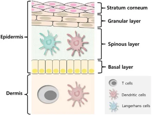
When AD occurs, it caused problems in the primarily skin epidermis. The epidermis is divided into four areas, namely stratum corneum, granular layer, spinous layer, and basal layer (Figure 1-1) 5,12.
5
Figure 1-1. Structure of skin and immune cell distribution.
The skin is largely divided into epidermis and dermis. Then the epidermis is divided into four regions such as stratum corneum, granular layer, spinous layer, and basal layer. Dendritic cells and langerhans cells is distributed in the spinous layer. Dendritic cells and T cells are distributed in the dermis.
6
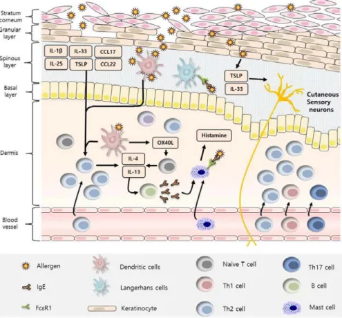
AD caused the increased of pH of the skin surface to make the skin dry, causing collapse of the stratum corneum 10. Then, granular layer is exposed to the outside, which allows various antigens such as S. aureus and allergens to infiltrate the skin 13. These antigens stimulate keratinocytes, which was constitutive cells of granular layer, to induce the production of chemokines such as CC chemokine ligand (CCL) 17 and CCL22, and interleukin (IL)-1β, IL-33 and thymic stromal lymphopoietin (TSLP). CCL17 and CCL22 bind to chemokine receptor (CCR) 4 and recruit the cells expressed CCR4, particularly T cells, to the skin. IL-1β, IL-33 and TSLP accelerated the differentiation and inflammatory responses of dendritic cells (DCs) and langerhans cells (LCs) in spinous layer 5. In particular, IL-33 and TSLP stimulated cutaneous sensory neurons directly to deepen the itch of the skin, promoting skin collapse 14. TSLP also induced and activated the expression of OX40 ligand (OX40L) in dermal DCs. Activated DCs bound to OX40L receptors on naïve CD4+ T cells and induced differentiation into Th2 cells, which were known to regulate skin inflammatory responses by producing IL-4 and IL-13 15,16. In particular, IL-4 activated signal transducer and activator of transcription (STAT) 6 to inhibit the differentiation of keratinocytes. IL-4 also induced B cell activation and proliferation, in accordance with induction of IgE class switching 17. IgE bound to immunoglobulin-ε receptors (FcεR1) of LCs to promote allergen uptake and initiated T cell-mediated delayed
7
hypersensitivity. IgE also bound to FcεR1 on mast cells and promoted the secretion of granules, especially histamine that promoted vasodilation and the recruitment of immune cells and exacerbated skin inflammation (Figure 1-2) 18.
8
Figure 1-2. Cellular and molecular immunologic mechanism of atopic dermatitis.
AD breaks down the skin, causing external antigens to penetrate, leading to the activation of skin cells and various immune cells. This worsens the symptoms of AD.
9
1.3. Animal models for atopic dermatitis
Various animal models were developed to characterize human AD for the better understanding of the causes and results of AD, and therefore providing therapeutic strategies. A mouse models for AD induced by genetic modification or repetitive skin irritation are well studied 19. Spontaneous mutation resulted in one of the genetically modified mice in which some genes related to AD are modified. Transgenic mouse was another type of genetically modified mouse that designed to overexpress selective cytokine (Table 1-1). Skin irritation was known to induce AD in mouse by repetitive treatment of skin irritants such as certain proteins, antigens and chemical reagents 19-21. These AD mouse models (Table 1-1) mostly induced excessive Th2 responses and developed AD-like skin changes in the dermis showing high density of mast cells and eosinophils 19-21.
10
Table 1-1. Mouse models for atopic dermatitis.
TSLP, thymic stromal lymphopoietin; Ova, ovalbumin; HDM, House dust mite; DNCB, 2,4-dinitrochlorobenzene.
Classification Characteristics Ref
Genetic modification Spontaneous mutant Nc/Nga mouse (Chromosome 9 mutant) 22 Transgenic
IL-4 overexpressing mouse 23 IL-18 overexpressing mouse 24 TSLP overexpressing mouse 25
Repetitive skin irritation
Ova (protein) stimulation 26 HDM (allergen) stimulation 27
DNCB (chemical reagents) stimulation
11
Among these, a model similar to the AD occurring in the general population without genetic diseases was a repetitive treatment of skin irritant. House dust mite (HDM) allergen was known as one of the most frequent factors that cause human AD 27. Mice subjected to repeated skin irritation by HDM allergen exhibited dermatitis due to epidermal hyperplasia. CD4+ and CD8+ T cells were infiltrated to the skin of these mice and systemic excessive Th2 responses were observed 27. Hapten such as 2,4-dinitrochlorobenzene (DNCB) were also commonly used to induce allergic contact dermatitis 28. When DNCB was exposed to mouse skin for 2 weeks or more, the skin inflammation migrated to a Th2 dominating response similar to human AD 28. Challenges to repetitive DNCB have resulted in increased epidermal hyperplasia and reduced expression of the skin differentiation proteins such as filaggrin1, loricrin2 and involucrin3 28. Studies of various AD mouse models have led to a better understanding of the pathogenesis of human AD and the potential for AD treatment.
1.4. Therapeutics for atopic dermatitis and their limitation
There are variety of therapies available to relieve the AD, including hydration and recovery of skin barriers by the use of emollients,
1
Filaggrin: Filament-related protein that binds to the keratinous fibers of epithelial cells
2
Loricrin : Protein component of the cornified cell envelope
3
12
microbial agents, and anti-inflammatory agents 29. The goals for the therapeutic were to reduce itching, inhibit inflammation, and restore skin barrier function 29. Emollients were widely recommended because they were not only effective but also safe for skin moisturization. The emollient was known to actively bind the epidermal barrier by tying or pulling the stratum corneum water, reducing the severity of the disease 30.
Topical corticosteroids (TCSs) had an anti-inflammatory effect and intermittent used reduces the recurrence of the disease 31. TCSs were reducing S. aureus levels and restoring skin barrier function. However, it was important to note that excessive use of TCS caused side effects such as skin thinning, purpura, telangiectasias, and growth retardation 32. Various inhibitors were also used in the treatment of AD. The Janus kinase (JAK) /STAT pathway is used by multiple cytokines and growth factors for signaling in AD 33. Tofacitinib, inhibiting JAK1 and JAK3, blocks Th2 response and induced Th1 response. Phosphodiesterase-4 (PDE4) inhibitors were also used as potential treatments for AD 34. PDE4 has been shown to directly attenuate inflammation by inhibiting degradation of cAMP resulting in a decrease in levels of tumor necrosis factor (TNF)-α, IL-12 and IL-21 35. Another topical application, SB011, contained the DNAzyme hgd40 targeting GATA3, a major regulator of Th2-induced immune responses, which effectively prevents the differentiation of Th2 cells 36. Most of the inhibitors are effective in short-term treatment, however they are not recommended because a
long-13
term use can cause side effects including immune irregularities 37. Some inhibitors were causing side effects such as drowsiness and edema 30. Therefore, for a fundamental atopic treatment, a particular strategy to maintain the continuous immune balance seems necessary. Recently, probiotics have been suggested and studied as such strategy for overcoming AD.
14
2. Probiotics
2.1. General characteristics
Probiotics are live microorganisms that had beneficial effects on host health and potential for disease prevention and treatment 38. Probiotics are usually commensal lactic acid bacteria that can be ingested through a variety of foods that found in dairy products and fermented foods. Among various probiotics, Lactobacillus, Bifidobacterium and
Saccharomyces have been widely studied and commonly used in humans
and animals 39. Microbial flora plays an important role as a part of intestinal mucosal immune system. Especially, probiotics competed with harmful microorganisms to prevent pathogens from adhering to the epithelium 40. For example, Lactobacillus rhamnosus and L. plantarum were able to inhibit the adherence of enteric pathogenic Escherichia coli in the gastrointestinal tract 41. Probiotics also enhanced the survival of intestinal epithelial cell (IEC) and improved the barrier function 42. The most important aspects of probiotics are beneficial to human health by activating and matured intestinal immune systems 42,43.
2.2. Probiotics as intestinal epithelial cell modulators
15
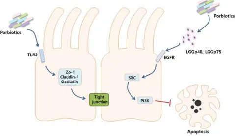
lumen and the lamina propria 44. Probiotics can modulate function of IECs in a variety of ways including directly stimulating the surface of IECs and indirectly producing soluble proteins. Furthermore, probiotics could improve the barrier function by controlling tight junctions of IECs 45. Various transmembrane proteins such as zonula occludens (ZO)-1, Claudin-1 and Occludin were involved in the formation of tight junctions 44. Probiotics could induce the expression of transmembrane proteins through direct toll-like receptor (TLR) stimulation. For example,
L. rhamnosus and L. plantarum stimulated TLR2 signaling, leading to
increased expression of ZO-1 and Occludin in IECs and epithelial permeability 46. In addition, L. casei activated p50/p50 NFκB homodimers through the activation of B-cell lymphoma 3-encoded protein (BCL-3) to prevent the production of inflammatory cytokines 47. It has been suggested that soluble proteins L. rhamnosus GG (LGG) p40 and LGGp75 of probiotics were involved in the survival of IECs. The LGGp40 and LGGp75 bound to epithelial growth factor receptor (EGFR) on IECs and stimulate serine-threonine kinase (Src) that activated phosphatidyl inositol 3-kinase (PI3K), resulting in the prevention of apoptosis 48. The ability of probiotics to regulate cell death and function of IECs could be a useful strategy to prevent immune collapse due to various inflammatory diseases (Figure 1-3).
16
Figure 1-3. Probiotics modulate the function of intestinal epithelial cells.
Probiotics induce the expression of ZO-1, Claudin-1 and Occludin on intestinal epithelial cells through TLR2 stimulation. In addition, probiotics produced LGGp40 and LGGp75, which activates SRC through EGFR and activates PI3K to block intestinal epithelial cells apoptosis.
17
2.3. Immunological roles of probiotics
Probiotics mainly interact directly with the IECs but some reach to the lamina propria through M cells and interact with immune cells to regulate gastrointestinal immune system 49. DCs in the lamina propria layer was known to be the main cell that recognizes probiotics 50. DCs are one of the antigen-presenting cells that can most effectively induce a primary immune response against pathogens as well as maintain tolerance to self-antigens 51. DCs are also known to played a key role in bridging innate and adaptive immune responses 52. DCs are stimulated through TLR signaling dependent on the type of stimulus. Several probiotic strains were known to regulate the function and characteristics of DCs 53. Certain probiotics, such as L. casei, induced the production of 12, IL-6 and TNF-α by DCs through the activation of STAT1 and STAT3, thereby supporting inflammatory responses 54. On the other hand, L.
reuteri inhibited the production of these cytokines and neutralized the
inflammatory response 55. In addition, IRT5, a mixture of probiotics consisting of L. casei, L. acidophilus, L. reuteri, B. bifidum, and
Streptococcus thermophiles, induces indoleamine 2,3-dioxygenase (IDO)
and cyclooxygenase (COX)-2 expression in DCs via TLR2 stimulation and helped to suppress inflammatory responses by producing IL-10 and transforming growth factor (TGF)-β 56. Specifically, DCs that were specialized for inhibiting inflammation, called tolerogenic DCs (tDCs), and CD103+ DCs played a similar role in the intestinal immunity 57.
18
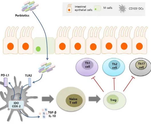
CD103+ DCs inhibited naive CD4+ T cell differentiation to Th2 cells and, at the same time, induced the differentiation of regulatory T cells (Tregs) through the production of IL-10 and TGF-β 57. B. breve induced the production of IL-10 in DCs through TLR2/MyD88 signal transduction and promoted the differentiation of Tregs 58. In addition, L. salivarius induced IL-10-dependent development of CD103+ DCs and Tregs 59. In conclusion, certain probiotic bacterial strains could regulate intestinal homeostasis by promoting the induction of Tregs through DCs (Figure 1-4).
19
Figure 1-4. Probiotics modulate the Dendritic cells and T cell differentiation.
Probiotics induced the expression of IDO and COX-2 through TLR2 stimulation and induced the expression of PD-L1 in CD103+ DC, while enhancing TGF-β and IL-10 production. These DCs and cytokines induced the differentiation of naive T cells into Tregs and prevented the differentiation of Th1, 2 and 17.
20
2.4. Probiotics in the treatment for atopic dermatitis
The collapse of immune balance due to excessive Th2 response is the major cause of AD. Certain probiotics are known to improve the symptoms of disease by controlling the Th2 immune response 1,2,4. There is a number of studies that have identified the potential efficacy of probiotics in the treatment of AD 60,61. L. plantarum showed prevention effect in AD symptom together with the reduction of IFN-γ and IL-4 level as well as proportion of eosinophil 62. L. salivarius showed the reduction of the Th2 cytokine production together with the maintenance of Th1 cytokine production relieved AD symptoms 63. Weissella cibaria induced a population of Tregs in mesenteric lymph node (mLN) and reduced the production of the Th2 cytokines, IL-4, IL-5 and IL-13 64. Moreover, W. cibaria attenuated epidermal thickening and AD-like skin lesions with reduction of serum IgE levels 64. Oral treatment of IRT5 induced tDC and Treg differentiation with up-regulation of IL-10 and TGF-β level in AD mice that eventually mitigates symptom of AD 56. These various probiotics also have shown clinical efficacy in AD. 8-weeks treatment of L. fermentum in 6-18 months old infants showed a significantly reduced severity of AD including Scoring of Atopic Dermatitis (SCORAD) 65. When L. acidophilus, B. lactis and fructo-oligosaccharide were supplemented to children, It showed the significant attenuation in SCORAD (33.7%), infant dermatitis quality of life (33%),
21
dermatitis family impact (35.2%) scores 66. ProBiotik® pur, a mixture of
L. salivarius, L. casei, L. acidophilus, and B. bifidum showed an effective
reduction in SCORAD, IgE, IL-6, IL-5, and IFN-γ levels with unchanged TNF-α, IL-10, IL-2, and IL-4 expression levels in children 65. It is important to note that not all probiotics had a such effect on AD. Importantly, even the same strain may have different efficacy depending on the combination or the target. The infant study with L. paracasei or B.
lactis showed no significant difference in SCORAD and other AD related
parameters compared with placebo groups 67. Although, L. rhamnosus showed alleviation of symptoms of AD syndrome including SCORAD score in IgE-sensitized infants, the cocktail of probiotic mixture containing the L. rhamnosus with Propionibacterium freudenreichii, B.
breve had no effect on the allergic diseases 68. Thus, previous studies suggested that probiotics are not always showing effective immunomodulatory response against AD. In other words, the efficacy of AD treatment of probiotics are depending on the selection and combination of strains. Therefore, to overcome atopy, it is necessary to study the exact immune mechanism induced by each probiotics. Moreover, research on the ideal combination of probiotics is essentially required.
22
3. Galectin
3.1. Characteristics and members of galectins
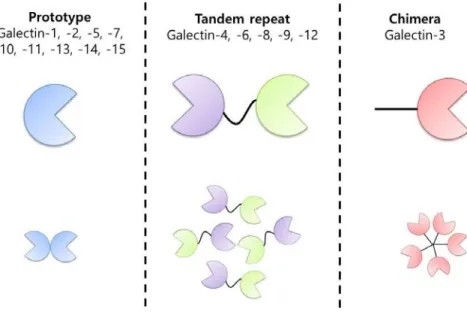
Galectin is non-glycosylated soluble proteins of lectin family that binds to glycan, especially β-galactosides 69,70. Galectins have carbohydrate-recognition domain (CRD) sequence motif and are widely distributed 69,70. There are mainly three types of mammalian galectins dependent on structural difference. Proto type galectins are galactin-1, -2, -5, -7, -10, -11, -13, -14, and -15, while tandem repeat types are galectins-4, -6, -8, -9 and -12. Galectin-3 is the only chimera type 70,71. Proto type galectins have single CRD and exist as monomers or homodimers. Tandem repeat types contained two different CRDs occur within a single polypeptide. Two different CRDs separated by a linker of up to 70 amino acids. Chimera type contained a single CRD connected to a non-lectin amino-terminal domain, which is rich in glycine and tyrosine residues. In humans, only Galectin-1, -2, -3, -4, -7, -8, -9, -10, and -12 are identified. Galectin-5 and -6 are found in mouse whereas galectin-11, -14 and -15 are found in sheep and goats (Figure 1-5) 70,71.
23
Figure 5. Three types of galectins.
Galectins are divided into proto type, tandem repeat type and chimera type based on the structure.
24
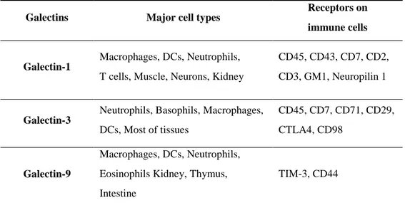
Galectins are synthesized in the cytoplasm of free ribosomes, and the synthesized galectins are detected in the cytoplasm and nucleus as well as extracellular space 72. Galectins caused cell adhesion through cross-linking glycan on neighboring cells 73. Galectins could also form galectin–glycoprotein lattices at the cell surface that can activate functional signaling pathways of endocytosis, host–pathogen interactions, and homeostasis of immune cells 72. Galectins are known to be expressed in a variety of cells and tissues. Among them, galectin-1, -3 and -9 were relatively well known and studied 74. Galectin-1 is secreted by various immune cells such as macrophages, DCs, neutrophils, T cells. It was also produced in thymus, muscles, neurons and kidneys 69,70. Galectin-1 bound to CD2, CD3, CD7, CD43 and CD45 on T cells and induced apoptosis 75. Galectin-1 also bound to neuropilin-1 on vascular endothelial cells, activating VEGFR-2 signaling and regulating migration 76. Galectin-3 is mainly produced in neutrophils, basophils, macrophages and DCs in immune cells and was produced in most of tissues 69,70. Galectin-3 bound to CD7, CD29, CD45 and CD71 on T cells and induced apoptosis 77. Galectin-3 also bound to CD98 on macrophages and differentiated it into alternative M2-type macrophages 78. Galectin-9 is mainly secreted by kidney, thymus and intestine 69,70. Galectin-9 bound to T cell immunoglobulin mucin-3 (TIM-3) on Th1 and Th17 cells and induced apoptosis 79. Galectin-9 also bounds to TIM-3 and CD44 on DCs and monocytes to regulate cell
25
maturation and cytokine release 80. Above all, Galectin-9, unlike other galectins, bounds to CD44 on Tregs and involved in immunosuppression (Table 1-2) 81.
26
Table 1-2. Main functional galectins for their source cells and known receptors.
Galectins Major cell types
Receptors on immune cells
Galectin-1
Macrophages, DCs, Neutrophils, T cells, Muscle, Neurons, Kidney
CD45, CD43, CD7, CD2, CD3, GM1, Neuropilin 1
Galectin-3
Neutrophils, Basophils, Macrophages, DCs, Most of tissues
CD45, CD7, CD71, CD29, CTLA4, CD98
Galectin-9
Macrophages, DCs, Neutrophils, Eosinophils Kidney, Thymus, Intestine
27
3.2. Immune function of galectin-9
Galectin-9 has been suggested to regulate immune function in allergy mouse 82. Galectin-9 is involved in the survival and differentiation of mouse T cells. T lymphocytes to be mature in the thymus, they should overcome positive and negative selection processes 83. Galectin-9 bound to TIM-3 on CD4-CD8- and CD4+CD8+ T cells and induces apoptosis from the thymus 83. Galectin-9 also bound to the TIM-3 on Th1 and Th17 cells inducing intracellular Ca2+ flux and promoting caspase-1 activation 84. Moreover, galectin-9 bound directly to CD44 on Tregs and phosphorylates Smad3 to stabilize Foxp3 85. Stabilized Foxp3, then, promoted Tregs differentiation and produced IL-10 and TGF-β to maximize immunosuppression 85.
Galectin-9 inhibited differentiation of naïve T cells to Th17 cells and at the same time induced differentiation of Tregs in vitro 86. Such ability to control T cell populations might be effective in attenuating autoimmune disease and prolonging allogeneic survival. Galectin-9 also affected the activity of DCs 87. Galectin-9 bound to CD44 on DCs induced phosphorylation of the MAPK p38 and extracellular signal–regulated kinases (ERK)1/2, preventing their maturation and activation 87. Galectin-9 also bound to TIM3 on DCs, preferentially producing IL-10, and initiated an adaptive immune response by synergizing with the activity of TLRs 80. Galectin-9 was known to bind directly to the
28
immunoglobulin. Galectin-9 bound to carbohydrate moieties of IgE to prevent IgE-antigen complex formation 88. Based on these immunomodulatory mechanism, galectin-9 has been recognized as a therapeutic to overcome in allergic diseases. Indeed, recombinant galactin-9 treatment improved clinical and immunological symptoms of allergic disease in a mouse model 89. Some studied have reported that galactin-9 is produced in the intestine by probiotics 90. When B. breve was fed to mouse with allergy induced with cow's milk, galectin-9 was produced in the intestinal cells and serum, leading to the differentiation of Th1 and Tregs 90. Thus, galectin-9 induced Treg activity differently from other galectins and appeared to be involved in immune tolerance. Galectin-9 also showed direct and indirect immunomodulatory effects in various allergy mouse models. However, research on probiotics involved in the production and immunoregulation of galectin-9 is still in its early stages. Therefore, it is necessary to investigate whether galectin-9 is involved in the immune regulation of probiotics.
29
This chapter comprises an article, published in Frontiers in Microbiology with a minor modification as a partial fulfillment of Han wool Kim’s Ph.D. program
Chapter 2
Dietary probiotic mixture, Duolac ATP
reduces the symptoms in mice with atopic
30
1. Introduction
Atopic dermatitis (AD) is a long lasting inflammatory skin disorder that affects the structural and barrier functions of the skin. Major symptoms of AD include excessive pruritus and eczema. Although the exact pathophysiology of AD is not yet fully understood, it can be caused by a combination of genetic, environmental, allergic, and microbial factors 91. Like other allergic diseases, AD is caused by an immune response primarily Th2 cell hypersensitivity. A balance between Th1 and Th2 cells is important for disease induction and tolerance. In AD patients, overexpression of Th2 cytokines, including IL-4, IL-5, and IL-13, upregulate IgE production, resulting in eosinophil accumulation within the dermis 92,93. Although steroid drugs are effective therapies, they have serious side effects, including dermal atrophy, acne, cataracts, growth retardation, and skin irritation. As such, long-term steroid treatment should generally be avoided 94,95. Other alternative therapeutic agents are being investigated, including herbs, phytochemicals, vitamins, and probiotics 96.
Probiotics, which are non-invasive, non-pathogenic, Gram-positive bacteria with known health-promoting effects, are primarily found in fermented food and feed products 97,98. An adequate quantity of probiotics can be helpful for host gut homeostasis, which is achieved through immune system modulation and the production of antimicrobial
31
agents that block the adhesion of pathogens and their toxins 99. Recent studies have reported that probiotics are also effective in preventing allergic disorders in mice and humans 100-102. Lactobacillus plantarum supplementation reduced the scoring atopic dermatitis (SCORAD) index in young atopic patients 62,100. In another study, L. casei supplementation prevented the development of AD in NC/Ng mice 103. However, the mechanism by which probiotics function is still not completely understood.
DCs are antigen-presenting cells that can effectively induce a primary immune response to pathogens as well as maintain tolerance to self-antigens 51. DCs play a key role in bridging innate and adaptive immune responses 51,52,104. Depending on the stimulus, DCs can secrete cytokines and induce naïve T-cell differentiation toward Th1, Th2, Th17, or Treg lineages. Therefore, much attention has focused on the impact of DC priming by probiotics to modulate T cell responses 105. Some probiotic strains, including Lactobacillus and Bifidobacterium, modulate the action of DCs to produce IL-10 and IL-12 along with the expression of co-stimulatory molecules 106,107. Several Lactobacillus strains have been shown to inhibit T cell proliferation, induce IL-10 and TGF-β production, and modify Th1 and Th2 cytokine production in various models of autoimmune diseases 108. In another study, B. lactis inhibited TGF-β production 109. These findings suggest that probiotics should be carefully selected so that the resultant immune response is appropriate for the
32
desired clinical application.
Duolac ATP is a probiotic preparation containing four probiotics strains (L. casei, L. plantarum, L. rhamnosus, and B. lactis) and additives (polydextrose, fructooligosaccharide, glucooligosaccharide, and magnesium stearate). Duolac ATP has been previously evaluated for anti-inflammatory activity in a trinitrobenzene sulfonic (TNBS)-induced colitis model 110 and a DNCB-induced AD model 111. However, these studies focused on the therapeutic effects of Duolac ATP through fragmentary indicators.
The aim of the present study was to evaluate the probiotics efficacy of Duolac ATP without additives. The effect of Duolac ATP on innate and adoptive immune systems was evaluated via BMDCs. Moreover, a transcription factor and cytokine are analysed from atopic mice to explore the mechanism by which Duolac ATP overcomes AD.
33
2. Materials and Methods
2.1. Animal
Female, 7-10-week old, BALB/c from Orient (Gapyeong, South Korea), or 4-week old NC/Nga mice from the Shizuoka Laboratory Animal Center (Tokyo, Japan) were purchased. The mice were randomized and housed in stainless steel cages in a controlled environment with a 12-h light-dark cycle. All the experimental procedures were carried out in accordance with the Animal Use and Care Protocol approved by the Institutional Animal Care and Use Committee (IACUC) at Seoul National University, Seoul, Korea (Approval No. SNU-170428-1).
2.2. Probiotics
Probiotic strains were obtained from Cell Biotech Co. Ltd (Gimpo, Korea) as a powder form, containing 1 × 10¹¹ CFU/g. In this study, minor modification of the Duolac ATP was used. These Duolac ATP is composed of four different strains of probiotics (Lactobacillus casei CBT LC5 (KCTC12398BP), L. plantarum CBT LP3 (KCTC10782BP), L.
rhamnosus CBT LR5 (KCTC12202BP), and Bifidobacterium lactis CBT
BL3 (KCTC11904BP)) without additives such as prebiotics.
34
Bone marrow (BM) cells were isolated from femurs of mice. Red blood cells were depleted using RBC-lysis buffer (Sigma-Aldrich, MO, USA) and BM cells were cultured in a complete RPMI with 20 ng/ml GM-CSF (Creagene, Korea). The complete RPMI was composed of RPMI-1640 supplemented with 10% fetal bovine serum, 20 mM HEPES, 1 mM sodium pyruvate, 220 nM 2-Mercaptoethanol, 100 μg/ml Gentamicin (all from Sigma-Aldrich). At day 0, BM cells were seeded at 3 × 10⁶ cells/well in 6-well plate in 3 ml media, and 2 ml of fresh media was added at day 3. At day 5, a half of the culture supernatant was discarded, and 3ml of fresh media was added. At days 7, suspended BM cells were harvested and sorted by CD11c MicroBeads UltraPour kit (Miltenyi Biotec Inc., CA, USA). Suspended CD11c+ BM cells that is BM-derived DCs (BMDCs). BMDCs were seeded at 2 × 10⁵ cells/well in 96-well plate and stimulated with Duolac ATP or 100 ng/ml lipopolysaccharide (LPS) in a complete RPMI. After the incubation for 24h, the supernatant was collected for cytokine concentration.
2.4. In vitro CD4
+T cell stimulation
CD4+ T cells were isolated from mesenteric lymph node (mLN) from wild type mice using mouse CD4 T lymphocyte enrichment Set (BD Biosciences, CA, USA). CD4+ T cells were labeled with CellTrace™ Violet (CTV) Cell Proliferation Kit (Thermo Scientific, Rockford, USA).
35
CD4+ T cells (2x10⁵ cells/well) were co-cultured with Duolac-treated BMDCs (2 × 10⁴ cells/well) incubated on anti-CD3/CD28 mAbs (BD Biosciences)-coated 96-well plate. After the incubation for 72h, the cells were examined for proliferation of Foxp3+CD4+ T cells and the supernatant was collected and examined for IL-10 and IFN-γ concentration.
2.5. Mouse AD model
The back of NC/Nga mice were shaved and dorsal skin and ears were sensitized with house dust mite (HDM) extracts (Biostir, Japan) or DNCB (Sigma-Aldrich). For the HDM-induced AD mouse model, 100 mg of HDM extracts were treated twice a week for 3 weeks. After following the last treatment, mice were administered with PBS (200 μL/day) or Duolac ATP (2 × 10⁹CFU/200 μL/day) every day for 28 days.
In case of DNCB-induced AD mouse model, 1% DNCB that was dissolved in acetone-olive oil (3:1) were treated twice a week for 3 weeks. After the last treatment, mice were administered with PBS (200 μL/day) or Duolac ATP (2 × 10⁹CFU/200 μL/day) three times a week for 28 days (Figure S2-3). At the end of the treatmentof the both types of AD mice models, the mice were anesthetized with CO2. Blood samples were
collected by heart puncture into heparinized tubes. The sera were then collected by centrifugation for 10 min at 3,000 rpm and stored at -80°C,
36
until further use for ELISA. The mice, at the end of experiment, were sacrificed, and dorsal skin and ear samples were collected for histological analysis and TUNEL assay, respectively. Peripheral blood mononuclear cells (PBMCs), purified from blood of mice treated with/without Duolac ATP, were purified by density gradient centrifugation using Histopaque®-1077 (sigma-Aldrich), and stored at -80°C, until further use for qPCR and western blot. mLN and PP were taken and the distribution of immune cells was measured by flow cytometric analysis.
2.6. Histology
The dorsal skin was removed and fixed in a 4% paraformaldehyde (Sigma-Aldrich, MO, USA). The paraffin-embedded skin sections were heat immobilized, deparaffinized by immersing in xylene (Sigma-Aldrich), rehydrated using a graded series of ethanol, and washed with distilled water. The dorsal skin samples were then cut and subjected to hematoxylin and eosin (H&E) (Sigma-Aldrich) staining. Samples were then examined under the light microscopy (Leica Microsystems, Wetzlar, Germany) for histological evaluation. All clinical and histological evaluations were performed in a blinded manner.
2.7. TUNEL assay
37
staining was performed using an In Situ Cell Death Detection Kit (Roche, Mannheim, Germany) according to the manufacturer’s protocol. The sections were post-fixed with ethanol-acetic acid (2:1) and rinsed. The sections were then incubated with proteinase K (100 mg/mL), rinsed, and incubated in 3% H2O2, permeabilized with 0.5% Triton X-100, rinsed
again, and incubated in the TUNEL reaction mixture. The sections were rinsed and visualized using Converter-POD with 0.03% 3,30-diaminobenzidine (DAB). Then, the sections counterstained with eosin were examined for TUNEL staining by using an optical microscope (Olympus BX53, Japan).
2.8. RNA isolation and qPCR
Total RNA was isolated from PBMCs by TRIzol® reagent (Life Technologies, Carlsbad, CA, USA). One microgram of RNA was reverse-transcribed in a 20μl reaction containing random primers (500 μg/ml), dNTP (10 mM), 5x first strand buffer, DTT (0.1M), Superscript III enzyme (200 U/μl) and RNase inhibitor (10 U/μl) (all from Invitrogen, Carlsbad, CA). RNA was reverse transcribed with HyperScript reverse transcription reagents (GeneAll Biotechnology, Seoul, Korea), and quantitative PCR (qPCR) was performed with the SYBR Green Supermix (iQ SYBR Green Supermix, Bio-Rad Laboratories, Hercules, CA, USA) on the LightCycler 480 Real-Time PCR System (Roche,
38
Indianapolis, IN, USA). This was then used to calculate the relative amounts of target mRNA in test samples. Quantities of all targets in test samples were normalized to the corresponding GAPDH levels. Primers for IL-2 (forward: 50-CCT GAG CAG GAT GGA GAA TTA CA-30, reverse: TCC AGA ACA TGC CGC AGA G-30), IL-4 (forward: 50-ACA GGA GAA GGG ACG CCA T-30, reverse: 50-GAA GCC CTA CAG ACG TCA-3 0), IL-5 (forward: 50-GGG CTT CCT GCT CCT ATC TA-30, reverse: 50-CAG TCA TGG CAC AGT CTG AT-30), IL-10 (forward: 50-CAA CAT
ACT GCT AAC CGA CTC CT-30, reverse: 50-TGA GGG TCT TCA GCT TCT CAC-30), IL-17 (forward: 50-TCT GAT GCT GTT GCT GCT G-30, reverse: 50-ACG GTT AGA GGT AGT CTG AGG-30), IFN-γ (forward: 50- CAG CAA CAA CAT AAG CGT CA-30, reverse: 50-CCT CAA ACT TGG CAA TAC TCA-30), TGF-β (forward: 50- GTG TGG AGC AAC ATG TGG AAC TCT-30, reverse: 50-TTG GTT CAG CCA CTG CCG TA-30), GAPDH (forward: 50-CAT GGC CTT CCG TGT TCC TA-30, reverse: 50-CCT GCT TCA CCA CCT TCT TGA T-30) were synthesized from Bioneer Inc (Daejeon, Korea).
2.9. Western blot
Total protein was isolated from PBMCs by using RIPA buffer (Abcam, Cambridge, UK). The amount of proteins was quantified by BCA Protein
39
assay kit (Thermo Scientific, Rockford, US A), with bovine serum albumin (BSA) as a standard. Each protein sample was loaded onto 10% SDS-polyacrylamide gel and transferred to nitrocellulosemembrane (Schleicher & Schuell BioScience, Germany) for 90 min at 4°C, and blocked with 5% skim milk in TBST (1M Tris-HCl, 5M NaCl, 10% Tween-20) for 1 h at room temperature. The blots were incubated with antiGATA3, Tbet, Cmaf, STAT1, PSTAT1, STAT4, PSTAT4 or -beta-actin (all from Abcam) antibodies overnight at 4°C, incubated with anti-GATA3, pics/biochemistry-generated with goat anti-rabbit IgG-HRP antibody (Santa Cruz Biotechnology, USA) for 1 h at room temperature. The target protein was visualized with enhanced chemiluminescence system (GE Healthcare Life Sciences, USA), followed by analysis using ChemiDoc XRS (Bio-rad).
2.10. Enzyme-linked immunosorbent assay (ELISA)
The immunological response of the mice following HDM extract or DNCB-induced AD was monitored by measuring the serum levels of mouse IL-10, IL-12p40, TGF-β, IFN-γ (all from R&D Systems, USA) and IgE (BD Biosciences). Mouse TGF-β, IL-10 and IL-12p40 were measured from the supernatants taken after the BMDC cultured with Duolac ATP by using ELISA DuoSet kits (all from R&D Systems). Mouse IFN-γ was measured from supernatants taken after the CD4+ T
40
cells treated with BMDCs supernatants by using ELISA DuoSet kit (R&D Systems). Briefly, 96-well microplate (Nunc) was pre-coated with 100 µl/well of capture antibody. After blocking with 1% BSA for 1 h at room temperature, 100 µl/well of supernatant along with the standard solution diluted in diluent buffer was added and incubated for 2 h at room temperature. After the wash PBS for three times, 100 µl/well of detection antibody was added and incubated for 2 h at room temperature, followed by addition of the Streptavidin-HRP in PBS. After the incubation for 20 min at room temperature, tetrame thyl benzidine (TMB, Millipore) was added to develop the color and then the reaction was stopped by adding 50 μl of 2M H2SO4. The absorbance at wavelength 450 nm was
measured by a microplate reader (Molecular Devices)
2.11. Phenotypic and functional examination of immune
cells by using flow cytometry analysis
In order to examine activation status of the cells, BMDCs were treated with Duolac ATP or LPS for 24h at 37°C. The cells were stained with anti-mouse CD86-FITC, PD-L1-PE, MHC II-PE-cy7, CD11c-APC (all from BD Biosciences) for 20 min at 4°C in the dark. To test the increase of Treg, CTV labeled CD4+ T cells cultured with Duolac-treated BMDCs for 3 days were stained with anti-mouse CD4-PE (BD Biosciences). After surface staining, CD4+ T cells were fixed and stained with anti-mouse
41
Foxp3-APC mAb (Biolegend, Dedham, MA) using FOXP3 Fix/Perm Buffer Set (Biolegend). In vivo examination, single cells from mLNs and PP were isolated from AD mice. Population changes of DCs and Tregs were examined as aforementioned. To analyze for subpopulation of Th cells, total mLN and PP cells were stimulated with PMA and ionomycin (Sigma-Aldrich) in the presence of brefeldin A for 4h. After the stimulation, the cells were stained with appropriate combination of anti-mouse CD11c-APC, CD4-bv605, Foxp3-APC, IFN-γ-PE, IL-4-bv605 and IL-17-APC-cy7 mAb (all from Biolegend). The cells were washed and the expression was examined using a FACSCanto II (BD Biosciences). All flow cytometric data acquired were analyzed with FlowJo software (Tree Star, Ashland, OR).
2.12. Statistical analysis
The levels of significance for comparison between samples were determined by Tukey's multiple comparison test by using GradPad InStat software (Ver 5.01, GraphPad). The data in the graphs were expressed as the mean ±SEM.
42
3. Results
3.1. Duolac ATP effectively induces regulatory immune
responses by BMDCs
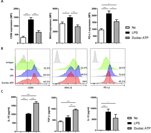
First, the harmful effects, if any, of Duolac ATP on BMDCs were examined. BMDCs were treated with various concentrations of Duolac ATP and their components. Regardless of the probiotic type, BMDC survival was significantly reduced when treated with over 2 × 10⁶ CFU (Figure S2-1), suggesting that the optimal concentration is less than 2 × 10⁶ CFU. Based on these results, Duolac ATP concentration was determined to be used at 2 × 10⁵ CFU. BMDCs treated with Duolac ATP showed similar surface expressions of the co-stimulatory molecules CD86 and MHC II compared with untreated cells within the same intensity (Figure 2-1A) and percentage of cells (Figure 2-1B and Figure S2-2). However, Duolac ATP-treated BMDCs demonstrated a significantly higher expression of PD-L1 compared with untreated cells within the same intensity (Figure 2-1A) and percentage of cells (Figure 2-1B and Figure S2-2). Next, immunomodulatory cytokines were examined in the culture supernatant from BMDC treated with Duolac ATP. These results showed that Duolac ATP induced a significant amount of IL-10, an anti-inflammatory cytokine, at a rate 1.5-fold greater than cells treated with LPS (Figure 2-1C). TGF-β production was also increased when BMDCs were treated with Duolac ATP, whereas
IL-43
12p40 production was slightly lower than that of those treated with LPS (Figure 2-1C). Taken together, these results indicate that Duolac ATP effectively induced a regulatory immune response in BMDCs.
44
Figure 2-1. Duolac ATP induced regulatory molecules in BMDCs.
BMDCs were treated with LPS and/or 2 × 10⁶ CFU of Duolac ATP for 24 hours. (A) The expression of surface markers and (B) a representative histogram of CD86, MHC II, and PD-L1 on BMDCs were measured by flow cytometry. (C) The expression of cytokines in the supernatants was measured by ELISA. Data are representative of at least three experiments. *P < 0.05, **P < 0.01, ***P < 0.001 using one-way ANOVA with Tukey’s multiple comparison test. Bars indicate mean ± SEM.
45
3.2.
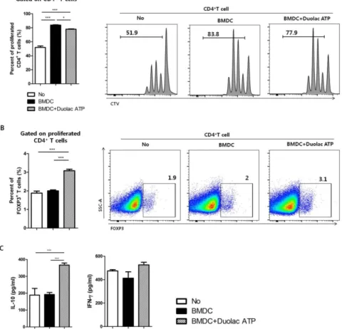
BMDCs treated with Duolac ATP promote
proliferation of Tregs in vitro
DC is known to induce the differentiation of Tregs and to suppress excessive immune responses. Thus, whether Duolac ATP-treated DCs could induce Treg differentiation was investigated. CD4+ T cells were co-cultured with Duolac ATP-treated BMDCs on an anti-CD3/CD28 mAbs-coated plate for 3 days. The results showed that BMDC-induced CD4+ T cell proliferation was higher than that in the control group, but it was slightly reduced when co-cultured with Duolac ATP-treated BMDCs (Figure 2-2A). The ratio of Foxp3+ Tregs in the proliferated CD4+T cells was also significantly increased in the Duolac ATP-treated BMDC group compared to the group treated with BMDC alone or controls (Figure 2-2B). Immunomodulatory cytokines were then examined in the culture supernatant from CD4+ T cells co-cultured with Duolac ATP-treated BMDCs. IL-10 was increased in a similar fashion to Foxp3+ Treg proliferation. However, the expression of IFN-γ was not affected by Duolac ATP treatment (Figure 2-2C). Taken together, these results indicate that Duolac ATP-treated BMDCs were able to induce Treg proliferation with a unique profile of cytokine induction.
46
Figure 2-2. BMDC treated with Duolac ATP promotes proliferation of Tregs in vitro.
BMDCs treated with Duolac ATP were co-cultured with CD4+T cells for 3 days on an anti-CD3/CD28 mAbs-coated plate. The percentage of the proliferated (A) total CD4+T cells and (B) proportion of CD4+Foxp3+ T cells among CD4+T cells were analyzed by flow cytometry. At the same time, supernatants were harvested and examined for (C) the production of IL-10 and IFN-γ in the CD4+ T cells using ELISA. Data are representative of at least three experiments. *P< 0.05, ***P < 0.001 using one-way ANOVA with Tukey’s multiple comparison test. Bars indicate mean ± SEM.
47
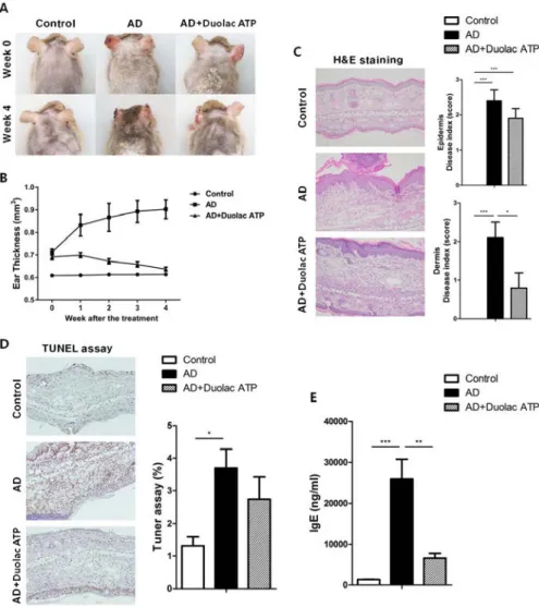
3.3. Amelioration of AD in mice treated with Duolac ATP
To determine the therapeutic properties of Duolac ATP in vivo, a mouse model for AD-like skin lesions was established. The NC/Nga mice were sensitized with HDM extract twice a week for 3 weeks. The mice were then administered phosphate-buffered saline (PBS) (200 μL/day) or Duolac ATP (2 × 10⁹ CFU/day) for 4 weeks, as shown in Figure S2-3. No significant changes were found in the mice’s physical habitus or spleen weight during the feeding period (Figure S2-4). Compared to the control group, the AD group showed severe atopic symptoms, such as itching, erythema/hemorrhage, edema, excoriation/erosion, and scaling/dryness, at week 4 (Figure 2-3A). Interestingly, however, the atopic symptoms of AD mice treated with Duolac ATP were less severe. In addition, weekly examination of ear thickness was performed after the induction of AD. Compared to the control group, the ear thickness of the AD group increased in a time-dependent manner. However, the AD group treated with Duolac ATP showed an attenuated increase in ear thickness (Figure 2-3B). AD is a chronic inflammatory skin disease that not only causes the aforementioned clinical symptoms but also increases epidermal and dermal thickness via activation and infiltration of immune cells 112. To investigate the increase in dermal thickness, the skin was stained and measured the epidermis and dermis based on known disease indices. Epithelial hypertrophy and hyperkeratosis coincident with immune cell
48
infiltration were observed in the AD group (Figure 2-3C, left panel). Furthermore, some mice showed epidermal collapse with bleeding and extensive cartilaginous ulceration. The AD group treated with Duolac ATP had fewer histopathologic anomalies (Figure 2-3C right panel). Furthermore, by conducting a TUNEL assay, the degree of apoptosis could be examined. In the AD group, several apoptotic cells were observed, whereas this was less obvious in the AD group treated with Duolac ATP (Figure 2-3D). Serum IgE levels were significantly increased in the AD group compared to the control group, whereas the AD group treated with Duolac ATP showed a significant decrease (Figure 2-3E). These results suggest that Duolac ATP efficiently ameliorates the symptoms of AD and decreases overall serum IgE levels.
49
Figure 2-3. Amelioration of AD symptoms in mice treated with Duolac ATP.
NC/Nga mice were sensitized by exposing them to HDM extract twice a week for 3 weeks. PBS or Duolac ATP was then orally administered for 4 weeks. (A) Atopic symptoms and (B) ear thickness were scored every week for the last 4 weeks. At the end of the experiment, (C) the results of the histological analysis of cell infiltration by H&E staining (left panel, one representative from 10 samples) and the epidermis and dermis index
50
score (right panel) were examined. (D) TUNEL assay on dermis samples (left panel, one representative from 10 samples) and the percentage of the plot (right panel) are shown. (E) Blood samples were acquired, and serum IgE levels were measured by ELISA. *P < 0.05, **P < 0.01, ***P < 0.001 using one-way ANOVA with Tukey’s multiple comparison test. Bars indicate mean ± SEM.
51
3.4. Maintenance of systemic T cell balance in AD mice
treated with Duolac ATP
To investigate the transcription factors associated with Th1 and Th2 cell differentiation in AD mice treated with Duolac ATP. T-bet (Figure 2-4A and B), STAT1 (Figure 2-2-4A and C), and STAT4 (Figure 2-2-4A and D) are factors that induce Th1 cell differentiation; these were all expressed at a significantly higher rate in PBMCs from AD mice treated with Duolac ATP compared to untreated AD mice. It was further noted that Th2 differentiation factors GATA3 (Figure 4A and E) and C-maf (Figure 2-4A and F) were increased in PBMCs from AD mice. These results suggested that Duolac ATP led to the balance between Th1 and Th2 cells in AD mice by preferentially increasing the release of Th1 differentiation factors.
52
Figure 2-4. Expression changes on transcriptional factors involved in the maintenance of T cell balance in AD mice administered Duolac ATP.
Nc/Nga mice (n=6 per group) were sensitized by exposing them to HDM extracts twice a week for 3 weeks. They were then subjected to oral administration of Duolac ATP. PBMCs collected at week 4 were used to make lysates, which were used to examine the expression of transcriptional factors using Western blotting. (A) The expression of T-bet, STAT1, p-STAT1, STAT4, p-STAT4. GATA3, C-maf, and beta-actin was used as an internal control. Density was measured using a densitometer for semi-quantitation for (B) T-bet, (C) STAT1, (D) STAT4, (E) GATA3, and (F) C-maf. **P < 0.01, ***P < 0.001 using one-way ANOVA with Tukey’s multiple comparison test. Bars indicate mean ± SEM.
53
In addition, the mRNA expression of Th1 and Th2 cytokines was examined in PBMCs from AD mice given Duolac ATP. Higher rates of IL-4 (Figure 2-5A) and IL-5 (Figure 2-5B) expression was found in AD mice, as was expected from a typical Th2 response. Furthermore, IL-17 appeared to be increased in AD mice (Figure 2-5C). However, AD mice treated with Duolac ATP showed a decrease in Th2 and Th17, with a concurrent increase in IL-2 (Figure 2-5D) and IFN-γ (Figure 2-5E and Figure S2-6A). mRNA expression of IL-10 was also increased in AD mice treated with Duolac ATP, whereas there was no change in TGF-β (Figure 2-5F and G). These results indicate that Duolac ATP can induce Th1- but not Th2- or Th17-driven responses.