Depth-Dose Measurement System for Proton Beam Using PMMA Phantom
Seok-Ki Lee, Young Kyung Lim, Kye-Ryung KimProton Engineering Frontier Project, Korea Atomic Energy Research Institute, [email protected]
1. Introduction
Recently, a variety of researches for proton applications have been performed in the fields of nano-technology, biological nano-technology, information technology, medical science, etc.[1,2] The energetic proton loses its kinetic energy as it passes through the target material. In this process the large energy loss happens in a very local region before it stops. This property is characterized by Bragg peak. By using the Bragg peak the energetic proton beam can be utilized for cancer therapy or mutation breeding.[3]
In order to measure the Bragg peak, a depth-dose measuring system using PMMA phantom was designed and fabricated.
2. Depth-dose measurement system
2.1 Design and fabrication
The depth-dose measurement system is composed of two main parts. One part is for protons with 18MeV or below, and the other is for protons with the energies of 15-45MeV. The two parts don’t disturb each other when one of them is used. In the first 6 PMMA shutters are installed in downstream and their thicknesses are 0.05, 0.1, 0.2, 0.4, 0.8, 1.6mm. So we can combine the PMMA sheets in 0.05mm step up to 3.15mm thick. The second consists of two inclined PMMA blocks, one is fixed and the other is movable by a motor. Therefore the PMMA thickness can be continuously controlled. Figure 1 shows the designed depth-dose measurement
Fig. 1 The designed depth-dose measurement system.
system.
In addition, one collimator with 20mm aperture and 6mm Al shutter is installed. The collimator limits the beam size in order to prevent the scattered beam from affecting to the measurement. Al shutter controls the beam exposure time. Figure 2 shows the photographs of the fabricated depth-dose measurement system.
Fig. 2 The photographs of the depth-dose measurement system.
2.2 Control system
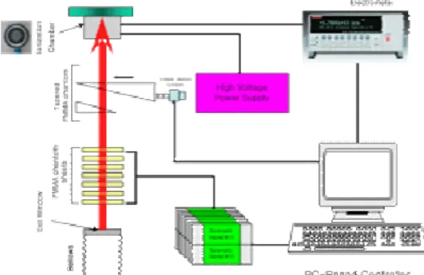
The depth-dose measurement system is controlled by a PC-based Labview program. To control the system and to acquire the measured data a motion controller and a DIO board is used. The PMMA shutters are controlled by the same number of air cylinders and solenoid valves. An inclined PMMA block can be moved continuously forward and backward by an AC servo motor. Therefore, the thickness of the PMMA block can be controlled finely. This fine control enables us to measure the precise position of a Bragg peak and the continuous Bragg curve. For fast measurement of dose an ion chamber and an electrometer is installed. Figure 3 shows the schematic diagram for the system
Figure 3. the schematic diagram for the system control and the data acquisition control and the data acquisition.
Transactions of the Korean Nuclear Society Autumn Meeting Busan, Korea, October 27-28, 2005
3. Experimental Results
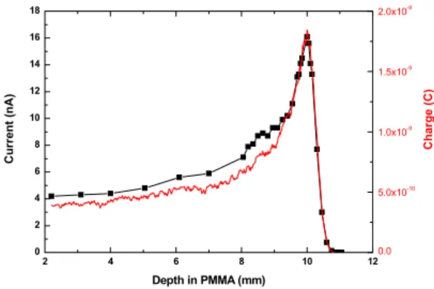
We tested the depth-dose measurement system with the control system by measuring the Bragg curves. These are shown in Figure 4, 5, and 6. In Figure 4 we compared two Bragg curves measured in two different operation modes. In one mode one datum is acquired when the PMMA block stops to a specific position. In the other mode all data are acquired continuously when the PMMA block is moving. Two positions of the Bragg peaks coincide definitely. Figure 5 shows the various Bragg curves for various proton energies. In a small energy region the mean proton energy decreases linearly when the thickness of degrader increases. In figure 6 we can see that the mean proton energy decreases as the measuring position is further, and also varies linearly in small distances.
2 4 6 8 10 12 0 2 4 6 8 10 12 14 16 18 0.0 5.0x10⁻¹⁰ 1.0x10⁻⁹ 1.5x10⁻⁹ 2.0x10⁻⁹ Ch arg e (C) Cu rr en t ( n A) Depth in PMMA (mm)
Fig. 4 The comparison of the two Bragg curves.
2 4 6 8 10 12 14 0.00E+000 5.00E-010 1.00E-009 1.50E-009 2.00E-009 0 1 2 3 4 5 15 20 25 30 35 40 Pr o to n En er g y (m eV) Degrader Thickness (mm) B Linear Fit of Data1_B
Ch a rg e (C) Depth in PMMA (mm) Al 0mm (E~35.5MeV) Al 1mm (E~31.6MeV) Al 2mm (E~27.7MeV) Al 3mm (E~23.1MeV) Al 4mm (E~17.9MeV)
Fig. 5 The depth-dose distributions for various beam energies. 2 4 6 8 10 12 0.0 5.0x10⁻¹⁰ 1.0x10⁻⁹ 1.5x10⁻⁹ 2.0x10⁻⁹ 80 100 120 140 160 180 200 32 33 34 35 36 Pr o to n En erg y ( M e V) Distance (cm) B Linear Fit of Data1_B
z= 95cm (E~35.5MeV) z=150cm (E~34.3MeV) z=200cm (E~33.5MeV) Ch ar g e ( C ) Depth in PMMA (mm)
Fig. 6 The depth-dose distributions for the several measurement positions.
4. Conclusion
The depth-dose measurement system using PMMA phantoms was fabricated and tested. The continuous Bragg curves could be measured by varying the thickness of PMMA block continuously. The mean proton energy could be estimated by measuring the position of a Bragg peak. The expectable energy range is 5–45MeV.
We expect that the depth-dose measurement system may offer more precise depth-dose distributions comparing to the water phantom dosimeters.[4] In addition it is possible to measure the depth-dose distribution for low proton energy, such as 10MeV more or less. Also, it is convenient to treat and use.
REFERENCES
[1] F. H. Ruddy, A. R. Dulloo, J. G. Seidel, F. W, Hantz, and L. R. Grobmyer, Nuclear Reactor Power Monitoring Using Silicon Carbide Semiconductor Radiation Detectors, Nuclear Technology, Vol.140, p. 198, 2002.
[2] F. H. Ruddy, A. R Dulloo, J. G Seidel, J.W.Palmour, and R. Singh, The Charged Particle Response of Silicon Carbide Semiconductor Radiation Detector, Nuclear Instruments and Methods In Physics Research, Vol.505, p.159, 2003.
[3] J. F. Ziegler, J. P. Biersack, “SRIM-2000, 40: The Stopping and Range of Ions in Matter”, IBM-Research, Yorktown, NY 2000.
[4] M. R. Fard, T. E. Blue, D. W. Miller, SiC Semiconductor Detector Power Monitors for Space Nuclear Reactors, Proceedings of the Space Technology and Applications International Forum(STAIF-2004), Feb.8-12, 2004, Albuquerque, NM.