www.nature.com/scientificreports
Sol narae (Sona) is a Drosophila
ADAMTS involved in Wg signaling
Go-Woon Kim
*,†, Jong-Hoon Won
*, Ok-Kyung Lee, Sang-Soo Lee, Jeong-Hoon Han,
Orkhon Tsogtbaatar, Sujin Nam, Yeon Kim & Kyung-Ok Cho
ADAMTS (a disintegrin and metalloproteases with thrombospondin motif) family consists of secreted proteases, and is shown to cleave extracellular matrix proteins. Their malfunctions result in cancers and disorders in connective tissues. We report here that a Drosophila ADAMTS named Sol narae (Sona) promotes Wnt/Wingless (Wg) signaling. sona loss-of-function mutants are lethal and rare escapers had malformed appendages, indicating that sona is essential for fly development and survival. sona exhibited positive genetic interaction with wntless (wls) that encodes a cargo protein for Wg. Loss of sona decreased the level of extracellular Wg, and also reduced the expression level of Wg effector proteins such as Senseless (Sens), Distalless (Dll) and Vestigial (Vg). Sona and Wg colocalized in Golgi and endosomal vesicles, and were in the same protein complex. Furthermore, co-expression of Wg and Sona generated ectopic wing margin bristles. This study suggests that Sona is involved in Wg signaling by regulating the level of extracellular Wg.
Proteases were originally started out as simple destructive enzymes in order to digest proteins and to provide amino acids to ancient organisms, but many proteases evolved in later times are specialized to change activity, localization, and binding properties of proteins and thereby affect many cellular functions. More than four hun-dred thousand proteases in all organisms can be classified into 9 categories and numerous subfamilies1,2. Among
these proteases, ADAMTS family as a subclass of ADAM (a disintegrin and metalloproteases) family constitutes a group of zinc-dependent secreted proteases widely expanded during metazoan evolution, including 6 members in flies, 5 members in nematodes, and 19 members in mammals3.
These ADAMTSs are involved in many biological actions by processing mostly ECM and some non-ECM sub-strates. For example, ADAMTS-1 cleaves versican and aggrecan, and plays a key role in the ovulation process4,5.
ADAMTS-2, 3, and 14 cleave procollagen I 6–8, and mutations in ADAMTS-2 cause Ehlers-Danlos syndrome, a
connective tissue disorder7,9. ADAMTS-7 and 12 are significantly upregulated in arthritic patients10. Besides ECM
proteins, ADAMTS-13 cleaves von Willebrand factor (vWF) in blood, and mutations in ADAMTS-13 result in thrombotic thrombocytopenic purpura (TTP)11. In addition, ADAMTSs either enhance or inhibit cancer
devel-opment. The level of ADAMTS-7 is upregulated in carcinoma12 and ADAMTS-1 promotes tumor development
through the induction of stromal reaction13. In contrast, ADAMTS-9 suppresses the formation of carcinoma by
inhibiting angiogenesis14, and stable expression of ADAMTS-16 decreases proliferation of cancer cells15. Loss of
ADAMTS-12 in mammals also increased tumor growth and progression16,17.
ADAMTS is synthesized as a zymogen and has a relatively long prodomain. The physical interaction between the prodomain and the metalloprotease domain is essential for the latency of enzyme activity18,19. Removal of
pro-domain in most ADAMTSs is mediated by furin, a proprotein convertase, in the secretory pathway20,21. However,
prodomains of ADAMTS-9, -10, -15 are processed by furin in ECM22–24. In case of MIG-17 that is involved in
male gonadal formation in C. elegans, the prodomain is cleaved autocatalytically25,26. Thus, the activation
mecha-nism of ADAMTS family appears to be diverse and may be tightly controlled in order to ensure the generation of active forms at the right time and place.
We report here that an ADAMTS encoded by the CG9850 gene in Drosophila melanogaster is capable of pro-moting Wnt/Wg signaling. Wnt family proteins are conserved morphogens for growth, development and adult homeostasis in all metazoans27,28. CG9850 was named sol narae (sona) meaning ‘small wing’ in Korean, based
on the small wing phenotype of mutant escapers. Fly Wg, a homolog of mammalian Wnt-1, is a prototype of Wnt family proteins essential for the development of all fly appendages, and the wing imaginal disc has been an Department of Biological Sciences, Korea Advanced Institute of Science and Technology, 291 Daehak-ro, Yuseong-gu Daejeon, Korea. †Present address: Center for Theragnosis, Biomedical Research Institute, Korea Institute of Science
and Technology, Seoul, Korea. *These authors contributed equally to this work. Correspondence and requests for
materials should be addressed to K.-O.C. (email: kocho@kaist.ac.kr) Received: 12 April 2016
accepted: 28 July 2016 Published: 18 August 2016
excellent system to study Wg signaling. Wg is known to be secreted from Wg-producing cells at the dorsal-ventral (DV) midline in the wing pouch and forms a concentration gradient in extracellular matrix (ECM). Wg binding to Frizzled receptors on the plasma membrane of Wg-responding cells activates Wg signaling cascade, and Wg effector proteins including Sens, Dll and Vg are expressed in different regions of wing pouch29–31.
In this study, we focused on answering the following questions. Where and when is sona transcribed and translated? Where is the active form of Sona present? Which gene shows genetic interaction with sona? What are the in vivo roles of sona? We report here that sona exhibits a positive genetic interaction with wntless (wls) whose function is essential for secretion of Wg, and sona is positively involved in Wg signaling. Based on data provided in this report, we propose that Sona may modify proteins involved in Wg signaling.
Results
Sona is an essential ADAMTS.
Drosophila has six ADAMTSs: Stall, Papilin, CG4096, CG6232,ADAMTS-A and CG9850 (Sona)32–35. Sona described in this study is Sona-PA, one of five Sona isoforms
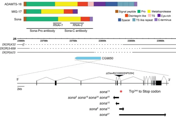
described in Flybase. Sona-PA protein shows ~45% similarity to mammalian ADAMTSs, and is composed of four domains: prodomain, protease domain, disintegrin-like domain and cysteine-rich domain. Members of ADAMTS family differ in the number of thrombospondin (TS) motifs, and some have unique carboxyl terminal domains. Sona is most similar to Caenorhabditis elegans MIG-17 involved in male gonadal formation25 (Fig. 1a;
Supplementary Fig. S1), and Bombyx mori BmADAMTS-like that is induced in wing discs during pupal ecdysis36.
The sona gene is present in the 59F7-60A1 region at the tip of right arm in the second chromosome. P{GawB} CG9850[NP5284] flies used as a ‘sona-Gal4’ line in this study has a P element in an intron of the sona gene and was homozygous viable (Fig. 1b). However, homozygous larvae of P{GawB}CG9850 were occasionally lethal and much larger with extended larval stage at a low frequency (< 1%). These larvae had tumorous imaginal discs as seen in lethal giant larvae (lgl) or discs large (dlg) mutants. To test if some P{GawB}CG9850 flies had lgl or
dlg mutation, they were crossed with dlgm30, dlgm52, and lgl4 mutants for complementation test. Cross between
P{GawB}CG9850 flies and lgl4 but not dlg yielded giant larvae, indicating that minor fraction of P{GawB}CG9850
flies had lgl mutation. To remove lgl and any other potential mutations, P{GawB}CG9850 was crossed with w1118
flies for four generations. The cleaned P{GawB}CG9850 line was homozygous viable without lgl phenotype. This line was crossed to Δ2–3 for P element excision, and about 200 white-eyed flies were obtained. These white-eyed flies were crossed with deficiencies (Df(2R)X32, Df(2R)23, Df(2R)3-659) that uncover all or part of the genomic region containing the sona gene. Nine lines that failed complement these deficiencies were identified (Fig. 1b).
The regions deleted in the nine lines were identified by both RT-PCR and genomic PCR. All lines except sona13
had deletion in the exons of the sona gene, and sona13 had a point mutation that changed Trp344 to stop codon
Figure 1. Domain structure of Sona and generation of sona mutants. (a) Domain structures of mammalian ADAMTS16, C. elegans MIG-17 and Drosophila Sona. The regions used to generate RNAi lines and those used to generate Sona-Pro antibody and Sona-C antibody were marked with black lines. (b) Three difficiency lines were shown with the genomic map of the region including the sona gene. The dashed lines represent the deleted regions in the difficiencies, and the sona gene is drawn as a pointed blue bar. Red asterisk indicates a mutation site of premature truncation in sona13 mutant, which was not produced by P element excision but by
www.nature.com/scientificreports/
and incomplete excision of the P element. On the contrary, ten other white-eyed lines that were complemented by the deficiency lines did not have deletion in the sona coding region. We further cleaned up sona deletion mutants by crossing them with w1118 for four generations in order to remove any unwanted mutations that might have been
generated during the process of imprecise excision of the P element.
Sona is essential for survival and development of flies.
All sona mutants exhibited lethality through-out larval and pupal stages. Three sona mutants, sona13, sona18, and sona47, were chosen for further analysis. Rareescapers from these sona mutants could reach adulthood at a very low frequency (< 1%), and exhibited various developmental defects. The percentage of escapers and their morphological defects were overall similar in the
sona mutants. Branches in their arista were partially missing or malformed (Fig. 2a,b), and the interommatidial
bristles were disoriented in dorsal eye (n = 31; Fig. 2c,d; Supplementary Fig. S2). 40% of the legs had kinks in the femur (n = 36; Fig. 2e,f), and loss of tarsal claw was occasionally observed (not shown). Their wings were crumpled and ~25% smaller than sona heterozygous wings (n = 26; Fig. 2g,h,r). Their wing margin bristles were missing in 80% of wings with average 2.7 anterior bristles (n = 26) (Fig. 2i,j).
We also generated two UAS-sona RNAi lines by using two different regions of the sona cDNA (Fig. 1a; Supplementary Fig. S1). Expression of these two lines by various Gal4 lines induced similar phenotypes such as lethality and malformation of appendages (Supplementary Table S1). Thus, the sona RNAi phenotypes were due to the loss of sona function but not off-target effects. For instance, expression of sona RNAi-111-4 (sona RNAi-1
hereafter) driven by nubbin (nub)-Gal4 at 18 °C and 25 °C resulted in small and crumpled wings, and similar wing phenotype was induced by expression of sona RNAi-223-2 (sona RNAi-2 hereafter) at 29 °C (Fig. 2k–n). When
sona RNAi-1 and sona RNAi-2 were driven by engrailed (en)-Gal4 at 18 °C and 25 °C, respectively, rarely obtained
adults had smaller wings (Fig. 2o–q). In summary, sona RNAi-1 induced stronger phenotype than sona RNAi-2, and Gal4 lines that drive expression of sona RNAi during embryonic stages induced lethality while those during later stages of development induced loss or malformation of appendages (Supplementary Table S1).
The lethality of sona mutants can be rescued by overexpression of Sona.
Similar to the pheno-types induced by sona RNAi expression, overexpression of Sona resulted in lethality or defects in appendages (Supplementary Table S1). This indicated that protease activity of Sona must be tightly regulated for proper devel-opment and survival. To test whether the protease activity of Sona is essential for its role in vivo, we generated Figure 2. Sona is essential for the development of fly appendages. These phenotypes were observed from escapers of sona47, sona13, and sona18 as well as transheterozygotes. (a,b) Arista in w1118 as a wild-type (wt)control (a), and missing and malformed branches of arista in sona47 escapers are marked with arrow and
arrowhead, respectively (b). (c,d) Disrupted ommatidial bristles in sona (d) compared to control (c). (e,f) Kinked femur of sona (arrowhead in f) compared to control (e). (g,h) Wings of sona47 (h) are smaller than
those of sona heterozygotes (g). (i,j) Missing bristles at the anterior wing margin of sona47 (j) compared to the
heterozygous wing (i). (k–n) Wing size becomes smaller by expression of sona RNAi-1 (l,m) and sonaRNAi-2 (n) by nub-Gal4. Wings become crumpled in severe cases (m). (o–q) Wings become smaller by expression of
sona RNAi-1 (p) and sonaRNAi-2 (q) by en-Gal4. (r) Quantitative analysis of the wing size (n = 10 each) shown
UAS-sonaE475A, which encodes the protease-dead form of Sona by substituting glutamic acid to alanine at 475th
residue in the conserved site for protease activity (Supplementary Fig. S1). Expression of SonaE475A resulted in neither lethality nor any developmental defects, demonstrating that the protease activity of Sona is essential for the role of Sona in vivo (Supplementary Fig. S3 and Table S1) unlike five mammalian ADAMTS-like (ADAMTSL) proteins that have no protease activity3,37.
To prove that the deletion in the CG9850 was responsible for phenotype of sona mutants, we checked if the lethality of sona47 can be rescued by expressing UAS-sona with various Gal4 drivers (Supplementary Table S2).
When UAS-sona sona47/CyO was crossed with nub-Gal4 sona47/CyO-GFP and cultured at 18 °C, about 36%
of UAS-sona sona47/nub-Gal4 sona47 were obtained as normal adults. When UAS-sona sona47/CyO-GFP were
crossed with ptc-Gal4 sona47/CyO-GFP and cultured at 18 °C, 23% of UAS-sona sona47/ptc-Gal4 sona47 adults were
obtained as normal adults. Two important conclusions were obtained from this rescue experiment. First, CG9850 is the sona gene. Second, the Sona-PA form is sufficient to rescue the lethal phenotype of sona mutants.
Transcription and translation of Sona are dynamically regulated during development.
As a first step toward understanding the role of Sona during development, we carried out both Northern and Western analyses to examine the expression pattern of sona mRNA and protein. Multiple sona transcripts were expressed during development, and a 3.4 kb transcript in the late 3rd instar larval stage was most abundant (Fig. 3a). Theamino acid sequence of Sona-PA reported in Flybase was identical to those deduced from a 2.97 kb sona cDNA that had been isolated from a late 3rd instar larval cDNA library, so the 3.4 kb transcript may encode the Sona-PA
form. We focused on the Sona-PA form in this study.
We generated two antisera, ‘Sona-Pro’ with the prodomain of Sona as an antigen in rabbit and ‘Sona-C’ with the carboxyl region of Sona as an antigen in mouse as marked in Fig. 1a, and purified them by affinity purifica-tion. All analyses were carried out with the purified antibodies. Western analysis with Sona-Pro antibody showed that multiple forms of Sona are dynamically expressed during development (Fig. 3b). For instance, the level of Sona was highly increased in the late third instar stage, which was corresponding to the increased level of sona transcripts in the same stage. 70 kDa Sona protein was most abundant in the late 3rd instar larval stage (arrow in
Fig. 3b), imlying that 70 kDa band is most likely the Sona-PA form (Fig. 3a).
To test whether multiple bands in the Western blot were authentic Sona proteins, UAS-sona RNAi-1 and
UAS-sona RNAi-2 were crossed with actin-Gal4, cultured at 18 °C to the third instar stage, shifted to 29 °C for
24 hours, and the larval extracts were prepared. No protein bands were identified with Sona-Pro antibody in the larval extracts. Thus, the bands in the control extract are either different isoforms or processed forms that contain all or some portion of the prodomain (Fig. 3c). Furthermore, full-length Sona was absent in homozygous sona13,
sona18 and sona47 larvae (Supplementary Fig. S4). The sona47 extract had a smaller fragment that may be the
trun-cated Sona protein. Sona-C antibody was not suitable for Western analysis.
Active Sona form is present in both intra- and extra-cellular regions.
Because ADAMTSs are secreted proteases, we examined if active form of Sona is secreted. An S2 cell line that constitutively expressessona cDNA was generated (Materials and Methods) and used to obtain cellular extract (CX) and conditioned
media (CM) for Western analysis. Sona-Pro antibody detected both full-length 70 kDa Sona and smaller frag-ments in CX but only 22 kDa prodomain-containing fragfrag-ments in CM, indicating that the full-length Sona is not secreted (Fig. 3d). To detect the active form of Sona, we constructed a sona cDNA tagged with HA in front of the stop codon and established S2 sona-HA cell line. Both CX and CM from the culture of this cell line were analyzed with anti-HA antibody (Fig. 3e). The 37 kDa form was detected in both fractions as an active form devoid of the prodomain. This demonstrated that active Sona is present in both intracellular and extracellular regions.
Sona is expressed in discrete regions and can diffuse far from the expressed site in imaginal
discs.
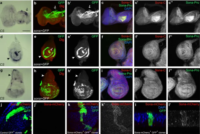
We checked expression pattern of sona transcripts in imaginal discs by in situ hybridization and the pattern of GFP driven by sona-Gal4. In the eye-antenna disc of sona > GFP, GFP was expressed at a high level in dorsal peripo-dial epithelium (arrow in Fig. 4b) and in photoreceptor clusters (Supplementary Fig. S5a–d). In the leg disc, GFP was expressed in the presumptive region of claw, tibia, and femur38 (arrow and arrowheads in Fig. 4e). In the pouch of wingdiscs, sona was expressed in a complicated mosaic pattern (Fig. 4h). Although the expression pattern of sona transcripts was not at high resolution, it was in accordance with the pattern of sona > GFP (arrows and arrowheads in Fig. 4a,d,g). We then examined the expression pattern of Sona protein in imaginal discs with both Sona-Pro and Sona-C antibodies that had been tested for their specificity (Supplementary Figs S5 and S6). Sona was more or less evenly distributed except some regions with a higher level of Sona (Fig. 4c,f,i). At higher magnification, Sona was highly enriched in the apical region of photoreceptor clusters (Supplementary Fig. S5a–d) and in the disc proper of wing discs, and was present at a negligible level in sona mutants (Supplementary Fig. S5e–h).
Because Sona protein was ubiquitously present in entire discs in contrast to the localized pattern of sona tran-scripts, we thought that Sona may be efficiently diffused from the expressed site. To check how far Sona can diffuse, we generated UAS-sona-HA and UAS-sona-HA-mCherry (hereafter ‘UAS-sona-mCherry’) flies using sona cDNA tagged with HA or HA-mCherry in front of the stop codon. These UAS lines driven by various Gal4 lines could induce lethality and structural defects, indicating that the tags did not compromise the protease activity of Sona (Supplementary Fig. S7). We generated clones expressing Sona-mCherry by the flp-out method39 and detected both
intra- and extra-Sona-mCherry proteins far from the GFP+ Sona-mCherry+ clone (Fig. 4k). The cross-section view
of the same clone also showed the diffusion of Sona-mCherry proteins away from the clone (Fig. 4l). mCherry signals were genuine because no signal was detected in and around a control GFP+ clone at the same level of laser
intensity (Fig. 4j). We proved that the mCherry signal is not from the mCherry tag cleaved off from Sona by Western analysis, in which Anti-HA antibody recognized full-length and active form of Sona-HA and Sona-HA-mCherry in
www.nature.com/scientificreports/
CX and CM, respectively, but not any other smaller bands (Supplementary Fig. S8). Thus, our data demonstrated that Sona can diffuse far from the source. How Sona can diffuse and which form of Sona can diffuse need further studies. Figure 3. Transcriptional and translational expressions of Sona during development and active form of Sona in S2 cell culture. (a) Expression of sona transcripts during development detected with the probe representing 732th to 1920th nucleotides of the sona-RA cDNA. sona-RA transcript is marked with an arrow.
E1, AEL (After egg laying) 0~12 hr; E2, AEL 12~24 hr; L1,2, first and second instar larvae; L3, late third instar larvae; P1, early pupae; P2, mid pupae; P3, late pupae; AB, adult body; AH, adult head. (b) Expression of Sona protein during development. E1, AEL 0.5~11 hr; E2, AEL 16~24 hr; L1, first instar larvae; L3, late third instar larvae; P, late pupae; AM, adult male; AF, adult female. The arrow indicates the full-length Sona-PA form. (c) The cellular extract was obtained from the late 3rd instar larvae raised at 18 °C and then at 29 °C for 24 hrs until
dissection. C is actin-Gal4 (act) > Gal80ts, (1) is act > sona RNAi-1; Gal80ts, and 2 is act > sona RNAi-2; Gal80ts.
(d,e) Sona proteins in the cellular extract (CX) or conditioned media (CM) of sona (d) or sona-HA (e) S2 cells. Red arrows and black arrows mark full-length Sona and activated Sona, respectively. Asterisk marks the 22 kDa prodomain fragments. Arrowheads indicate intracellular fragments of Sona.
Carboxyl region and prodomain of Sona are not colocalized in ECM.
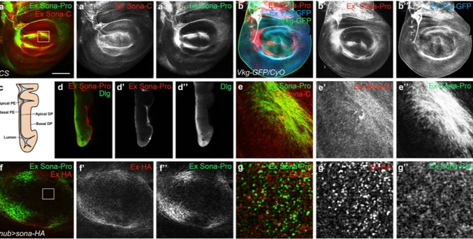
To confirm that the active form but not the full-length Sona are secreted in vivo, Canton S (CS) wing discs were stained to visualize extra-cellular Sona with Sona-Pro and -C antibodies, as described previously (see Materials and Methods)40. Bothanti-bodies recognized extracellular Sona in the presumptive region of wing blade in the same region as lip-shaped but not in the DV midline region on the basal side of the disc proper (Fig. 5a). To prove that the pattern of extracel-lular Sona is authentic but not due to non-specific binding of Sona antibodies to ECM, Viking-GFP, Collagen IV protein fused to GFP commonly used as an ECM marker41, was also examined at the same confocal level. To
distinguish intracellular Viking-GFP from extracellular Viking-GFP, the extracellular Viking-GFP was differ-entially stained with the extracellular staining protocol (Fig. 5b). At a glance, extracellular Viking-GFP was more evenly distributed than extracellular Sona-Pro, which was confirmed by the image at higher magnification (Supplementary Fig. S9).
A cross-section view of a wing disc also showed that extracellular Sona is present in the basal ECM of both disc proper and peripodial epithelium (Fig. 5c,d). However, there were clear differences between the structures recognized by the two Sona antibodies. The Sona-C antibody recognized some particulate structures that were not recognizable by the Sona-Pro antibody (Fig. 5e). These particulate structures may have active Sona that is devoid of the prodomain. In contrast, Sona-Pro antibody recognized a bunch of string-like structures (Fig. 5e”). Similar pattern was also observed in nub > sona-HA wing discs visualized with both Sona-Pro and HA antibodies (Fig. 5f). A magnified image revealed that a Sona form recognized by only anti-HA antibody, probably an active Sona, was localized in ECM as a separate identity (Fig. 5g).
Loss of sona decreases the level of extracellular Wg.
To understand the in vivo role of sona, we carried out a genetic screen with ethyl methanesulfonate (EMS) as a mutagen to obtain suppressors that could overcome the lethality by Sona overexpression. One of suppressors turned out to have a mutation in the wls gene. This study will be addressed in detail elsewhere (J.-H. W. and K.-O. C., manuscript in preparation). Wls is a transmem-brane protein that is required for the secretion of Wnt/Wg42–44 and interacts with retromer complex for cyclingfrom Golgi to the plasma membrane45–49. Consistent with the positive genetic interaction between sona and wls,
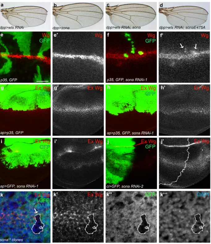
dpp > wls RNAi flies had notched wing phenotype (n > 50 each), but dpp > wls RNAi sona flies had no notching
Figure 4. Expressions of sona transcripts and Sona proteins in flies and S2 cells. Eye-antenna discs (a–c), leg discs (d–f) and wing discs (g–i) from late third instar larvae were shown. (a,d,g) in situ hybridization with sona cDNA as a probe. Arrows and arrowheads indicate the region with high level of sona transcripts. (b,e,h) The pattern of GFP expressed by sona-Gal4. Discs-large (Dlg) was used as membrane marker. (c,f,i) The localization pattern of Sona visualized with two Sona antibodies, Pro- and C-antibodies. (j–l) Patterns of Sona-mCherry produced from a Sona-mCherry+ clone in a wing disc (k) or that of control (j). The Sona-mCherry+ clone is
outlined in (k’). A cross section view of the same clone in (k) at the DV boundary (l). Black and white images of GFP, Sona-C, Sona-Pro or Sona-mCherry are shown. Scale bars: (a–i) 100 μ m; (j,k) 30 μ m; (l) 9.5 μ m.
www.nature.com/scientificreports/
(n = 20) at 18 °C (Fig. 6a–d). dpp > wls RNAi sonaE475A flies had notched wings, demonstrating that protease activity of Sona is essential to suppress the wls RNAi phenotype. The penetrance was 100% in all cases.
The result above prompted us to examine whether secretion of Wg is compromised in the clones expressing
sona RNAi-1. Because loss of sona causes cell death (O. T. and K.-O. C., manuscript in preparation), the flp-out
clones that coexpressed sona RNAi and caspase inhibitor p35 were generated50. The level of intracellular Wg was
increased in some clones compared to that of control clones (Fig. 6e,f). Not all clones showed the same pheno-type, which suggests that only certain cells express Sona and need the function of Sona. This idea is consistent with the mosaic expression pattern of sona > GFP in wing pouch (Fig. 4h). When sona RNAi-1 was expressed by the apterous (ap)-Gal4 driver in the dorsal wing pouch, the level of intracellular Wg was also increased in the dorsal region regardless of coexpression with p35 (Supplementary Fig. S10). On the contrary, the level of extra-cellular Wg was overall decreased in ap > sona wing discs regardless of coexpression with p35 (Fig. 6g–i). When the sona RNAi-2 was expressed by cubitus interruptus (ci)-Gal4 in the anterior region of the wing disc, the extra-cellular level of Wg was lower in the anterior region than the posterior region (Fig. 6j; Supplementary Fig. S11). We also generated sona13 clones by the FLP-FRT method, and some of them exhibited the decreased level of
extracellular Wg (Fig. 6k). Taken together, loss of sona increased the level of intracellular Wg but decreased that of extracellular Wg.
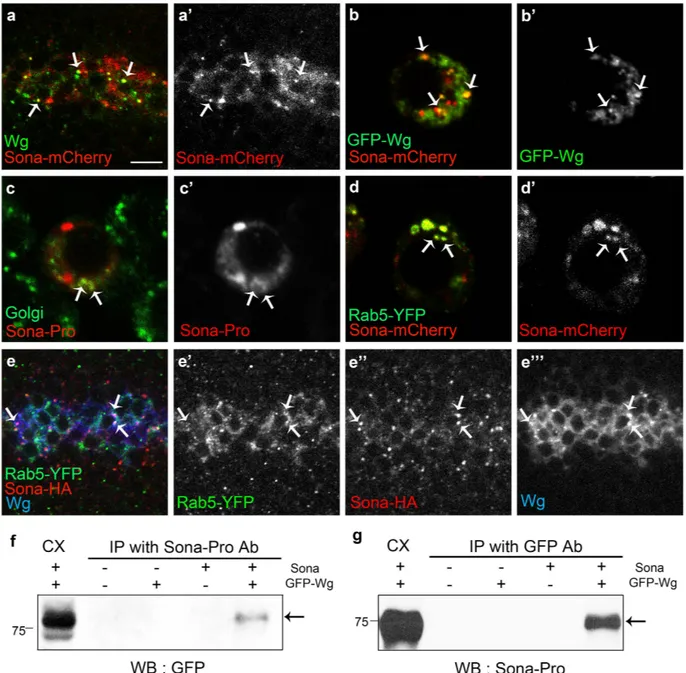
Sona and Wg colocalize in Golgi and endosomal vesicles.
Because both Sona and Wg are secreted proteins, and loss of sona decreased the level of extracellular Wg, we examined whether intracellular Sona and Wg are colocalized in S2 cells and wing discs. Both Wg and Sona-mCherry were enriched in the apical region of the wg > sona-mCherry wing disc (Supplementary Fig. S12), and about 60% (46/75, n = 4) of Sona-mCherry+vesicles contained Wg and vice versa (Fig. 7a). Vesicles containing both Wg and Sona were also observed in S2 cells cotransfected with GFP-wg and sona-mCherry cDNAs (Fig. 7b). To address the nature of the vesicles con-taining both Wg and Sona, we checked whether Sona+ vesicles corresponded to Golgi vesicles in both wing discs
and S2 cells. We found that some Sona+ vesicles are visualized with a Golgi marker P12051,52 in S2 cells (Fig. 7c).
Vesicles containing both Rab5-YFP and Sona-mCherry were frequently observed inside of S2 cells (Fig. 7d). About 30% (16/53) of Sona+ vesicles in the DV boundary region also contained Rab5-YFP in wg > rab5-YFP wing
discs (Fig. 7e). Furthermore, Rab5-YFP+ Sona+ vesicles also contained Wg (Fig. 7e”’). Rab5, a small GTPase that
regulates endocytic vesicle formation and early endosome fusion, is known to significantly co-localize with Wg and is involved in activation of Wg signaling53. Co-immunoprecipitation analysis showed that Sona and Wg are
Figure 5. Active Sona is present in ECM. (a) Extracellular patterns of Sona in the basal ECM of disc proper. Sona-Pro and -C antibodies detected overlapping structures. The region marked with a square was magnified in (e). (b) Extracellular patterns detected by Sona-Pro antibody in Collagen IV GFP-trap line, vikingG454
(Vkg-GFP/CyO). GFP signal (green) was detected as representing both intracellular and extracellular Viking-GFP. To detect only the extracellular Viking-GFP, rabbit anti-GFP antibody and Cy5-conjugated secondary antibody were used with extracellular staining method in (b”). (c) A diagram of peripodial epithelium (PE) and disc proper (DP) layer of a wing disc. (d) A cross-section image of a wing disc with the extracellular pattern shown with Sona-Pro antibody. Intracellular Dlg that is enriched at the septate junction was counterstained to show the structure of the wing disc. (e) The magnified image of the marked region in (a), showing the difference in the pattern recognized by Sona-Pro and Sona-C antibodies. (f,g) Extracellular patterns of HA-tagged Sona and Sona prodomain visualized with HA and Sona-Pro antibody in a nub > sona-HA disc. The region marked with a square in (f) was magnified in (g). Scale bars: (a,b,d) 100 μ m; (e) 18.8 μ m; (f) 40 μ m; (g) 6.5 μ m.
present in the same protein complex (Fig. 7f,g). Taken together, Sona and Wg may be secreted together in the same secretory pathway.
Figure 6. Loss of Sona increases the level of intracellular but decreases extracellular Wg. (a–d) Notched wing phenotype of dpp > wls RNAi flies (a) was rescued by expression of sona (c) but not sonaE475 protease mutant form (d). dpp > sona wings had no phenotype (b). (e,f) Intracellular Wg in sona RNAi clones. hs-Flp,
P[Actin > yellow > Gal4; w + ]/+; UAS-p35, UAS-GFP/+ wing disc as a control (e). sona RNAi-1 clones in hs-Flp, P[Actin > yellow > Gal4; w +]/+; UAS-p35, UAS-GFP/+; UAS-sona RNAi-1/+ (f). Arrows indicate the sona RNAi-1 clones with the increased level of intracellular Wg (f′). (g-i) Changes in the level of extracellular Wg by sona RNAi expression. UAS-p35, UAS-GFP/apterous (ap)-Gal4; +/TM6 Tb as a control (g), UAS-p35, UAS-GFP/ap-Gal4; UAS-sona RNAi/+ (h), and UAS-GFP/ap-UAS-GFP/ap-Gal4; UAS-sona RNAi/+ (i). (j) Decrease in the extracellular
level of Wg in the anterior region of ci > sona RNAi-2 discs. The control is shown in Supplementary Fig. S11. (k) Decrease in the level of extracellular Wg in sona13 clones near the DV boundary. Scale bar: (e,f) 40 μ m; (g–j)
www.nature.com/scientificreports/
Sona positively regulates Wg signaling.
To further test whether Sona is required for Wg signaling, we checked the effect of sona RNAi-1 on the expression of Sens, Dll and Vg in wing discs. P35 was coexpressed to prevent cell death by sona RNAi expression. Expression level of Sens was reduced in 80% (n = 9), and those of Dll (n = 46) and Vg (n = 25) were reduced in 100% of discs expressing sona RNAi-1 (Fig. 8b,d,f), compared to the control discs (n > 10 for each) (Fig. 8a,c,e). Same results were obtained with sona RNAi-2 driven by ci-Gal4 (Supplementary Fig. S13). The expression level of Wg-LacZ was not decreased upon sona RNAi expression (Supplementary Fig. S14). Hence, Sona positively regulates Wg signaling by post-transcriptional regulation.We then examined adult wings whether Sona can enhance Wg signaling. Since prolonged overexpression of either Sona or Wg caused lethality and developmental defects, we used Gal80ts system to transiently express
Figure 7. Sona and Wg colocalize in Golgi and endosomal vesicles. (a,b) Expression patterns of Sona-mCherry driven by wg-Gal4 and endogenous Wg in the DV midline of wing discs (a) and those of GFP-Wg and Sona-mCherry in S2 cells transfected with GFP-wg and sona-mCherry cDNAs (b). Arrows point to a few representative vesicles containing both Wg and Sona-mCherry. (c) S2 cells transfected with sona cDNA were visualized for Sona (red) and P120 (green), a Golgi marker. Some vesicles containing both Sona and P120 are marked with arrows. (d) In S2 cells transfected with rab5-YFP and sona-mCherry cDNAs, Rab5-YFP+
Sona-mCherry+ vesicles were frequently present in the cytoplasm (arrows). (e) Vesicles containing
Rab5-YFP and Sona-HA expressed by wg-Gal4 in addition to endogenous Wg are marked with arrows. (f,g) Co-immunoprecipitation (IP) with cell extracts obtained from GFP-wg and sona transfected S2 cells. Cell extracts were IPed with Sona-Pro antibody, and GFP-Wg was detected with GFP antibody (arrow in f). Cell extracts were IPed with GFP antibody, and Sona was detected with Sona-Pro antibody (g). Scale bars: (a,e) 7.5 μ m; (b–d) 5 μ m.
GFP-Wg and Sona54. Transient expression of either GFP-wg or sona with nub-Gal4 during the late third larval
and early pupal stage for 18 hours had no effect on the number of anterior bristles in adult wings or on lethality (n > 40 each; Fig. 8g,h). However, coexpression of GFP-wg and sona in the same culture condition caused lethality in about 85% of animals, and the survivors had multiple ectopic bristles near the anterior wing margin (n > 50; Fig. 8i,j). These phenotypes demonstrated that Sona promotes Wg signaling.
Discussion
ADAMTSs are secreted metalloproteases that are known to be involved in mainly ECM remodeling. Among six ADAMTSs in the fly, Papilin is essential for the formation of basement membrane and fly development32, Stall
functions in ovarian follicle formation and exhibits positive genetic interaction with Delta35, and ADAMTS-A is
important for cell migration, especially in detaching cells from the apical ECM in salivary gland34. In this report, we
have shown that Sona is a fly ADAMTS essential for fly development and survival. Transient coexpression of Sona and Wg increased the number of wing margin bristles, indicating that Sona is positively involved in Wg signaling. Figure 8. Sona enhances Wg signaling. (a–f) UAS-p35, UAS-GFP/CyO-GFP; UAS-sona RNAi/TM6 Tb and
ap-Gal4/CyO-GFP flies were crossed and the larval progeny UAS-p35 UAS-GFP/ap-Gal4; +/TM6 Tb and UAS-p35, UAS-GFP/ap-Gal4; UAS-sona RNAi/+ were used as control and experimental animals, respectively. Patterns of
Sens, Dll and Vg in wing discs. Anterior is left, and dorsal is up. Images of control discs (a,c,e) and experimental discs (b,d,f) are shown. Black and white images of Sens, Dll or Vg in (a’–f’). The expression region of ap-Gal4 is marked by GFP (green). (g–j) Wing phenotypes of flies transiently expressing GFP-Wg (g), Sona (h) or both (i,j) by nub-Gal4 driver. The anterior margins of wings in (g–j) are magnified in (g’–j’). Ectopic bristles are marked with arrows (i’,j’). Genotypes of discs and adult wings are indicated at lower left. Scale bar: (a–f) 100 μ m.
www.nature.com/scientificreports/
Accordingly, loss of sona decreased the level of Wg effector proteins as well as the level of extracellular Wg. Based on these results, we propose that Sona, as an ADAMTS, modifies yet unidentified protein(s) essential for Wg signaling. During fly development, sona was transcribed at a high level in discrete regions in imaginal discs, which cor-responded to the malformed regions in adult appendages of sona escapers (Figs 2 and 4). For instance, dorsal eye disc, the center of antenna disc, and outer ring of leg disc expressed the high level of sona transcripts, and sona escapers accordingly had disoriented ommatidial bristles in the dorsal eye, malformed arista, and kinked femur (Figs 2a–f and 4a–f). Wing disc also exhibited the complicated mosaic pattern of sona transcription, and adult wings of sona escapers were small and abnormally shaped (Figs 2g–j and 4g–i). Involvement of Sona in modulat-ing the level of extracellular Wg may explain why these malformed adult structures are generated in sona escapers because Wg is specifically expressed in eye, wing and leg discs and determines the fate of organs55–58.
The genetic link between Sona and Wg signaling was identified in a genetic screen in which a wls allele could rescue the lethal phenotype caused by the overexpression of Sona. Likewise, wing notching by the loss of wls was rescued by overexpression of sona (Fig. 6c). Furthermore, the loss of sona decreased the level of extracellular Wg (Fig. 6g–i). Taken together, these results raised a possibility that Sona may be involved in Wg signaling by affecting Wg secretion. How may Sona positively regulate Wg secretion? To act on Wg secretion, Sona has to be activated intracellularly, and function in secretory pathways. It has been shown that the prodomains of most ADAMTSs are cleaved in trans-Golgi network to become active. Thus, activated intracellular Sona may cleave unidentified pro-teins involved in Wg secretion and thereby promote the secretion of Wg. Indeed, intracellular Sona was enriched in the apical region while extracellular Sona are more enriched in the basolateral region. Similarly, intracellular and extracellular Wg are enriched in the apical and basolateral regions, respectively40 (Fig. 5d; Supplementary Fig. S12).
It has been recently shown that Wg is secreted to the apical side and then reentered cells by endocytosis, and then moves to the basal side and secreted by transcytosis59. It will be interesting to figure out whether Sona and Wg
may be secreted together by transcytosis.
Besides the function of intracellular Sona for Wg secretion, presence of active Sona in conditioned medium of S2 cell culture suggests that extracellular active Sona may be involved in Wg signaling by modifying unknown ECM components (Fig. 3e). Immunocytochemical analysis of Sona confirmed that the active form of Sona devoid of the prodomain is present in basal ECM of wing discs (Fig. 5e–g). Therefore, active Sona may cleave ECM pro-teins that affect stability or activity of Wg. Well-studied ECM propro-teins essential for Wg signaling and formation of Wg gradient are Heparan sulfate proteoglycans (HSPG) such as Division abnormally delayed (Dally) and Dally-like (Dlp)60–67. These HSPGs can be modified by proteins such as Notum and Matrix metalloprotease 2
(Mmp2). Notum blocks Wg activity as α /β -hydrolase by modifying Dally and Dlp68, and Mmp2 cleaves Dlp to
inhibit the interaction between Dlp and Wg69. Thus, Sona may act on these HSPGs or related ECM proteins to
affect the stability or activity of extracellular Wg.
Extracellular Sona was highly localized in the presumptive region of wing blade in the basal ECM near the Collagen-IV containing region, but was present at lower level in the DV midline region where Wg is synthesized (Fig. 5). This data suggests that secreted extracellular Sona may not be diffused freely but restricted to a defined region by interacting with some ECM components. Another component of Wg signaling, Frizzled2 (Fz2), is also localized in the presumptive region of wing blade, but Frizzled3 (Fz3) is expressed exclusively in the DV midline64,70–72. Interestingly, Fz2 promotes but Fz3 attenuates Wg signaling71,73. Thus, these Fz proteins are
stra-tegically localized to bind the extracellular Wg in order to regionally regulate strength of Wg signaling. Similarly, Dally promotes but Dlp decreases Wg signaling in the DV midline63,64, and extracellular Dlp is present at a lower
level in the DV midline region60. Taken together, specific localization of these Wg signaling components in ECM
may be essential for modulating Wg signaling with regional specificity in wing discs.
Involvement of Sona in Wg signaling raises a possibility that some mammalian ADAMTSs may also be involved in Wnt signaling. Some mammalian ADAMTSs are known to function as positive factors for tumor invasion and progression74–76. Overexpression of Wnts or downstream components of Wnt signaling also induces
various tumors such as colon cancer, breast cancer, and leukemia77,78. Wnt signaling is also essential for the
growth and remodeling of bones and connective tissues79–82. Overlapping functions of ADAMTSs and Wnt
sig-naling supports our view that some mammalian ADAMTSs may be linked to Wnt sigsig-naling. Further work on identifying the intracellular or extracellular substrate(s) of Sona is required to fully understand how Sona is pos-itively involved in Wg signaling.
Materials and Methods
Drosophila strains, transgenic lines and generation of ectopic clones.
We carried out imprecise excision of a P element, P{GawB}CG9850[NP5284], and obtained 9 sona deletion mutants by screening about 200 flies. Deficiency lines (Df(2R)X32, Df(2R)23, Df(2R)3-659) that uncover the sona gene were used to confirmsona deletions. We generated two sona RNAi lines targeting different regions of the sona coding region (Fig. 1b;
Supplementary Fig. S1) and several UAS transgenic flies: sona, sona-HA, sonaE475A, and
UAS-sona-mCherry. UAS-GFP-wg83, wg-Gal464 and viking-GFP 41 were kindly provided. All other lines were obtained
from Bloomington stock center.
For generation of ectopic clones, hs-Flp, UAS-sona-mCherry/TM6 Tb males and y w ; P[Actin > CD2 > Gal4;
w + ]/Cyo-GFP ; UAS-GFP/TM6 Tb females were crossed, their progeny were raised at 18 °C until 48 hr AEL, and
then heat shocked at 37 °C for 1 hour as described3. They were then kept at 18 °C until dissection.
DNA constructs.
sona cDNA was originally obtained from a two-hybrid cDNA library screening84 using thefirst and second PDZ domains of Discs-Large (Dlg) as a bait85,86. Out-of-frame fusion between the Gal4 activating
domain and the portion of sona gene that encodes the carboxyl terminus fortuitously generated a perfect but gratuitous PDZ binding motif, which let Sona to be identified in the screen.
To generate sona-HA construct, HA tag was attached at Gly638 by removing stop codon with HpaI digestion and ligating amplified HA tag. pUAST-sona-HA-mCherry was generated by tagging mCherry at the downstream of
HA tag in the pUAST-sona-HA plasmid. pUAST-sonaE475A was generated by changing GAA (glu) to GCA (ala)
by site-directed mutagenesis. pAc-GFP-wg was constructed by recombining the pAc5.1 vector and GFP-wg that was derived from MK33-GFP-wg (a gift from J.P. Vincent, unpublished).
Northern blot and in situ hybridization.
For Northern analysis, RNA was prepared as described and 20 μ g of RNA each was loaded in 2.2 M formaldehyde containing agarose gel87. The RNAs were transferred tonitrocel-lulose and probed with 32P-labeled 1.9 kb XhoI fragment that covers the 3′ half of the sona cDNA for Northern
analysis. The same 1.9 kb XhoI fragment was linearlized and amplified by polymerase chain reaction using one primer to make digoxigenin-labelled single-stranded DNA probe for in situ mRNA hybridization as described88.
Cell culture and transfection.
Drosophila S2 cells were grown in M3 media (Sigma-Aldrich) supplementedwith 10% IMS (Sigma-Aldrich) at 25 °C. Transfections were carried out with transfection reagents effectene (Qiagen) or cellfectin (Invitrogen) according to the manufacturers’ instructions. For each transfection, a total of 1–2 μ g DNA was used.
To establish S2 sona-HA cell line that stably expresses sona-HA, Hygromycin B selection system was used according to the manufacturers’ instructions (Invitrogen life technologies). Briefly, 4 × 10⁶ S2 cells were
cotrans-fected with total 1 μ g of two plasmids, pAC sona-HA and pCoHygro (19:1 ratio), for 3 days. Then, the culture medium was changed to selective medium containing 150 μ g/ml of hygromycin B (Invitrogen). The selective medium was replaced every 5 days, and the sona-HA cell lines were established after 3 weeks. The established
sona-HA cell line was maintained in selective medium containing Hygromycin B.
Antibodies and immunocytochemistry.
We generated Sona-Pro antisera in rabbits using GST fusion pro-tein with 245 amino acid residues representing 20th to 264th, and Sona-C antisera in mice using 378 amino acid residues representing 262th to 639th of Sona protein as antigens. Both Sona-Pro and Sona-C antisera were puri-fied by affinity purification using MBP- Pro and GST-C, respectively. Following dilutions were used for Western analysis: Sona-Pro, 1:5,000; HA (Santa Cruz, rabbit), 1: 250, and for immunocytochemistry: Sona-Pro, 1:300~500; Sona-C, 1:300~500; Golgi (Calbiochem, mouse), 1:200; GFP (Abd Serotec, sheep), 1:100; Senseless (gift from H. Bellen, guinea pig), 1:1,000; HA (Roche, rat), 1:150; HA (Santa Cruz, rabbit), 1:100; Dlg (rabbit), 1:500; Dlg (DSHB, mouse), 1:100; Wg (DSHB, mouse), 1:100; Dll (Santa Cruz, goat), 1:100; Vg, 1:100 (gift from S. Carroll, rabbit).Fly larvae were cultured at 25 °C unless stated otherwise. For intracellular staining, imaginal discs were dis-sected and stained as described85. The samples were incubated with primary antibodies in washing buffer (50 mM
Tris pH6.8, 150 mM NaCl, 0.5% NP-40, 1 mg/ml BSA) for overnight at 4 °C or room temperature and washed with washing buffer several times. Then, samples were incubated with secondary antibodies and washed several times before mounting. For extracellular staining of Wg and Sona, we followed the method as described87. The
wing imaginal discs were dissected in M3 media at 4 °C. The samples were incubated with about 3–4 fold more primary antibodies than for intracellular staining in cold M3 media for 2 hrs. Then, samples were washed with cold M3 and PBS twice, fixed for 40 minutes in 4% paraformaldehyde/PBS, and then socked in non-detergent blocking buffer (60 mM Tris pH6.8, 150 mM NaCl, 5 mg/ml BSA) for 3 hrs at 4 °C. Samples were then incubated with secondary antibodies with non-detergent washing buffer (50 mM Tris pH6.8, 150 mM NaCl, 1 mg/ml BSA) and washed with non-detergent blocking buffer four times and detergent blocking buffer twice before mounting. For HA and GFP extracellular staining, the following antibodies, HA (Roche, rat, 1:30) and GFP (Abcam, rabbit, 1:30) were used. Fluorescent images were captured using Zeiss LSM laser scanning confocal microscope and presented using Adobe Photoshop.
Western analysis and co-immunoprecipitation.
For Western analysis, samples were mixed with 4 × SDS sample buffer and boiled at 95 °C for 10 min. Samples were then separated by 10–12% SDS-PAGE and transferred to the nitrocellulose membrane (Millipore). Membranes were blocked with 5% nonfat milk in TBST buffer (10 mM Tris pH 7.4, 0.8% NaCl, 0.1% Tween-20), and probed with the antibody. After washing membranes with TBST several times, membranes were incubated with horseradish peroxidase (HRP)-conjugated secondary antibody in TBST with 5% nonfat milk. After washing, protein bands were visualized using the ECL system (AbFrontier).For co-immunoprecipitation, cells were lysed in HEPES buffer (20 mM HEPES, 70 mM KCl, 2 mM DTT, 0.1% NP40, 8% Glycerol, 1 mM PMSF, 10 mM EDTA, 10 mM EGTA, and protease inhibitor cocktail (Roche)) on ice. The lysates were precleared by incubating with protein G-sepharose beads (Amersham Bioscience) for 30 min at 4 °C. A new set of G-sepharose beads were incubated with anti-GFP (Abcam, rabbit) or Sona-Pro (rabbit) for cou-pling at room temperature for 2 hrs. The precleared lysates were then incubated with coupled protein G-sepharose beads for overnight at 4 °C. The protein G-sepharose beads were washed with HEPES buffer and Western blots were performed as described.
References
1. Page, M. J. & Di Cera, E. Evolution of peptidase diversity. J Biol Chem 283, 30010–30014, doi: 10.1074/jbc.M804650200 (2008). 2. Rawlings, N. D., Barrett, A. J. & Bateman, A. Using the MEROPS Database for Proteolytic Enzymes and Their Inhibitors and
Substrates. Curr Protoc Bioinformatics 48, 1 25 21-21 25 33, doi: 10.1002/0471250953.bi0125s48 (2014).
3. Porter, S., Clark, I. M., Kevorkian, L. & Edwards, D. R. The ADAMTS metalloproteinases. Biochem J 386, 15–27, doi: BJ20040424 10.1042/BJ20040424 (2005).
4. Kuno, K., Iizasa, H., Ohno, S. & Matsushima, K. The exon/intron organization and chromosomal mapping of the mouse ADAMTS-1 gene encoding an ADAM family protein with TSP motifs. Genomics 46, 466–471, doi: S0888-7543(97)95064-3 10.1006/ geno.1997.5064 (1997).
www.nature.com/scientificreports/
5. Russell, D. L., Doyle, K. M., Ochsner, S. A., Sandy, J. D. & Richards, J. S. Processing and localization of ADAMTS-1 and proteolytic cleavage of versican during cumulus matrix expansion and ovulation. J Biol Chem 278, 42330–42339, doi: 10.1074/jbc.M300519200 (2003).
6. Colige, A. et al. cDNA cloning and expression of bovine procollagen I N-proteinase: a new member of the superfamily of zinc-metalloproteinases with binding sites for cells and other matrix components. Proc Natl Acad Sci USA 94, 2374–2379 (1997). 7. Jones, G. C. & Riley, G. P. ADAMTS proteinases: a multi-domain, multi-functional family with roles in extracellular matrix turnover
and arthritis. Arthritis Res Ther 7, 160–169, doi: ar1783 10.1186/ar1783 (2005).
8. Wang, W. M. et al. Transforming growth factor-beta induces secretion of activated ADAMTS-2. A procollagen III N-proteinase. J
Biol Chem 278, 19549–19557, doi: 10.1074/jbc.M300767200 M300767200 (2003).
9. Colige, A. et al. Human Ehlers-Danlos syndrome type VII C and bovine dermatosparaxis are caused by mutations in the procollagen I N-proteinase gene. Am J Hum Genet 65, 308–317, doi: S0002-9297(07)62047-3 10.1086/302504 (1999).
10. Liu, C. J. The role of ADAMTS-7 and ADAMTS-12 in the pathogenesis of arthritis. Nat Clin Pract Rheumatol 5, 38–45, doi: 10.1038/ ncprheum0961 (2009).
11. Dong, J. F. et al. ADAMTS-13 rapidly cleaves newly secreted ultralarge von Willebrand factor multimers on the endothelial surface under flowing conditions. Blood 100, 4033–4039, doi: 10.1182/blood-2002-05-1401 2002-05-1401 (2002).
12. Roy, R. et al. Tumor-specific urinary matrix metalloproteinase fingerprinting: identification of high molecular weight urinary matrix metalloproteinase species. Clin Cancer Res 14, 6610–6617, doi: 10.1158/1078-0432.CCR-08-1136 (2008).
13. Rocks, N. et al. Emerging roles of ADAM and ADAMTS metalloproteinases in cancer. Biochimie 90, 369–379, doi: S0300-9084(07)00222-2 10.1016/j.biochi.2007.08.008 (2008).
14. Lo, P. H. et al. Extracellular protease ADAMTS9 suppresses esophageal and nasopharyngeal carcinoma tumor formation by inhibiting angiogenesis. Cancer Res 70, 5567–5576, doi: 10.1158/0008-5472.CAN-09-4510 0008-5472.CAN-09-4510 (2010). 15. Surridge, A. K. et al. Characterization and regulation of ADAMTS-16. Matrix Biol 28, 416–424, doi: 10.1016/j.matbio.2009.07.001
(2009).
16. El Hour, M. et al. Higher sensitivity of Adamts12-deficient mice to tumor growth and angiogenesis. Oncogene 29, 3025–3032, doi: 10.1038/onc.2010.49 onc201049 (2010).
17. Moncada-Pazos, A. et al. The ADAMTS12 metalloprotease gene is epigenetically silenced in tumor cells and transcriptionally activated in the stroma during progression of colon cancer. J Cell Sci 122, 2906–2913, doi: 10.1242/jcs.050468 jcs.050468 (2009). 18. Cal, S. et al. Cloning, expression analysis, and structural characterization of seven novel human ADAMTSs, a family of
metalloproteinases with disintegrin and thrombospondin-1 domains. Gene 283, 49–62, doi: S0378111901008617 (2002). 19. Tortorella, M. D. et al. ADAMTS-4 (aggrecanase-1): N-terminal activation mechanisms. Arch Biochem Biophys 444, 34–44, doi:
S0003-9861(05)00401-7 10.1016/j.abb.2005.09.018 (2005).
20. Longpre, J. M. & Leduc, R. Identification of prodomain determinants involved in ADAMTS-1 biosynthesis. J Biol Chem 279, 33237–33245, doi: 10.1074/jbc.M313151200 (2004).
21. Wang, P. et al. Proprotein convertase furin interacts with and cleaves pro-ADAMTS4 (Aggrecanase-1) in the trans-Golgi network. J
Biol Chem 279, 15434–15440, doi: 10.1074/jbc.M312797200 (2004).
22. Koo, B. H. et al. Cell-surface processing of pro-ADAMTS9 by furin. J Biol Chem 281, 12485–12494, doi: M511083200 10.1074/jbc. M511083200 (2006).
23. Somerville, R. P., Jungers, K. A. & Apte, S. S. Discovery and characterization of a novel, widely expressed metalloprotease, ADAMTS10, and its proteolytic activation. J Biol Chem 279, 51208–51217, doi: 10.1074/jbc.M409036200 M409036200 (2004). 24. Dancevic, C. M. et al. Biosynthesis and Expression of a Disintegrin-like and Metalloproteinase Domain with Thrombospondin-1
Repeats-15: A Novel Versican-Cleaving Proteoglycanase. J Biol Chem 288, 37267–37276, doi: 10.1074/jbc.M112.418624 M112.418624 (2013).
25. Nishiwaki, K., Hisamoto, N. & Matsumoto, K. A metalloprotease disintegrin that controls cell migration in Caenorhabditis elegans.
Science 288, 2205–2208, doi: 8628 (2000).
26. Ihara, S. & Nishiwaki, K. Prodomain-dependent tissue targeting of an ADAMTS protease controls cell migration in Caenorhabditis elegans. EMBO J 26, 2607–2620, doi: 7601718 10.1038/sj.emboj.7601718 (2007).
27. Clevers, H. & Nusse, R. Wnt/beta-catenin signaling and disease. Cell 149, 1192–1205, doi: 10.1016/j.cell.2012.05.012 S0092-8674(12)00586-7 (2012).
28. Logan, C. Y. & Nusse, R. The Wnt signaling pathway in development and disease. Annu Rev Cell Dev Biol 20, 781–810, doi: 10.1146/ annurev.cellbio.20.010403.113126 (2004).
29. Zecca, M., Basler, K. & Struhl, G. Direct and long-range action of a wingless morphogen gradient. Cell 87, 833–844, doi: S0092-8674(00)81991-1 (1996).
30. Neumann, C. J. & Cohen, S. M. Long-range action of Wingless organizes the dorsal-ventral axis of the Drosophila wing. Development
124, 871–880 (1997).
31. Nolo, R., Abbott, L. A. & Bellen, H. J. Senseless, a Zn finger transcription factor, is necessary and sufficient for sensory organ development in Drosophila. Cell 102, 349–362, doi: S0092-8674(00)00040-4 (2000).
32. Kramerova, I. A. et al. Papilin in development; a pericellular protein with a homology to the ADAMTS metalloproteinases.
Development 127, 5475–5485 (2000).
33. Meyer, H. et al. Drosophila metalloproteases in development and differentiation: the role of ADAM proteins and their relatives. Eur
J Cell Biol 90, 770–778, doi: 10.1016/j.ejcb.2011.04.015 S0171-9335(11)00088-4 (2011).
34. Ismat, A., Cheshire, A. M. & Andrew, D. J. The secreted AdamTS-A metalloprotease is required for collective cell migration.
Development 140, 1981–1993, doi: 10.1242/dev.087908 dev.087908 (2013).
35. Ozdowski, E. F., Mowery, Y. M. & Cronmiller, C. Stall encodes an ADAMTS metalloprotease and interacts genetically with Delta in Drosophila ovarian follicle formation. Genetics 183, 1027–1040, doi: 10.1534/genetics.109.107367 genetics.109.107367 (2009). 36. Ote, M., Mita, K., Kawasaki, H., Kobayashi, M. & Shimada, T. Characteristics of two genes encoding proteins with an ADAM-type
metalloprotease domain, which are induced during the molting periods in Bombyx mori. Arch Insect Biochem Physiol 59, 91–98, doi: 10.1002/arch.20059 (2005).
37. Tsutsui, K. et al. ADAMTSL-6 is a novel extracellular matrix protein that binds to fibrillin-1 and promotes fibrillin-1 fibril formation.
J Biol Chem 285, 4870–4882, doi: 10.1074/jbc.M109.076919 (2010).
38. Couso, J. P. & Bishop, S. A. Proximo-distal development in the legs of Drosophila. Int J Dev Biol 42, 345–352 (1998). 39. Pignoni, F. & Zipursky, S. L. Induction of Drosophila eye development by decapentaplegic. Development 124, 271–278 (1997). 40. Strigini, M. & Cohen, S. M. Wingless gradient formation in the Drosophila wing. Curr Biol 10, 293–300, doi:
S0960-9822(00)00378-X (2000).
41. Morin, X., Daneman, R., Zavortink, M. & Chia, W. A protein trap strategy to detect GFP-tagged proteins expressed from their endogenous loci in Drosophila. Proc Natl Acad Sci USA 98, 15050–15055, doi: 10.1073/pnas.261408198 (2001).
42. Banziger, C. et al. Wntless, a conserved membrane protein dedicated to the secretion of Wnt proteins from signaling cells. Cell 125, 509–522, doi: S0092-8674(06)00446-6 10.1016/j.cell.2006.02.049 (2006).
43. Bartscherer, K., Pelte, N., Ingelfinger, D. & Boutros, M. Secretion of Wnt ligands requires Evi, a conserved transmembrane protein.
Cell 125, 523–533, doi: S0092-8674(06)00451-X 10.1016/j.cell.2006.04.009 (2006).
44. Goodman, R. M. et al. Sprinter: a novel transmembrane protein required for Wg secretion and signaling. Development 133, 4901–4911, doi: dev.02674 10.1242/dev.02674 (2006).
45. Belenkaya, T. Y. et al. The retromer complex influences Wnt secretion by recycling wntless from endosomes to the trans-Golgi network. Dev Cell 14, 120–131, doi: S1534-5807(07)00480-7 10.1016/j.devcel.2007.12.003 (2008).
46. Eaton, S. Retromer retrieves wntless. Dev Cell 14, 4–6, doi: 10.1016/j.devcel.2007.12.014 S1534-5807(07)00491-1 (2008). 47. Franch-Marro, X. et al. Wingless secretion requires endosome-to-Golgi retrieval of Wntless/Evi/Sprinter by the retromer complex.
Nat Cell Biol 10, 170–177, doi: 10.1038/ncb1678 ncb1678 (2008).
48. Port, F. et al. Wingless secretion promotes and requires retromer-dependent cycling of Wntless. Nat Cell Biol 10, 178–185, doi: 10.1038/ncb1687 ncb1687 (2008).
49. Yang, P. T. et al. Wnt signaling requires retromer-dependent recycling of MIG-14/Wntless in Wnt-producing cells. Dev Cell 14, 140–147, doi: S1534-5807(07)00481-9 10.1016/j.devcel.2007.12.004 (2008).
50. Clem, R. J., Fechheimer, M. & Miller, L. K. Prevention of apoptosis by a baculovirus gene during infection of insect cells. Science 254, 1388–1390 (1991).
51. Yano, H. et al. Distinct functional units of the Golgi complex in Drosophila cells. Proc Natl Acad Sci USA 102, 13467–13472, doi: 0506681102 10.1073/pnas.0506681102 (2005).
52. Cho, K. O., Kim, G. W. & Lee, O. K. Wolbachia bacteria reside in host Golgi-related vesicles whose position is regulated by polarity proteins. PLoS One 6, e22703, doi: 10.1371/journal.pone.0022703PONE-D-11-00756 (2011).
53. Marois, E., Mahmoud, A. & Eaton, S. The endocytic pathway and formation of the Wingless morphogen gradient. Development 133, 307–317, doi: dev.02197 10.1242/dev.02197 (2006).
54. McGuire, S. E., Le, P. T., Osborn, A. J., Matsumoto, K. & Davis, R. L. Spatiotemporal rescue of memory dysfunction in Drosophila.
Science 302, 1765–1768, doi: 10.1126/science.1089035 302/5651/1765 (2003).
55. Baker, N. E. Transcription of the segment-polarity gene wingless in the imaginal discs of Drosophila, and the phenotype of a pupal-lethal wg mutation. Development 102, 489–497 (1988).
56. Wilder, E. L. & Perrimon, N. Dual functions of wingless in the Drosophila leg imaginal disc. Development 121, 477–488 (1995). 57. Couso, J. P., Knust, E. & Martinez Arias, A. Serrate and wingless cooperate to induce vestigial gene expression and wing formation
in Drosophila. Curr Biol 5, 1437–1448 (1995).
58. Treisman, J. E. & Rubin, G. M. wingless inhibits morphogenetic furrow movement in the Drosophila eye disc. Development 121, 3519–3527 (1995).
59. Yamazaki, Y. et al. Godzilla-dependent transcytosis promotes Wingless signalling in Drosophila wing imaginal discs. Nat Cell Biol
18, 451–457, doi: 10.1038/ncb3325 (2016).
60. Baeg, G. H., Selva, E. M., Goodman, R. M., Dasgupta, R. & Perrimon, N. The Wingless morphogen gradient is established by the cooperative action of Frizzled and Heparan Sulfate Proteoglycan receptors. Dev Biol 276, 89–100, doi: S0012-1606(04)00577-9 10.1016/j.ydbio.2004.08.023 (2004).
61. Han, C., Belenkaya, T. Y., Wang, B. & Lin, X. Drosophila glypicans control the cell-to-cell movement of Hedgehog by a dynamin-independent process. Development 131, 601–611, doi: 10.1242/dev.00958 (2004).
62. Khare, N. & Baumgartner, S. Dally-like protein, a new Drosophila glypican with expression overlapping with wingless. Mech Dev 99, 199–202, doi: S0925477300005025 (2000).
63. Kirkpatrick, C. A., Dimitroff, B. D., Rawson, J. M. & Selleck, S. B. Spatial regulation of Wingless morphogen distribution and signaling by Dally-like protein. Dev Cell 7, 513–523, doi: S1534580704002795 10.1016/j.devcel.2004.08.004 (2004).
64. Kreuger, J., Perez, L., Giraldez, A. J. & Cohen, S. M. Opposing activities of Dally-like glypican at high and low levels of Wingless morphogen activity. Dev Cell 7, 503–512, doi: S1534580704002801 10.1016/j.devcel.2004.08.005 (2004).
65. Lin, X. & Perrimon, N. Dally cooperates with Drosophila Frizzled 2 to transduce Wingless signalling. Nature 400, 281–284, doi: 10.1038/22343 (1999).
66. Tsuda, M. et al. The cell-surface proteoglycan Dally regulates Wingless signalling in Drosophila. Nature 400, 276–280, doi: 10.1038/22336 (1999).
67. Yan, D., Wu, Y., Feng, Y., Lin, S. C. & Lin, X. The core protein of glypican Dally-like determines its biphasic activity in wingless morphogen signaling. Dev Cell 17, 470–481, doi: 10.1016/j.devcel.2009.09.001 S1534-5807(09)00383-9 (2009).
68. Giraldez, A. J., Copley, R. R. & Cohen, S. M. HSPG modification by the secreted enzyme Notum shapes the Wingless morphogen gradient. Dev Cell 2, 667–676, doi: S1534580702001806 (2002).
69. Wang, X. & Page-McCaw, A. A matrix metalloproteinase mediates long-distance attenuation of stem cell proliferation. J Cell Biol
206, 923–936, doi: 10.1083/jcb.201403084 (2014).
70. Cadigan, K. M., Fish, M. P., Rulifson, E. J. & Nusse, R. Wingless repression of Drosophila frizzled 2 expression shapes the Wingless morphogen gradient in the wing. Cell 93, 767–777, doi: S0092-8674(00)81438-5 (1998).
71. Sato, A., Kojima, T., Ui-Tei, K., Miyata, Y. & Saigo, K. Dfrizzled-3, a new Drosophila Wnt receptor, acting as an attenuator of Wingless signaling in wingless hypomorphic mutants. Development 126, 4421–4430 (1999).
72. Sivasankaran, R., Calleja, M., Morata, G. & Basler, K. The Wingless target gene Dfz3 encodes a new member of the Drosophila Frizzled family. Mech Dev 91, 427–431 (2000).
73. Bhanot, P. et al. A new member of the frizzled family from Drosophila functions as a Wingless receptor. Nature 382, 225–230, doi: 10.1038/382225a0 (1996).
74. Held-Feindt, J. et al. Matrix-degrading proteases ADAMTS4 and ADAMTS5 (disintegrins and metalloproteinases with thrombospondin motifs 4 and 5) are expressed in human glioblastomas. Int J Cancer 118, 55–61, doi: 10.1002/ijc.21258 (2006). 75. Kessenbrock, K., Plaks, V. & Werb, Z. Matrix metalloproteinases: regulators of the tumor microenvironment. Cell 141, 52–67, doi:
10.1016/j.cell.2010.03.015 S0092-8674(10)00288-6 (2010).
76. Cal, S. & Lopez-Otin, C. ADAMTS proteases and cancer. Matrix Biol 44-46, 77–85, doi: 10.1016/j.matbio.2015.01.013 (2015). 77. Anastas, J. N. & Moon, R. T. WNT signalling pathways as therapeutic targets in cancer. Nat Rev Cancer 13, 11–26, doi: 10.1038/
nrc3419 nrc3419 (2013).
78. Reya, T. & Clevers, H. Wnt signalling in stem cells and cancer. Nature 434, 843–850, doi: 10.1038/nature03319 (2005).
79. Mercurio, S., Latinkic, B., Itasaki, N., Krumlauf, R. & Smith, J. C. Connective-tissue growth factor modulates WNT signalling and interacts with the WNT receptor complex. Development 131, 2137–2147, doi: 10.1242/dev.01045 (2004).
80. Luo, Q. et al. Connective tissue growth factor (CTGF) is regulated by Wnt and bone morphogenetic proteins signaling in osteoblast differentiation of mesenchymal stem cells. J Biol Chem 279, 55958–55968, doi: 10.1074/jbc.M407810200 (2004).
81. Macsai, C. E., Foster, B. K. & Xian, C. J. Roles of Wnt signalling in bone growth, remodelling, skeletal disorders and fracture repair.
J Cell Physiol 215, 578–587, doi: 10.1002/jcp.21342 (2008).
82. Minear, S. et al. Wnt proteins promote bone regeneration. Sci Transl Med 2, 29ra30, doi: 10.1126/scitranslmed.3000231 (2010). 83. Pfeiffer, S., Ricardo, S., Manneville, J. B., Alexandre, C. & Vincent, J. P. Producing cells retain and recycle Wingless in Drosophila
embryos. Curr Biol 12, 957–962, doi: S0960982202008679 (2002).
84. Durfee, T. et al. The retinoblastoma protein associates with the protein phosphatase type 1 catalytic subunit. Genes Dev 7, 555–569 (1993).
85. Woods, D. F. & Bryant, P. J. The discs-large tumor suppressor gene of Drosophila encodes a guanylate kinase homolog localized at septate junctions. Cell 66, 451–464, doi: 0092-8674(81)90009-X (1991).
86. Lee, O. K. et al. Discs-Large and Strabismus are functionally linked to plasma membrane formation. Nat Cell Biol 5, 987–993, doi: 10.1038/ncb1055 ncb1055 (2003).
www.nature.com/scientificreports/
87. Choi, K. W. & Benzer, S. Rotation of photoreceptor clusters in the developing Drosophila eye requires the nemo gene. Cell 78, 125–136 (1994).
88. Sambrook, J., Fritsch, E. F. & Maniatis, T. Molecular Cloning: A Laboratory Manual, Second Edition (Cold Spring Harbor, New York: Cold Spring Harbor Laboratory Press) (1989).
89. McLean, I. W. & Nakane, P. K. Periodate-lysine-paraformaldehyde fixative. A new fixation for immunoelectron microscopy.
J Histochem Cytochem. 22, 1077–1083 (1974).
Acknowledgements
We are grateful to K.-W. Choi and colleagues in our lab and Y.-K. Bae at KRISS for discussion and comments on manuscript. We are in debt to J. P. Vincent, K. Basler, S. Eaton, S. Hayashi, S. M. Cohen, R. Holmgren and H. Kramer for fly lines, K. Basler, J. P. Vincent and S.-T. Hong for DNA constructs, and H. Bellen and S. Carroll for antibodies. We thank Bloomington Stock Center, Drosophila Genetic Resource Center and Developmental Studies Hybridoma Bank for fly strains and antibodies. This research was supported by a KAIST Grant G04080077, N10100001, National Research Foundation R1A1A301573, and National Research Council of Science and Technology Grant DRC-14-2-KRISS.
Author Contributions
G.-W.K., J.-H.W. and K.-O.C. designed experiments, analyzed the data and wrote the paper. G.-W.K., J.-H.W., O.-K.L., S.-S.L., J.-H.H., O.T., S.N. and Y.K conducted the experiments.
Additional Information
Supplementary information accompanies this paper at http://www.nature.com/srep Competing financial interests: The authors declare no competing financial interests.
How to cite this article: Kim, G.-W. et al. Sol narae (Sona) is a Drosophila ADAMTS involved in Wg signaling.
Sci. Rep. 6, 31863; doi: 10.1038/srep31863 (2016).
This work is licensed under a Creative Commons Attribution 4.0 International License. The images or other third party material in this article are included in the article’s Creative Commons license, unless indicated otherwise in the credit line; if the material is not included under the Creative Commons license, users will need to obtain permission from the license holder to reproduce the material. To view a copy of this license, visit http://creativecommons.org/licenses/by/4.0/