2 0 13 년
2 월
석 사 학 위논 문
Bulkfill
복 합 레 진 의 중 합 수 축 평 가
장 슬 기
2013년 2월 석사학위논문
Bulk fill 복합레진의 중합 수축 평가
조선대학교 대학원
치 의 학 과
장 슬 기
[UCI]I804:24011-200000263518
Bulk fill 복합레진의 중합 수축 평가
Evaluation of polymerization shrinkage in bulk fill composites
2013년 2월 25일
조선대학교 대학원
치 의 학 과
장 슬 기
Bulk fill 복합레진의 중합 수축 평가
지도교수 민 정 범
이 논문을 치의학 석사학위신청 논문으로 제출함
2012년 10월
조선대학교 대학원
치 의 학 과
장슬기의 석사학위논문을 인준함
위원장 조선대학교 교 수 김 병 훈 인 위 원 조선대학교 교 수 황 호 길 인 위 원 조선대학교 교 수 민 정 범 인
2012년 11월
조선대학교 대학원
목 차
영문초록 ···iv
Ⅰ. 서 론 ···1
Ⅱ. 실험재료 및 방법 ···3
Ⅲ. 실험결과 ···7
Ⅳ. 총괄 및 고안 ···12
Ⅴ. 결 론 ···14
참고 문헌 ···15
표 목 차
Table 1. Resin filling and light curing methods for each group
···5 Table 2. Criteria for degree of microleakage ···6
Table 3. Means, standard deviations and test significance on poly- merization shrinkage strain (㎛/m) of resin composites (Mean ± SD, ㎛/m) ···8 Table 4. Microleakage scores for each group ···8
도 목 차
Fig. 1. The test setup used for post-gel shrinkage strain measure- ment ···4
Fig. 2. Polymerization shrinkage strain versus time curves for resin composites. ···7
Fig. 3. SEM images of venus bulk fill group
···9
Fig. 4. SEM images of Z250 bulk group
···10 Fig. 5. SEM images of Z250 increments group
···11
ABSTRACT
Evaluation of polymerization shrinkage in bulk fill composites
Jang Seul-Gi
Advisor : Prof. Min Jeong-Bum, D.D.S., M.S.D., Ph.D.
Department of Dentistry,
Graduate School of Chosun University
Tooth-colored composites have established themselves worldwide as the restorative material of choice. The composite resin generates contraction stresses which affect the cavity margin.
In the clinical situation, these stresses are responsible for the composite pulling away from the margin, creating a marginal gap. Bulk fill resin-based composites were developed for posterior restorations; fast and easy placement and durability.
These bulk filling materials need excellent low shrinkage stress values as the bulk layering does not allow a compensation of the cavity’s C-factor by layering in increments. The aim of this study was to compare a bulk fill resin-based composite (Venus Bulk Fill) and a hybrid composite (Filtek Z250), in terms of polymerization shrinkage strain and microleakage.
The strain gauge method was used for the determination of polymerization shrinkage strain. Specimens were divided by 2 groups according to composite materials. Filtek Z-250 (Z250, 3M ESPE) was used a conventional methacrylate-based composite and Venus Bulk Fill (VB, Heraeus Kulzer) was used as a bulk fill resin-based composite. Measurements were recorded at each 1 second for the total of 800 seconds including the periods of light application.
For the microleakage test, class I cavity with enamel margins prepared in human molar teeth (5⨯3⨯3㎜) were restored in bulk (VB and Z250) and incremental layer (Z250), applying the same energy density used in the contraction strain test. The specimens underwent 500 thermocycles 5℃ and 55℃. After imme- rsion in 2% methylen blue for 24hours, specimens were sectioned twice and dye penetration score was recorded. Polymerization shrinkage strain and microleakage were analyzed by one-way ANOVA/Tukey’s test.
The polymerization shrinkage strain of VB was higher than Z250, but did not show significant difference (p > 0.05). In the microleakage test, there were not significantly different among groups (p > 0.05).
Within the limitation of this study, significant differences between VB and Z250 were not found for polymerization shrinkage strain and microleakage.
Ⅰ. 서 론
치과용 레진 수복물은 1950년대 처음 소개된 이후, 화학적 조성의 개발로 인 해 물리적 성질이 많이 발전되었다. 최근의 변화는 중합 수축 응력을 줄이고자 재료의 중합체 기질 개발에 초점을 두고 있다.1 또한 단순화된 접착제의 개발과 더불어 레진 수복물 또한 임상적으로 치료 시간을 단축할 수 있는 방향으로 개발 되고 있다.
최근 Bulk fill 복합레진은 중합 수축 응력을 줄이고, 광중합 깊이를 증가시켜 4 mm 단일층 충전이 가능하다고 소개되었다. 이 중 Surefil SDR (Dentsply, Caulk, USA)은 중합 조절제(polymerization modulator)가 포함되어 중합 수축을 줄였고, Venus bulk fill (Heraeus Kulzer, Hanau, Germany) 은 탄성 계수를 낮춰 중합 수축 응력을 줄였다.2-4
중합 수축의 양은 레진의 조성, 모노머 사이에 형성된 공유결합의 수, 모노머 크기, 무기 필러의 종류와 양, 강성도(stiffness), 그리고 적용 방법, 와동 형태 (C-factor)에 의해 영향을 받는다.5-8 중합 시 겔 화 이전에 발생한 수축 (pre-gel polymerization shrinkage)은 응력을 발생시키지 않고, 레진의 흐 름성에 의해 완화되지만, 겔 화 이후에 발생한 수축(post-gel polymerization shrinkage)은 탄성계수가 급격히 증가함에 따라 완화되지 못하고 응력을 발생 시킨다.9-12 복합 레진의 수축은 치아, 접착 계면, 또는 수복물 자체에 손상을 주 는 응력을 발생시켜 접착 실패와 미세누출, 술 후 민감증, 이차 우식, 법랑질 미 세 파절, 변연 착색 등을 일으키므로 임상적으로 중요하다.13-15
기존의 중합 수축에 대한 연구는 미세누출과 연관된 변연 접합성과 결합 강도 를 측정하여 간접적으로 접근하거나, 중합 수축, 수축응력을 직접 측정하는 방법 으로 행해졌다.16 직접 측정법 중 주로 사용되었던 팽창계(dilatometes)를 이 용한 방법은 매우 정교하지만, 온도변화와 저장 시간, 실험실 온도에 민감하여 실험 결과에 영향을 미치는 단점이 있었다.17 스트레인 게이지(strain gauge)
를 이용한 방법은 임상적으로 의미가 있는 겔 화 이후의 중합 수축(post-gel polymerization shrinkage)을 측정하며, 시간에 따른 레진의 중합 수축 변형 량을 측정할 수 있는 장점이 있었다.18 본 연구에서는 임상적으로 의미가 있는 겔 화 이후의 중합 수축 변형량을 측정하기 위해 스트레인 게이지를 이용한 직접 측정법을 사용하였고, 더불어 1급 와동에서 염색을 이용한 미세누출 평가를 시행 하여 간접적으로도 중합 수축을 평가하였다.
본 연구의 목적은 새로운 bulk fill 복합 레진의 중합 수축을 기존의 met- hacrylate 계열의 복합레진과 비교하여 평가하는 것이었고, 이를 위해 복합레 진의 중합 수축 변형량과 미세누출을 측정하였다.
Ⅱ. 실험 재료 및 방법
1. 중합 수축 변형량(polymerization shrinkage strain) 측정 (1) 시편 제작
본 연구에서는 대조군으로 수복용 methacrylate 기질의 복합레진 Filtek Z250 (Shade A2, 3M ESPE, St. Paul, MN, USA)을 사용하였고, 실험 군으로 bulk fill 복합레진인 Venus bulk fill (Universal shade, Heraeus Kulzer, Hanau, Germany)을 사용하였다. 각 군당 10개의 시편 을 준비하였다(n=10).
외경 10 ㎜, 내경 7 ㎜, 높이 3 ㎜의 아크릴릭 주형을 준비하고 주형의 내면 은 Microetcher (Denville engineering Inc., USA)을 이용하여 50 ㎛ aluminum oxide powder로 5초간 sandblasting 처리한 후, 30초간 32%
인산으로 산부식 시행하였다. 주형의 외면은 cyanoacrylate adhesive (SOKKI, Japan)로 스트레인 게이지(Strain Gauges, Kyowa Electronic Instruments Co, LTD, Tokyo, Japan, Lot No.Y4387S)를 부착하였다. 게이지의 길이 는 1㎜, 전기저항은 119.6 ± 0.4Ω, 게이지 인자는 2.09 ± 1.0% 였다.
주형의 내면과 복합레진을 접착하기 위한 접착제로 Single Bond (3M ESPE, St. Paul, MN, USA)를 적용하였고 할로겐 광조사기 Spectrum 800 (Dentsply Caulk, Milford, DE, USA)를 사용하여 400 ㎽/㎠의 광강 도로 10초간 광중합하였다.
(2) 복합레진 충전 및 광중합
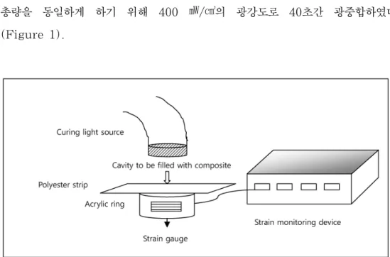
시편에 부착된 스트레인 게이지를 스트레인 모니터링 장비(Strain-Meter PCD-300A Kyowa-Electronic Instruments Co, LTD, Tokyo, Japan) 에 연결시키고 광중합 전의 초기값을 설정하였다. 중합시간은 모든 군의 에너지
총량을 동일하게 하기 위해 400 ㎽/㎠의 광강도로 40초간 광중합하였다 (Figure 1).
Figure 1. The test setup used for post-gel shrinkage strain measurement.
(3) 중합 수축 변형량 측정
광중합 시점부터 0.1초 간격으로 800초 간의 스트레인 값을 측정한 후 분석을 위해 Strain meter sofrware-PCD 30A (Kyowa-Electronic Instruments Co, LTD, Tokyo, Japan)를 통해 컴퓨터에 데이터를 전송하였다. 데이터는 저장 후 엑셀 프로그램으로 변환, 그래프화하여 분석하였다.
(4) 통계분석
각 군의 중합개시 40초, 800초에 각 군 간의 수축 변형량 차이를 비교, 평가 하기 위해 one-way ANOVA를 사용하였고, Tukey test로 사후검정을 하였다.
2. 미세누출(Microleakge) 측정 (1) 치아 준비 및 와동 형성
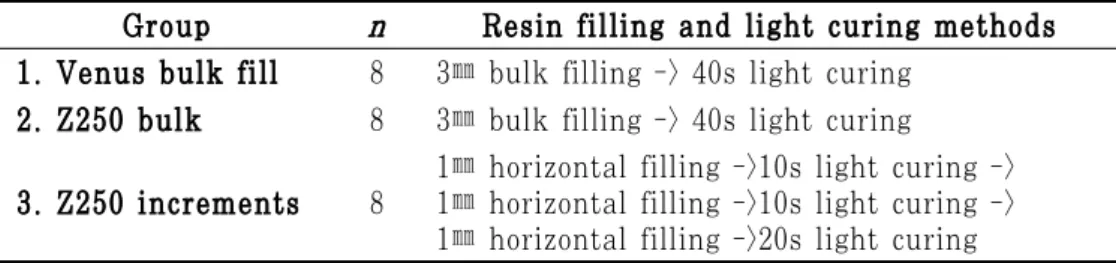
충전물이나 미세균열, 치아 우식이 없는 건전한 24개의 갓 발거된 성인 대구치 를 실온의 생리식염수에 보관하였다. 초음파 스케일러와 큐렛으로 치근 표면에 부착된 연조직과 치석을 제거하였다. 주수 하에 고속 엔진용 #245 bur를 이용 하여 교합면에 근원심 길이 5 ㎜, 협설 길이 3 ㎜, 깊이 3 ㎜의 1급 와동을 형 성하였다. 와동이 형성된 치아를 레진 종류와 충전 방법에 따라 무작위로 3 그룹 (n=8)으로 나누었다.
(2) 복합레진 충전 및 광중합
와동이 형성된 치아를 32% 인산으로 30초간 산부식 시행한 후 10초간 수세, 습윤, 건조 시행하였다. 접착제로 Single Bond (3M ESPE, St. Paul, MN, USA)를 적용하였고, 400 ㎽/㎠의 광강도로 20초간 광중합 하였다. 복합 레진 충전 및 광중합은 Table 1과 같이 시행하였다. 중합 후 복합레진 표면은 yellow diamond resin finishing bur로 마무리와 연마를 시행하였다.
Table 1. Resin filling and light curing methods for each group Group n Resin filling and light curing methods 1. Venus bulk fill 8 3㎜ bulk filling -> 40s light curing
2. Z250 bulk 8 3㎜ bulk filling -> 40s light curing
3. Z250 increments 8 1㎜ horizontal filling ->10s light curing ->
1㎜ horizontal filling ->10s light curing ->
1㎜ horizontal filling ->20s light curing
(3) 미세누출 측정
충전 후, 레진 수복물의 수화작용을 위해 치아를 7일 간 37℃ 증류수에 보관 하였다. 그 후 5-55℃ 사이의 물에서 500회 열순환 처리하였고, 치근단을 교정 용 자가 중합 레진으로 밀봉한 후 복합레진 수복물 변연에서 1 ㎜를 제외한 모
든 표면을 nail varnish를 이용하여 2회 도포, 건조하였다. 2% Methylene blue 염색 용액에 24시간 동안 담가둔 후 시편을 흐르는 물에 세척하였다.
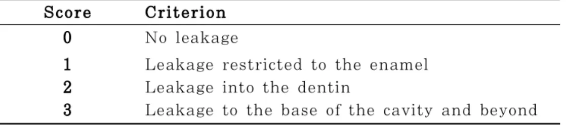
시편은 저속 다이아몬드 디스크를 사용하여 물로 냉각시키면서, 협설 방향으로 치아 장축을 따라 절단하여 2개의 절단면을 얻었다. 각 절단면에서 협측, 설측의 치아-레진간 계면을 미세광학현미경(OPMI® pico, Carl Zeiss, Germany)으 로 관찰하고, 염색 용액의 침투도를 다음과 같은 기준에 의하여 점수를 기록하였 다(Table 2).
각 군을 대표하는 시편을 에탄올로 단계적 탈수과정(70%, 80%, 90%, 100%)을 거친 후 건조하고, 백금으로 코팅하여 FE-SEM (S4700, Hitachi, Japan)으로 각 수복물과 치아 경조직 사이의 계면을 관찰하였다(15㎸, ×30, ×1000).
Table 2. Criteria for degree of microleakage Score Criterion
0 No leakage
1 2 3
Leakage restricted to the enamel Leakage into the dentin
Leakage to the base of the cavity and beyond
(4) 통계분석
각 군의 미세누출 점수를 평가하기 위해 one-way ANOVA를 사용하였고, Tukey test로 사후검정을 하였다.
Ⅲ. 실험결과
1. 중합 수축 변형률 (polymerization shrinkage strain)
광중합 직후에는 일시적으로 팽창되었다가 중합 과정이 진행될수록 수축하였고, 시간에 따라 수축률은 감소하는 경향을 보였다. 광중합 후 초기 최대 팽창률에 도달하는 평균 시간은 Venus bulk fill이 17.08초, Z250이 9.57초 였다 (Figure 2).
Figure 2. Polymerization shrinkage strain versus time curves for resin composites.
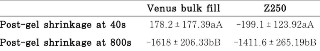
중합 후 40초, 800초에서 복합레진의 중합 수축 변형량을 Table 3에 표시하 였다. 40초에서 Venus bulk fill의 중합 수축 변형률은 178.2177.39 ㎛/m로 팽창되었고, Z250은 -199.1 123.92 ㎛/m로 팽창 후 수축상태로 전환되었다.
그러나 두 그룹간에 통계학적으로 유의한 차이는 없었다(p > 0.05). 800초에서 중합 수축 변형량은 Venus bulk fill이 -1618206.33 ㎛/m, Z250이 -1411.6265.19 ㎛/m였다. Venus bulk fill의 수축 변형량이 Z250보다 컸지 만, 유의적인 차이는 없었다(p > 0.05).
Table 3. Means, standard deviations and test significance on polymerization shrinkage strain (㎛/m) of resin composites (Mean ± SD, ㎛/m)
Venus bulk fill Z250 Post-gel shrinkage at 40s 178.2 ± 177.39aA -199.1 ± 123.92aA Post-gel shrinkage at 800s -1618 ± 206.33bB -1411.6 ± 265.19bB Analysis per row = differences between groups are identified with different
lower case letters (p < 0.05 ).
Analysis per column = differences between post-gel shrinkage times are identified with different upper case letters (p< 0.05 ).
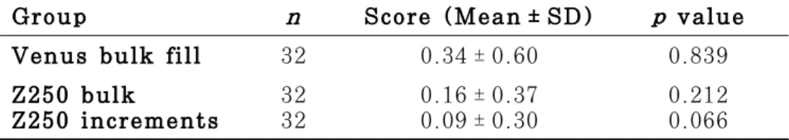
2. 미세누출(Microleakge)
각 군의 미세누출 점수는 다음과 같았다(Table 4). 각 군간 미세누출 비교에서 Venus bulk fill 군이 가장 높았고, Z250 bulk 군, Z250 increments 군 순으로 나타났다. 그러나 군 사이에 통계학적으로 유의한 차이는 나타나지 않았 다(p > 0.05).
Table 4. Microleakage scores of each group
Group n Score (Mean ± SD) p value
Venus bulk fill 32 0.34 ± 0.60 0.839
Z250 bulk
Z250 increments 32
32 0.16 ± 0.37
0.09 ± 0.30 0.212
0.066
레진수복물과 치아 사이 계면을 SEM으로 관찰한 결과는 Figure 3과 같다.
전반적으로 양호한 접합을 보였으나 Venus bulk fill 군은 와동 상부에서 (Figure 3B), Z250 bulk 군은 와동 중, 하부에서(Figure 4C and 4D),
Figure 3. SEM images of Venus bulk fill group
A: Whole cavity B: Upper cavity C: Middle cavity D: Lower cavity (A: Original magnification ×30, B,C,D: Originalmagnification ×1000)
Figure 4. SEM images of Z250 bulk group
A: Whole cavity B: Upper cavity C: Middle cavity D: Lower cavity (A: Original magnification ×30, B,C,D: Originalmagnification ×1000)
Figure 5. SEM images of Z250 increments group
A: Whole cavity B: Upper cavity C: Middle cavity D: Lower cavity (A: Original magnification ×30, B,C,D: Originalmagnification ×1000)
Ⅳ. 총괄 및 고찰
응력 집중이 높은 1급 와동에서 응력을 완화하고 수복재료의 접합성을 향상시 키기 위한 방법으로 적층 충전, 접착력이 우수한 상아질 접착제의 사용, 저점도 레진 이장재의 사용, 광원의 강도를 조절하는 방법(soft-start light polymeriza- tion)등이 보고되었다.19-24 적층 충전법은 변연부의 중합수축으로 인한 간극을 25%까지 감소시킨다고 하였다.25 기존의 구치부 수복용 복합 레진은 필러 함량 이 높아 수축이 적지만, 높은 탄성 계수를 갖고 흐름성이 거의 없어 레진-상아질 계면에 응력이 집중되었다.26 이를 완화시키기 위해 유동형 복합레진을 탄성 이 장재로 사용하여 와동 내부에서 간극 형성을 최소화하였다.27-29
기존의 유동형 복합레진(flowable composite)은 일반적으로 전통형 혼합형 복합레진(hybrid composite)에 비해 낮은 탄성 계수와 필러 함량, 낮은 기계적 물성을 갖고, 높은 수축 응력, 수축률을 보였다.2,30 그러나 최근에 개발된 bulk fill 복합레진은 기계적 물성이 혼합형 복합레진보다 낮지만 기존의 유동형 복합 레진보다 향상되었다.31 또한 기존 methacrylate 계열의 유동형 복합레진보다 수축 응력 및 수축률이 낮았고, 혼합형 복합레진과 유사하였다.2 Surefil SDR은 silorane 계열의 복합레진보다 중합 수축 응력이 낮다는 연구 결과도 있었으며, 가능한 기전으로 중합 조절제 첨가로 인해 겔 화 되는 시점을 늦춰 응력을 줄인 다고 하였다.2 그러나 Venus bulk fill에 대한 중합 수축 연구는 거의 없어 기 존의 유동형 복합레진과 비교가 어려웠으며, 향후 연구가 필요한 부분이다.
Venus bulk fill은 6㎜까지 충전이 가능하다고 시판되었지만, 최근 연구에 의하면 4 ㎜ 충전까지는 물성의 저하가 관찰되지 않지만 6 ㎜ 충전시에는 경도 가 80% 정도로 저하된다고 보고되었다.4 따라서 와동 하부는 bulk fill 복합레 진을 최대 4 ㎜까지 충전하여 응력을 완화시키고, 와동 상부는 혼합형 복합레진 을 2 ㎜ 정도 충전하여 교모나 마모에 대한 저항성을 갖도록 하는 것이 바람직
반면, 다른 적층법에 비해 바람직하지 않은 C-factor를 보이는 것으로 보고되었 다.32 따라서 대조군 설정 시 낮은 C-factor를 갖는 적층법을 사용한다면 연구 결과가 달라질 수 있을 것으로 생각된다.
본 연구에서는 strain gauge를 사용하여 중합 수축 변형량을 측정하였다.
Strain gauge는 임상적으로 문제를 일으키는 겔 화 이후의 중합 수축을 측정 하고, 시간에 따른 수축률 변화를 관찰할 수 있는 장점이 있었다. 그러나 중합 발열에 의한 열, 광원에서 발생한 열에 의해 레진과 gauge의 팽창이 일어나므로, 실제 레진의 변형량보다 낮게 측정되므로 이를 고려하여야 한다.10,16,33
또한 광중합 후 초기에 발생하는 이 열팽창은 광원의 강도와 레진의 투과성 및 열 전달능에 영향을 받는다.33 Venus bulk fill 군이 Z250군보다 초기 팽창량 이 높게 측정된 이유도 Venus bulk fill의 색조가 투명하여 광 투과성이 높기 때문일 것이다.
미세누출에 영향을 미칠 수 있는 인자로 중합 수축, 변형에 저항하는 능력, 탄 성계수, 와동 형태, 노출된 법랑질과 상아질의 양, 수복 과정 등이 있다.34-37 수 복물의 미세누출을 평가하는 방법으로 주사전자현미경법, 공기압력 이용법, 방사 선 동위원소 이용법, 중성자활성 분석법, 색소 침투법 등이 있다.38 이 중 수복재 의 변연누출 평가에 주로 사용되는 색소 침투법을 사용하여 평가하였으며, 변연 간극의 염색에 적합한 methylene blue 용액을 사용하였다. 본 연구의 미세누 출 측정 결과 Venus bulk fill, Z250 bulk, Z250 increments 순으로 미세 누출이 높게 나타났지만, SEM 관찰 시 Venus bulk fill 군은 와동 상부에서, Z250 bulk 군은 와동 중, 하부에서, Z250 increments 군은 와동 하부에서 간극이 관찰되었다. 색소 침투법을 이용한 측정법은 와동 상부 변연 간극을 통한 미세누출만을 측정하게 되므로, 향후 와동 내부 간극에 대한 평가가 필요할 것으 로 사료된다.
본 연구 결과로 미루어 볼 때 깊은 1급 와동 수복 시 bulk fill 복합레진을 와동 하부에 3-4 ㎜ 단일층 충전시행하고, 와동 상부 2 ㎜ 정도를 기존 metahcry-late 계열의 혼합형 복합레진으로 수복하는 것이 와동 상부와 하부 간극을 방지하면서 기계적 물성을 유지할 수 있는 방법이 될 수 있을 것이다.
Ⅴ. 결 론
본 연구에서는 새로운 Bulk fill 복합 레진의 중합 수축을 기존의 metha- crylate 계열의 복합레진과 비교하여 평가하였다. 이를 위해 복합레진의 중합 수축 변형량과 미세누출을 측정한 결과 Venus bulk fill 군이 Z250 군보다 높 은 중합 수축 변형량과 미세누출을 보였다. 그러나 통계학적으로 유의한 차이는 없었다.
참 고 문 헌
1. Jack L. Ferrancane. Resin composite-state of the art. Dent Mater 2011;27:29-38.
2. Ilie N, Hickel R. Investigations on an methacrylate-based flowable composite based on the SDR™ technology. Dent Mater 2011;27:348-55.
3. Rullmann I, Schattenberg A, Marx M, Willershausen B, Ernst CP. Photoelastic determination of polymerization shrinkage stress in low-shrinkage resin composites. Schweiz Monatsschr Zahnmed 2012;122:294-299.
4. Czasch P, Ilie N. In vitro comparison of mechanical properties and degree of cure of bulk fill composites. Clin Oral Investig 2012 Mar 14.
5. Hansen EK. Visible light-cured composite resin: polymerization contraction, contraction pattern and hygroscopic expansion.
Scand J Dent Res 1982;90:329-335.
6. Calais JG, Soderholm KJ. Influence of filler type and water exposure on flexural strength of experimental resin composites.
J Dent Res 988;67:836-840.
7. Kinomoto Y, Torii M, Takeshige F, Ebisu S. Comparison of polymerization contraction stresses between self- and light- curing composites. J Dent 1999;27:383-389.
8. Feilzer AJ, de Gee AJ, Davidson CL. Setting stress in compo- site resin in relation to the configuration of the restoration. J Dent Res 1987;66:1636-1639.
9. Davidson CL, de Gee AJ. Relaxation of polymerization contraction stresses by flow in dental composites. J Dent Res 1984;63:
146-148.
10. Davidson CL, de Gee AJ, Feilzer A. The competition between the composite-dentin bond strength and the polymerization contraction stress. J Dent Res 1984;63:1396-1399.
11. Hansen EK. Visible light-cured composite resins: polymerization contraction, contraction pattern and hygroscopic expansion.
Scand J Dent Res 1982;90:329-335.
12. van Noort R, Cardew GE, Howard IC. A study of the interfacial shear and tensile stresses in a restored molar tooth. J Dent 1988;16:286-293.
13. Sakaguchi RL, Wiltbank BD, Shah NC. Critical configuration analysis of four methods for measuring polymerization shrinkage strain of composites. Dent Mater 2004;20:388-396.
14. Eick JD, Welch FH. Polymerization shrinkage of posterior composite resins and its possible influence on postoperative sensitivity. Quintessence Int 1986;17:103-111.
15. Yamazaki PC, Bedran-Russo AK, Pereira PN, Wsift EJ Jr.
Microleakage evaluation of a new low-shrinkage composite restorative material. Oper Dent 2006;31:670-676.
16. Ryu SJ, Cheon JH, Min JB. Evaluation of polymerization shrinkage stress in silorane-based composites. J Korean Acad Conserv Dent 2011;36:188-195.
18. Sakaguchi RL, Sasik CT, Bunczak MA, Douglas WH. Strain gauge method for measuring polymerization contraction of composite restoratives. J Dent 1991;19:312-316.
19. Uno S, Asmussen E. Marginal adaptation of a restorative resin polymerized at reduced rate. Scand J Dent Res 1991;99:
440-444.
20. Opdam NJ, Roeters JJ, Peters TC, Burgersdijk RC, Teunis M.
Cavity wall adaptation and voids in adhesive CI I resin composite restorations. Dent Mater 1996;12:383-389.
21. Feilzer AJ, Dooren LH, De Gee AJ, Davidson CL. Influence of light intensity on polymerization shrinkage and integrity of restoration-cavity interface. Eur J Oral Sci 1995;103:322-326.
22. Lutz E, Krejci I, Oldenburg TR. Elimination of polymerization stresses at the margins of posterior composite resin resto- rations: a new restorative technique. Quintessence Int 1986;17:
777-784.
23. Watts DC, Hinde AA. Intrinsinc “Soft-Start” polymerization shrinkage-kinetics in an acrylate-based resin-composite. Dent Mater 1999;15:39-45.
24. Kubo S, Yokota H, Hayashi Y. Effect of low viscosity resin based composite on the microleakae of cervical restorations.
Am J Dent 2003;16:244-248.
25. Torstenson B, Oden A. Effects of bonding agent types and incremental techniques on minimizing contraction gaps around resin composite. Dent Mater 1989;5:218-223.
26. Dauvillier BS, Aarnts MP, Feilzer AJ. Developments in shrinkage control of adhesive restoratives. J Esthet Dent 2000;12:291-299.
27. Kemp-Scholte CM, Davidson CL. Marginal sealing of curing contraction gaps in Class V composite resin restorations. J Dent Res 1988;67:841-845.
28. Kemp-Scholte CM, Davidson CL. Complete marginal seal of Class V resin composite restorations effected by increased flexibility. J Dent Res 1990;69:1240-1243.
29. Abedian B, Millstein P. An effective method for spreading flowable composites in resin-based restorations. Oper Dent 2006;
30:151-154.
30. Bayne SC, Thompson JY, Swift EJ, Stamatiades P, Wilkerson M. A characterization of first-generation flowable composites.
J Am Dent Assoc 1998;129:567-577.
31. Frauscher KE, Ilie N. Depth of cure and mechanical pro- perties of nano-hybrid resin-based composites with novel and conventional matrix formulation. Clin Oral Investig 2012;16:
1424-1434.
32. Bortolotto T, Onisor I, Krejci I. Proximal direct composite restorations and chairside CAD/CAM inlays: marginal adapation of a two-step self-etch adhesive with and without selective enamel conditioning. Clin Oral Investig 2007;11:35-43.
33. Sakaguchi RL, Versluis A, Douglas WH. Analysis of strain gage method for measurement of post-gel shrinkage in resin composites. Dent Mater 1997;13:233-239.
34. Davidson CL, de Gee AJ. Light-curing units, polymerization,
Dent Mater 1989;5:346-349.
36. Lu H, Lee YK, Oguri M, Powers JM. Properties of a dental resin composite with a spherical inorganic filler. Oper Dent 2006;31:734-740.
37. Yamazaki PC, Bedran-Russo AK, Pereira PN. The effect of load cycling on nanoleakage of deproteinized resin/dentin interfaces as a function of time. Dent Mater 2008;24:867-873.
38. Déjou J, Sindres V, Camps J. Influence of criteria on the results of in vitro evaluation of microleakage. Dent Mater 1996;12:
342-349.