Divergent and self-reactive immune responses in the CNS of COVID-19 patients with neurological
symptoms
Graphical abstract
Highlights
d Immune cell scRNA-seq showed divergent T cell activation in the CNS during COVID-19
d Individuals with COVID-19 had a compartmentalized cytokine response in the CNS
d All individuals with COVID-19 had anti-SARS-CoV-2 antibodies in their CSF
d Five of seven individuals with COVID-19 had antineural autoantibodies in their CSF
Authors
Eric Song, Christopher M. Bartley, Ryan D. Chow, ..., Samuel J. Pleasure, Michael R. Wilson, Shelli F. Farhadian
Correspondence
[email protected] (E.S.),
[email protected] (S.J.P.), [email protected] (M.R.W.), [email protected] (S.F.F.)
In brief
Neurological symptoms are frequent in hospitalized individuals with acute COVID-19. Song et al. find that, compared with control individuals, those with COVID-19 with neurologic symptoms have divergent immune responses between the CNS and periphery, including high rates of antineural autoantibodies in their CSF.
Song et al., 2021, Cell Reports Medicine2, 100288 May 18, 2021ª2021 The Author(s).
https://doi.org/10.1016/j.xcrm.2021.100288
ll
Article
Divergent and self-reactive immune responses in the CNS of COVID-19 patients
with neurological symptoms
Eric Song,1,*Christopher M. Bartley,2,3,4Ryan D. Chow,5Thomas T. Ngo,3,4Ruoyi Jiang,1Colin R. Zamecnik,3,6 Ravi Dandekar,3,6Rita P. Loudermilk,3,6Yile Dai,1Feimei Liu,1Sara Sunshine,7Jamin Liu,7,8Wesley Wu,9
Isobel A. Hawes,3,6,10Bonny D. Alvarenga,3,6Trung Huynh,3,6Lindsay McAlpine,11Nur-Taz Rahman,11Bertie Geng,13 Jennifer Chiarella,11Benjamin Goldman-Israelow,1,12Chantal B.F. Vogels,14Nathan D. Grubaugh,14
Arnau Casanovas-Massana,14Brett S. Phinney,15Michelle Salemi,15Jessa R. Alexander,3,6Juan A. Gallego,16,17,18 Todd Lencz,16,17,18Hannah Walsh,13Anne E. Wapniarski,3,6Subhasis Mohanty,13Carolina Lucas,1Jon Klein,1 Tianyang Mao,1Jieun Oh,1Aaron Ring,1Serena Spudich,11Albert I. Ko,13,14Steven H. Kleinstein,1,19,20John Pak,9 Joseph L. DeRisi,7,9Akiko Iwasaki,1,21,22Samuel J. Pleasure,3,6,*Michael R. Wilson,3,6,*and Shelli F. Farhadian11,13,23,*
1Department of Immunobiology, Yale School of Medicine, New Haven, CT, USA
2Hanna H. Gray Fellow, Howard Hughes Medical Institute, Chevy Chase, MD, USA
3Weill Institute for Neurosciences, University of California, San Francisco, San Francisco, CA, USA
4Department of Psychiatry, University of California, San Francisco, San Francisco, CA, USA
5Department of Genetics, Yale School of Medicine, New Haven, CT, USA
6Department of Neurology, University of California, San Francisco, San Francisco, CA, USA
7Department of Biochemistry and Biophysics, University of California, San Francisco, San Francisco, CA, USA
8University of California, Berkeley—University of California, San Francisco Gradate Program in Bioengineering, Berkeley, CA, USA
9Chan Zuckerberg Biohub, San Francisco, CA, USA
10Biomedical Sciences Graduate Program, University of California, San Francisco, San Francisco, CA, USA
11Department of Neurology, Yale School of Medicine, New Haven, CT, USA
12Bioinformatics Support Program, Cushing/Whitney Medical Library, Yale University School of Medicine, New Haven, CT, USA
13Department of Internal Medicine, Section of Infectious Diseases, Yale School of Medicine, New Haven, CT, USA
14Department of Epidemiology of Microbial Diseases, Yale School of Public Health, New Haven, CT, USA
15Proteomics Core Facility, UC Davis Genome Center, University of California, Davis, Davis, CA 95616, USA
16Institute for Behavioral Science, The Feinstein Institute for Medical Research, Manhasset, NY, USA
17Division of Psychiatry Research, The Zucker Hillside Hospital, Glen Oaks, NY, USA
18Department of Psychiatry, Zucker School of Medicine at Hofstra/Northwell, Hempstead, NY, USA
19Department of Pathology, Yale School of Medicine, New Haven, CT, USA
20Interdepartmental Program in Computational Biology and Bioinformatics, Yale University, New Haven, CT, USA
21Department of Molecular, Cellular, and Developmental Biology, Yale School of Medicine, New Haven, CT, USA
22Howard Hughes Medical Institute, Chevy Chase, MD, USA
23Lead contact
*Correspondence:[email protected](E.S.),[email protected](S.J.P.),[email protected](M.R.W.),shelli.farhadian@yale.
edu(S.F.F.)
https://doi.org/10.1016/j.xcrm.2021.100288
SUMMARY
Individuals with coronavirus disease 2019 (COVID-19) frequently develop neurological symptoms, but the biological underpinnings of these phenomena are unknown. Through single-cell RNA sequencing (scRNA-seq) and cytokine analyses of cerebrospinal fluid (CSF) and blood from individuals with COVID-19 with neurological symptoms, we find compartmentalized, CNS-specific T cell activation and B cell responses. All affected individuals had CSF anti-severe acute respiratory syndrome coro- navirus 2 (SARS-CoV-2) antibodies whose target epitopes diverged from serum antibodies. In an an- imal model, we find that intrathecal SARS-CoV-2 antibodies are present only during brain infection and not elicited by pulmonary infection. We produced CSF-derived monoclonal antibodies from an individ- ual with COVID-19 and found that these monoclonal antibodies (mAbs) target antiviral and antineural antigens, including one mAb that reacted to spike protein and neural tissue. CSF immunoglobulin G (IgG) from 5 of 7 patients showed antineural reactivity. This immune survey reveals evidence of a com- partmentalized immune response in the CNS of individuals with COVID-19 and suggests a role of autoimmunity in neurologic sequelae of COVID-19.
Cell Reports Medicine2, 100288, May 18, 2021ª2021 The Author(s). 1
ll
OPEN ACCESS
INTRODUCTION
The causative pathogen of pandemic coronavirus disease 2019 (COVID-19), severe acute respiratory syndrome coronavirus 2 (SARS-CoV-2), primarily causes respiratory illness. However, in some people, SARS-CoV-2 infection is associated with severe and debilitating neurological symptoms.1About a third of individ- uals with moderate to severe COVID-19 experience neurological sequelae, including anosmia, dysgeusia, headache, impaired consciousness, and seizures, only some of which are explained by systemic complications, including hypercoagulability.2 Rarely, SARS-CoV-2 RNA is detected in the cerebrospinal fluid (CSF) of individuals with COVID-19, and some studies have found evidence of SARS-CoV-2 protein in brain parenchyma.
However, there is little evidence that SARS-CoV-2 directly dam- ages neural tissue.3–7These observations suggest that mecha- nisms other than direct cytopathic effects of SARS-CoV-2 contribute to neurological symptoms. Therefore, a broad char- acterization of CNS immunity may provide further insight into the causes of neurologic impairment in COVID-19. In this explor- atory study, we profiled intrathecal and peripheral immune re- sponses in individuals with COVID-19 complicated by diverse neurological symptoms.
RESULTS Overview
Hospitalized individuals with COVID-19 with various neurological symptoms who underwent clinically indicated lumbar puncture consented to collection of surplus CSF to be used for research.
Six participants with acute COVID-19, based on positive SARS- CoV-2 qRT-PCR of nasopharyngeal swabs, were enrolled (Table S1). Neurological symptoms included encephalopathy, intrac- table headaches, and seizures. All participants donated paired blood and CSF, except for one individual who did not donate blood. Lumbar punctures were performed on median hospital day 12.5 (range, 2–43 days). Pre-pandemic CSF from age- and gender-matched healthy control individuals (n = 3) was obtained from a neuroinfectious disease biorepository at Yale. Because fresh CSF is required for single-cell transcriptomics, we recruited additional uninfected control participants during the COVID-19 pandemic (n = 3); 2 were healthy community-dwelling adults, and 1 was hospitalized for work-up of frequent falls. Additional blood and CSF single-cell sequencing data were included from publicly available data derived from healthy control individuals (n = 8).8 Recruited control individuals tested negative for SARS-CoV-2 by RT-PCR of nasopharyngeal swabs. Fresh CSF and blood samples were processed into CSF-resident cells, CSF supernatant, peripheral blood mononuclear cells (PBMCs), and plasma (Figure 1A). CSF (n = 5) and plasma (n = 6) samples from individuals with COVID-19 were negative for SARS-CoV-2 RNA by qRT-PCR using Centers of Disease Control and Preven- tion (CDC) primer-probe sets.9
Transcriptional analysis reveals a coordinated innate immune cell response to COVID-19 in the CNS
To investigate the effect of SARS-CoV-2 infection on host im- mune cell gene expression, we performed single-cell RNA
sequencing with the 10x Genomics platform on 76,473 immune cells from the CSF and blood of individuals hospitalized with acute COVID-19 and uninfected control individuals. To test for the presence of intracellular virus, open reading frames of SARS-CoV-2 (spike, ORF3a, envelope, membrane glycoprotein, ORF6, ORF7a, ORF8, nucleocapsid, and ORF10) were added to the reference genome before alignment of RNA sequencing data with CellRanger. Viral transcripts were not detected in any CSF immune cells or PBMCs. We performed unsupervised cluster analysis of CSF cells and PBMCs and identified distinct T cell, B cell, and myeloid cell populations (Figure 1B), characterized by expression patterns of canonical immune cell marker genes (Figure S1A). Next we identified genes that were differentially ex- pressed in CSF—but not peripheral blood—immune cells of indi- viduals with COVID-19 compared with control individuals (Data S2).
In the CSF of individuals with COVID-19, dendritic cells had an activated transcriptional profile; 57% and 47% of their upregu- lated genes were classified as type 1 and type 2 interferon-stim- ulated genes, respectively (Figure 1C). Genes associated with natural killer (NK) cell activation were also upregulated in the CSF of individuals with COVID-19 (Figure 1D). Although NK cells in the CSF and peripheral blood demonstrated comparable changes in the number of differentially expressed genes in indi- viduals with COVID-19 compared with control individuals, the affected genes were mostly unique to each compartment (Fig- ure S1C). Using CellphoneDB signaling network analysis,11,12 in individuals with COVID-19, CSF dendritic and NK cells were predicted to have significantly increased interactions with CSF CD8 and CD4 T cells relative to healthy controls, whereas inter- actions between CD4 T cells and monocytes were diminished, suggesting a dysregulated innate-to-adaptive immune interface (Figure 1E).
T cells in the CSF display increased cellular activation during COVID-19
Because signaling network analysis predicted that T cells were the main recipients of altered innate-adaptive cross-talk, we isolated and re-clustered CSF and peripheral T cells for tar- geted transcriptional analysis of T cell subsets (Figures 2A andS2A–S2C). Among peripheral T cells, there was a decrease in the frequency of naive CD4 T cells (mean: COVID-19, 9.65%;
healthy, 18.219%; p = 0.001) and an increase in effector CD8 T cells (mean: COVID-19, 30.9%; healthy, 16.245%; p = 0.02) (Figure 2B). In contrast, in CSF, the relative proportions of T cell populations were conserved in individuals with COVID- 19 compared with control individuals, but we found significant COVID-19-associated transcriptional changes in CSF T cells (Figure S2D). After excluding any genes that were also differen- tially expressed between T cells in the CSF and periphery of healthy control individuals, we identified genes that were upregulated in Th1 and Th2 CD4 T cells from COVID-19 CSF (Figure 2C). These genes were enriched for interleukin-1 (IL-1)- and IL-12-mediated signaling pathways and several ge- netic pathways important for T cell activation (Figure 2D).
Effector CD8 T cells in the CSF were similarly enriched for genes involved in canonical immune response pathways, including (1) increased motility and cell adhesion, (2)
Figure 1. Distinct immunological landscape of CSF and PBMCs in individuals with COVID-19 with neurological symptoms
(A) Schematic of the study design. CSF and blood were collected from individuals with COVID-19 and healthy control individuals. PBMCs and CSF cells were isolated, along with the CSF supernatant and plasma, for downstream analysis.
(B) UMAP (Uniform Manifold Approximation and Projection) projections of 10x single-cell RNA sequencing of CSF and PBMCs of individuals with COVID-19 and healthy control individuals.
(C) Venn diagram depicting upregulated interferon-stimulated genes (ISGs) and non-ISGs in dendritic cells in COVID-19 CSF compared with healthy control CSF based on the Interferome database.10
(D) Gene Ontology enrichment of genes upregulated in NK cells of individuals with COVID-19 in the CSF and peripheral blood.
(legend continued on next page)
Article
ll
OPEN ACCESS
differentiation/proliferation, and (3) effector programming (re- sponses to IL-12, IL-1, interferon-g, and T cell co-stimulation), indicating the presence of a coordinated T cell-based immune response in the CNS (Figures 2E and 2F). Transcrip- tional changes in individuals with COVID-19 were observed in CD4 and CD8 T cells in the CSF and predicted cell-cell interac- tions that were unique to the CSF in individuals with COVID-19, including T cell co-stimulation factors and trafficking interac- tions (Figures S3A and S3B). Analysis of T cell receptor (TCR) sequences in CSF and blood revealed clonal expansion of unique but not shared CD4 T cell clones in the CSF of individ- uals with COVID-19 (Figure S4), further suggesting a compart- mentalized T cell response to CNS antigen.
Unique cytokine profiles exist in CSF of individuals with COVID-19 compared with serum
To validate the transcriptional enrichment in IL-12 and IL-1 signaling in the CSF of individuals with COVID-19 with neurologic symptoms, we measured inflammatory cytokine levels in the CSF and plasma using a Luminex cytokine panel (Figures 2G and 2H). Consistent with the single-cell RNA sequencing results, IL-1b and IL-12 were elevated in the CSF of individuals with COVID-19 compared with healthy control individuals but were not elevated in the plasma of individuals with COVID-19.
Conversely, CCL2, CXCL9, and IL-8 were increased significantly in the plasma of individuals with COVID-19 compared with con- trol individuals but not in their CSF. Because IL-12 is thought to be produced by activated antigen-presenting cells to orches- trate Th1 responses through T and NK cell activation, we exam- ined the cellular source of IL-12 in individuals with COVID-19.
The innate immune cells with the highest IL12A expression were CSF NK and dendritic cells (Figure S1E). These data support the single-cell RNA sequencing analyses that identified IL-12 as differentially expressed in CSF but not blood innate im- mune cells of individuals with COVID-19. Moreover, they suggest a distinct effect of COVID-19 in the CNS on cytokines important for innate immunity and induction of cell-mediated immunity, including IL-1 and IL-12.
CNS B cell responses to SARS-CoV-2 differ from those in the periphery
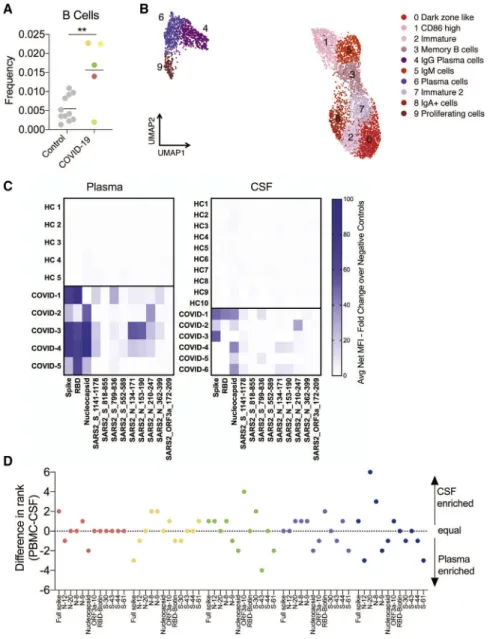
We found significant enrichment of B cells in the CSF of individ- uals with COVID-19 cases compared with CSF of healthy control individuals (Figure 3A). Single-cell RNA sequencing identified several subtypes among peripheral and CSF B cells (Figures 3B andS5A–S5C), including distinct CSF plasma cell clusters.
We therefore wanted to find out whether antibody-secreting B cells in the CSF exhibit a different anti-SARS-CoV-2 antibody profile than the those in the periphery. To do so, we utilized a recently developed SARS-CoV-2 epitope Luminex panel13to screen for anti-SARS-CoV-2 antibodies in the CSF and plasma of individuals with COVID-19 and control individuals. As ex- pected, anti-SARS-CoV-2 antibodies were not detected in any
control individuals. In contrast, all individuals with COVID-19 had anti-SARS-CoV-2 antibodies in the CSF and plasma. How- ever, although all individuals with COVID-19 developed anti- bodies to SARS-CoV-2 spike and nucleocapsid in the plasma and CSF, anti-receptor binding domain (RBD) antibodies were rare in CSF but uniformly present in the plasma (Figure 3C). In addition, we found that, in all individuals with COVID-19, the rela- tive prevalence (rank score: 12, most frequent; 1, least frequent;
Figure 3D) and levels of antibody (Figure S5D) diverged between the CSF and plasma, indicating a different anti-SARS-CoV-2 antibody profile between the CSF and plasma of the same individual.
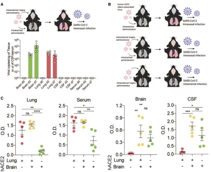
A mouse model of SARS-CoV-2 brain infection demonstrates compartmentalized CNS antibody secretion in response to CNS infection
Direct detection of SARS-CoV-2 in the CSF is extremely rare in reported cases of neurological complications of COVID-19,14 and SARS-CoV-2 RNA was not detected in the CSF our cohort.
However, we detected intrathecal antiviral antibodies in all cases. With some other encephalitis-causing viruses, including West Nile virus, Japanese encephalitis virus, and measles vi- rus,15–17the presence of antiviral antibodies is consistent with viral neuroinvasion even in the absence of viral nucleic acid. To determine whether CNS infection is sufficient to stimulate a CNS humoral response during COVID-19, we used a recently developed mouse model that reliably dissociates pulmonary and neurological infection of SARS-CoV-2.18
We used an adeno-associated virus (AAV) to express the hu- man ACE2 (hACE2) receptor in the lungs or brain or the lungs and brain, allowing us to target SARS-CoV-2 infection to spe- cific tissue. First, we used mice that express hACE2 in the lung and brain and administered SARS-CoV-2 intranasally (Fig- ure 4A). This permits SARS-CoV-2 to infect the lungs and brain.
In these mice, we detected increased titers of SARS-CoV-2 RNA in lung and brain tissue following inoculation. However, despite robust brain infection, we did not detect SARS-CoV-2 RNA in the CSF of these mice (Figure 4B). This suggests that direct detection of SARS-CoV-2 RNA in CSF at a single time point may be insensitive to parenchymal or short-lived SARS- CoV-2 neuroinvasion.
We next used the mouse model to evaluate whether detection of intrathecal anti-SARS-CoV-2 antibodies in our individuals with COVID-19 was more likely triggered by a local antigen (i.e., as a consequence of SARS-CoV-2 neuroinvasion) or reflected pas- sive transfer of antibody from the systemic circulation.14When SARS-CoV-2 was administered intranasally to mice expressing hACE2 only in the lungs (generating mice with pulmonary but not brain infection), we detected significantly elevated anti-spike SARS-CoV-2 immunoglobulin G (IgG) in the lungs and serum but not in the brain or CSF (Figure 4B, red). When SARS-CoV-2 was administered intranasally to mice expressing hACE2 in the brain and lungs (generating mice with pulmonary and brain infection),
(E) Heatmap depicting cell-cell interactions between innate immune cells and adaptive immune cells by clustering shown in (B). The difference in interaction strength (COVID-19 interaction minus control interaction) is color coded and derived from log-scaled interaction counts using the CellphoneDB repository of ligands, receptors, and their interactions.11Single-cell RNA-seq is derived from a total of 16 libraries plus 8 additional controls from Gate et al.8(n = 3 for control CSF and PBMCs, n = 5 for COVID-19 CSF and PBMCs, and n = 8 from Gate et al.8).
we detected increased anti-spike antibodies in all four compart- ments: lungs, serum, brain, and CSF (Figure 4B, orange). Finally, when hACE2 was expressed in the brain only and SARS-CoV-2 was administered intracranially (causing infection in the brain but not in the lungs), we detected increased anti-spike antibodies in the brain and CSF but not in the lungs or serum (Figure 4B, green). These data support the hypothesis that CSF antibodies do not solely reflect passive transfer of antibodies from the sys- temic circulation. Indeed, in these mice, anti-spike antibodies in the CSF and brain were only observed in the setting of brain infection, independent of whether there was an accompanying systemic SARS-CoV-2 infection.
Monoclonal antibodies from CSF B cells from individuals with COVID-19 are self reactive
Although our individuals with COVID-19 did not have detectable SARS-CoV-2 RNA in their CSF, CSF serology suggested that
CSF-expanded B cell populations might be reactive to SARS- CoV-2 antigen(s). To address this, we cloned individual mono- clonal antibodies from the affected individual with the largest number of clonally expanded B cell receptor (BCR) sequences in the CSF (n = 5) and blood (n = 4) (case 1; Data S1 and S5).
In this individual, the most prevalent BCR sequences comprised 25% and10% of total B cells in the blood and CSF, respec- tively (Figure 5A). Notably, the most prevalent BCR sequences detected in the CSF of case 1 did not overlap with the most prev- alent blood BCR sequences (Figure 5B), supporting the hypoth- esis that a subset of CSF antibodies targets antigens within the CNS.
We found that one of five CSF-derived (monoclonal antibody [mAb] C2) and two of four peripherally derived mAbs (mAbs P1 and P2) targeted the SARS-CoV-2 spike protein (Figure 5C).
None of the other mAbs recognized other SARS-CoV-2 antigens in the Luminex panel. Using biolayer interferometry, we Figure 2. Transcriptional characterization of T cells in CSF and PBMCs of individuals with COVID-19
(A) Reclustered UMAP projection of combined CSF and peripheral blood T cells, demonstrating CD4 and CD8 T cell subsets (two KLRG1+ clusters are distinguished by GZMB and IFNG expression;Figure S3).
(B) Pie charts depicting the relative population frequency of different T cell subtypes found in CSF and PBMCs of control individuals and those with COVID-19.
(C) Venn diagram depicting genes upregulated (adjusted p < 0.05) in CSF of individuals with COVID-19 compared with PBMCs of individuals with COVID-19 in Th1 and Th2 CD 4 T cells.
(D) Gene Ontology analysis of genes that are upregulated in Th1 and Th2 cells, as depicted in (C).
(E) Quad-Venn diagram of genes upregulated in CSF of individuals with COVID-19 compared with CSF of control individuals in CD8 T cells. Genes shared by the three effector CD8 T cell subtypes are circled.
(F) Gene Ontology analysis of genes shared between the three effector CD8 T cell subtypes in (F).
(G and H) Heatmap of Luminex-based cytokine profiling of CSF (G) and plasma (H) from individuals with COVID-19 and control individuals showing cytokines that were increased significantly increased in individuals with COVID-19 compared with control individuals (n = 6 CSF, n = 5 plasma). For each cytokine, two-tailed p values were calculated using Student’s t test. Data for each row were mean centered; each column shows data from one sample.
Article
ll
OPEN ACCESS
determined that all three anti-spike mAbs derived from individ- uals with COVID-19 bound 2P-stabilized spike protomers with high affinity (KD[mAb C2] = 2 nM, KD[mAb P1] = 0.2 nM, KD[mAb P2] = 2 nM) (Data S3). Because these binding affinities are similar to those of SARS-CoV-2-neutralizing antibodies, we tested each anti-spike mAb for neutralizing activity against wild-type SARS- CoV-2.19None of the mAbs exhibited neutralizing activity at con- centrations ranging from 2.5–25mg /mL (Figure S6).
Given reports of new-onset humoral autoimmunity in COVID-19, we wondered whether any of the mAbs from CSF-expanded B cells were autoreactive to neural tissue.
Therefore, we tested all mAbs using a standard and validated screening method for antineural autoreactivity: anatomic mouse brain tissue staining.20mAbs were used as a primary antibodies to immunostain mouse brain tissue and labeled with an anti-human IgG secondary antibody. An anti-influenza antibody targeting the hemagglutinin antigen (anti-HA) was
Figure 3. Localized central nervous system B cell responses in individuals with COVID- 19
(A) Frequency of B cells as a percentage of all CSF cells in control individuals and those with COVID- 19. Colors represent different individuals.
(B) Re-clustered UMAP projection of B cells from CSF and blood.
(C) Heatmap showing antibody binding in plasma (left) and CSF (right) to nine peptides from immu- nogenic regions of S, N, and ORF3a as well as whole S and N protein along with the RBD of the S protein. All data are represented as fold change of the fluorescent anti-IgG antibody signal over intra- assay negative controls. HC, healthy control.
(D) Epitope frequency was ranked in each sample individually, and a difference in rank number for each cluster was graphed to determine CSF-en- riched (positive) or plasma-enriched (negative) antibody epitopes. Two-tailed unpaired t test,
**p < 0.01.
used as a negative control. Similar to anti-HA, none of the PBMC-derived mAbs recognized mouse brain tissue.
In contrast, four of five CSF-derived mAbs exhibited some degree of anti- neural immunoreactivity, including the anti-spike mAb (mAb C2) (Figures 5D and 5E). Notably, mAb C2 produced a neuropil-predominant immunostaining pattern, suggesting that the antigen may be enriched in neuronal process or harbor an extracellular epitope.
Intrathecal humoral autoimmunity in individuals with COVID-19 with neurological symptoms
The emergence of inflammatory and hu- moral autoimmune disorders of the nervous system during the para- or post-infectious period in COVID-19 is increasingly recognized and includes acute disseminated encephalomyelitis (ADEM), autoimmune encephalitis associated with known autoantibodies, transverse myelitis, and Guillain- Barre´ syndrome and one of its variants, Miller Fisher syn- drome.21–25Given this literature and the autoreactivity of CSF- derived monoclonal antibodies from case 1, we hypothesized that our other individuals with COVID-19 might harbor intrathecal autoantibodies. To test this, we screened our cohort of individ- uals with COVID-19 for intrathecal antineural antibodies using a suite of complementary autoantigen detection platforms:
anatomic mouse brain tissue immunostaining, immunoprecipita- tion-mass spectrometry (IP-MS), and pan-human proteome phage display IP sequencing (PhIP-seq).26–28In these screens, we included one additional individual with post-COVID-19 sei- zures and cognitive impairment who had been recruited after completion of the transcriptomics and cytokine analyses were completed (case 7; Table S1; Data S1). Like the other six
individuals with acute COVID-19, this subject had SARS-CoV-2 antibodies in his CSF (Figure S7).
Anatomic mouse brain immunostaining demonstrates the presence of intrathecal antineural autoantibodies in most individuals with COVID-19
More COVID-19 CSF samples (5 of 7) were immunoreactive to mouse brain tissue at 1:10 dilution than control CSF (2 of 6) (Fig- ures 5A andS8A). Control CSF staining was not specific to any anatomic region, weakly pan-nuclear, or primarily subpial (Fig- ure S8B). None of the control CSF samples were immunoreac- tive beyond 1:10 dilution, indicating the absence of high-titer or
high-affinity antineural autoantibodies. In contrast, at 1:10 dilu- tion, COVID-19 CSF produced immunoreactive staining of spe- cific anatomic regions, including cortical neurons (n = 4), the ol- factory bulb (n = 3), the thalamus (n = 3), the CA3 field of the hippocampus (n = 3), the cerebellum (n = 3), the brain stem (n = 4), and cerebral vasculature (n = 2) (Figures 5B, 5C, and S8A). Four and three COVID-19 CSF samples showed continuing immunoreactivity at 1:25 and 1:50 dilution, respec- tively. The data indicate that an unexpectedly high proportion of CSF samples from individuals with COVID-19 with neurolog- ical impairment harbor high titers of antineural autoantibodies of unknown pathogenic significance.
Figure 4. CSF antibodies reflect localized CNS infection
(A) Mice were transduced with AAV-hACE2 intrathecally and intratracheally for expression in the brain and lungs, and SARS-CoV-2 was introduced intranasally to establish brain and lung infection. Mouse brains, lungs, CSF, and serum were collected on day 0 (before infection) and on days 3 and 7 after infection, and qPCR was performed to detect SARS-CoV-2 RNA.
(B) Schematic of the experimental procedure for (C). Mice were given localized AAV-hACE2 to overexpress human ACE2 in the lungs (top), brain and lungs (center), or brain only (bottom). After 2 weeks, mice were infected with SARS-CoV-2.
(C) ELISA against SARS-CoV-2 spike protein was performed with lung homogenates, serum, brain homogenates, and CSF. n = 5 for all three conditions. Two- tailed unpaired t test (*p < 0.05, **p < 0.01, ***p < 0.005, ****p < 0.0001) and one-way ANOVA were performed (lungs, p < 0.0001; serum, p = 0.002; brain, p = 0.0082; CSF, p = 0.0016).
Article
ll
OPEN ACCESS
IP-MS identifies intrathecal candidate autoantigens in a subset of individuals with COVID-19
To screen for the neural protein targets of intrathecal autoan- tibodies, we immunoprecipitated mouse whole-brain lysate
using CSF and plasma, trypsinized precipitated proteins, and analyzed the resulting peptides by MS. IPs was per- formed in technical replicates by different individuals using different mice as input. First we searched the resulting spectra Figure 5. Antigenic specificity of CSF- and PBMC-derived monoclonal antibodies
(A) Bar graph depicting the frequency of the top five most expanded clones in PBMCs and CSF of affected individual 1.
(B) Graph depicting overlap of clones found in CSF and PBMCs of an affected individual. Green indicates clones only found in CSF, orange clones shared between the CSF and PBMC, and red clones unique to PBMCs. The yellow box indicates clones that would fall under the top 10 most frequent clones in each compartment.
(C) Heatmap showing CSF-derived (mAbs C1–C5) and PBMC-derived (mAbs P1–P3 and P5) mAb binding to nine peptides from immunogenic regions of S, N, and ORF3a as well as whole S and N protein along with the RBD of the S protein. mAb numbers correspond to the clone numbers from (A) and (B); PBMC clone 4 (mAb P4) did not express well as a mAb and was not used for subsequent studies. mAbs were screened in technical replicates. Heatmap values are mean fold change of the fluorescent anti-IgG antibody signal over intra-assay negative controls.
(D) Sagittal mouse brain sections were immunostained with mAbs 1–9, and a representative whole-brain sagittal image is shown for PBMC-derived mAbs (mAb 7) and CSF-derived mAbs (mAb 4). An anti-hemagglutinin (anti-HA) antibody in the same IgG1 backbone was used as a negative control. Scale bars, 500mm.
(E) Select regions of immunostaining from mAbs 1–4. (i) mAb 1 immunostaining of cerebellar Purkinje cells (arrow) and the overlying molecular layer. (ii) mAb C2 immunostaining of cortical neuropil and occasional staining of neuron-like somata (arrow). (iii) mAb C3 immunostaining of large cells within the hilus of the hippocampus. (iv) mAb C4 immunostaining of mitral-like cells of the olfactory bulb (arrow). (v) mAb C4 immunostaining of pyramidal neurons (arrow) in CA3 of the hippocampus. (vi) mAb C4 immunostaining of neuronal cell bodies in layer II of the cortex (arrow). Scale bars, 10mm.
Figure 6. Autoantibodies in the CSF of individuals with COVID-19
(A) Sagittal mouse brain sections were immunostained with CSF at 1:10 dilution (green) and the nuclear stain DAPI (blue). Anti IgG secondary only antibody negative control (left) and 4/6 control CSF (example CTRL 6, center) were not immunoreactive. In contrast, 5 of 7 COVID-19 CSF samples were immunoreactive (example CASE 3, right). Scale bars, 100mm. CTRL, control.
(B) Binary matrix indicating anatomic immunoreactivity of COVID-19 CSF at 1:10 dilution.
(C) Select examples of COVID-19 CSF anatomic immunostaining of the hippocampus (n = 3; arrows, CA3; left column; scale bar, 100mm), cerebrovasculature (top panel, second column arrow indicates endothelial staining [scale bar, 50mm]; bottom panel arrow indicates a perivascular cell [scale bar, 10mm]), olfactory bulb (n = 3, two shown; third column, top panel shows neuron-like cells; bottom panel, mitral cells; scale bars, 10mM), and cortical neuron-like cells (n = 4, two cases shown; fourth column; scale bars, 10mM).
(D) Heatmaps of sequence-sharing peptides mapping to IFT88 (case 1, top) and THAP3 (case 3, bottom) that were enriched by CSF, shown with their corre- sponding enrichment by plasma. Rows, individual peptides; left two columns, technical replicates for case 1 (top) and case 3 (bottom). For COVID-19 and control columns, cell values represent the mean of log10(fold change enrichment) of technical replicates. For case 1 and case 3, candidate IFT88 and THAP3 peptides, respectively, were enriched more by CSF than plasma.
(legend continued on next page)
Article
ll
OPEN ACCESS
against the SARS-CoV-2 proteome (Uniprot SARS-CoV-2 reference proteome, June 2020). SARS-CoV-2 proteins were not detected. We then searched for human proteins. Consis- tent with circulating protein expression patterns29,30 and circulating prothrombotic autoantibodies31in COVID-19, com- plement, coagulation, and platelet degranulation pathway pro- teins of human origin were significantly overrepresented in the IgG-bound protein fraction of plasma from our individuals (Figure S9).
To specifically identify candidate antineural autoantibodies in the CSF, we searched for mouse proteins that were observed in both technical replicates and significantly en- riched by spectral counting and/or 1.53 enriched by MS1 peak area in COVID-19 CSF IP relative to controls.32Between 5 and 56 (median = 20) proteins were enriched by the CSF, but not by the plasma, of the same case or control CSF, indicating that they were unique to the CSF compartment of individuals with COVID-19. Gene Ontology pathway analysis indicated that COVID-19 CSF and plasma IP-MS fractions were en- riched for brain-enriched and synaptic proteins. In some cases, neural antigens were statistically enriched by CSF but not plasma (e.g., NEFM and NEFH by cases 3 and 4 and APP by case 4).
PhIP-seq identifies intrathecal candidate autoantigens in a subset of individuals with COVID-19
COVID-19 CSF was also screened for autoantibodies using a previously described PhIP-seq platform (T7 bacteriophage display) displaying730,000 overlapping 49-amino-acid pep- tides spanning all human proteins,26including all known and predicted isoforms.27,28To identify peptides that were signifi- cantly enriched by COVID-19 CSF compared with controls, we first established an empirical enrichment threshold using a validated commercial antibody targeting the protein GFAP (Data S4). For COVID-19 CSF samples, peptides with supra- threshold enrichment in both technical replicates were consid- ered candidate autoantigens. COVID-19 CSF enriched between 2 and 40 CSF-specific proteins (median = 18; Data S4). By Gene Ontology, synaptic proteins were also enriched by COVID-19 CSF (e.g., NRG3, SYNJ2, and DPYSL2; Bonfer- roni-corrected p = 1.6 3 101). COVID-19 plasma PhIP-seq candidates were enriched for transcriptional activators of catabolism (Bonferroni-corrected p = 2.63102) but not syn- aptic or brain-enriched proteins. COVID-19 CSF that was immunoreactive to mouse brain tissue at a 1:50 dilution was associated with greater enrichment of candidate autoantigens by PhIP-seq (19–40, median = 37) than CSF that immuno- stained at a lower dilution or was not immunoreactive (1–16, median = 6), suggesting a correlation between immunostaining status and the burden of CSF autoantibodies. In some in- stances, the same candidate autoantigen was detected in indi- viduals with COVID-19 by IP-MS and PhIP-seq: UHRF1BP1 (case 2), NUAK1 (case 3), and DBN1 (case 7).
Validation of two candidate autoantigens in the CSF of individuals with COVID-19
To validate autoantibodies identified in CSF, we selected two COVID-19 candidate autoantigens that were enriched more by CSF than plasma on PhIP-seq: intraflagellar transport protein 88 homolog (IFT88) and THAP domain-containing 3 (THAP3).
To validate IFT88, HEK293T cells were transfected with a plasmid encoding RFP-IFT88, methanol fixed, and immuno- stained with CSF from case 1. Case 1 CSF IgG, but not control CSF IgG, colocalized with an anti-RFP and an anti-IFT88 com- mercial antibody (Figures 6E andS10). To validate THAP3, lysate from HEK293T cells overexpressing FLAG-tagged THAP3 was separated by SDS-PAGE, transferred to a polyvinylidene fluoride (PVDF) membrane, and sequentially probed with case 3 CSF (1:60 dilution) and a commercial anti-FLAG antibody. CSF IgG and anti-FLAG recognized the same band of 25 kDa in the THAP3 overexpression lane but not in the untransfected lane (Figure 6F). Therefore, anti-IFT88 and anti-THAP3 arebona fide autoantibodies in the CSF of COVID-19 cases 1 and 3, respec- tively (Figures 6E, 6F, andS10).
DISCUSSION
Neurotropic viruses, such as herpes simplex virus, West Nile vi- rus, and HIV, are common pathogens of the CNS, but it is increasingly recognized that respiratory viruses, including influ- enza virus and human respiratory syncytial virus, also lead to neurological complications in a minority of individuals.33–39The biological underpinnings of neurological complications of respi- ratory viruses are diverse and include neuronal damage because of direct viral neuroinvasion as well as parainfectious processes, including elevation of pro-inflammatory cytokines, and post-viral autoimmune reactions In this exploratory study, we performed an extensive set of immunologic investigations to assess CNS- specific immune responses in a series of individuals with COVID-19 with neurological symptoms. Although systemic multi-organ dysfunction is almost certainly a driver of neurolog- ical complications in a proportion of individuals with COVID-19, here we identified innate and adaptive antiviral immune re- sponses as well as humoral autoimmunity, which appears to be unique to the CNS and may therefore contribute to COVID- 19 neuropathology.
CSF, although not identical to brain, is produced by the choroid plexus and bathes the CNS. It is the only CNS tissue sur- rogate that can be sampled readily in living humans. Analysis of CSF immune cells has shed light on immune mechanisms of neuronal injury during other infections, including HIV, neurosy- philis, and neuroborreliosis.40–42By assessing CSF and blood in individuals with acute COVID-19 and neurological symptoms, we find evidence of a compartmentalized CNS immune response to SARS-CoV-2. Through transcriptional and cytokine analyses, we find an increase in CSF but not plasma IL-12 and IL- 1b, factors that are central for coordinating innate and adaptive
(E) The HEK293 overexpression cell-based assay was performed in technical replicates. A representative example demonstrates that case 1 CSF is immuno- reactive to overexpressed RFP-IFT88 (CSF, green; anti-RFP, red; anti-IFT88 antibody, magenta). Scale bars, 10mM.
(F) Western blot validation of anti-THAP3 autoantibodies in CSF of case 3. CSF IgG (green) and anti-FLAG (red) recognize the same25-kDa band in THAP3- overexpressing (OE) lysate but not untransfected (UN) lysate (arrow).
immune responses to invading pathogens. Notably, neuroinva- sion of mouse hepatitis virus, a coronavirus of laboratory mice, also leads to IL-12 production by astrocytes and microglia.43
Our data identified increased and divergent humoral re- sponses in the CNS. This humoral response included indirect ev- idence of neuroinvasion of SARS-CoV-2 through the presence of antiviral antibodies in the CNS during acute SARS-CoV-2 infec- tion. Using an animal model, we show that SARS-CoV-2 infec- tion in the CNS stimulates production of intrathecal antibodies and that an isolated systemic infection is not sufficient to do so. These data suggest that the presence of anti-SARS-CoV-2 antibodies in the CSF of individuals with COVID-19 may similarly reflect viral infection of the CNS. Further supporting this conten- tion, we generated a mAb from the CSF of an individual with COVID-19 (case 1) with marked B cell clonal expansion in the CSF that was specific for the SARS-CoV-2 spike protein. This B cell clone was not detected in the peripheral blood of the same individual. Notably, the other CSF-derived mAbs were var- iably immunoreactive to mouse brain tissue, which motivated and expanded our search for autoantibodies in the CSF of our other individuals with COVID-19.
Our extended studies of the CNS humoral response suggest that a subset of individuals with COVID-19 with neurological symptoms has an elevated burden of autoreactive antibodies in the CSF. As seen by anatomic immunostaining, an unexpect- edly high proportion of our individuals with COVID-19 harbored intrathecal autoantibodies, including antineural autoantibodies.
Hippocampal immunostaining from three individuals was pri- marily restricted to the CA3 region, similar to a recent report of SARS-CoV-2-associated encephalitis.44Other immunoreactive anatomic regions included the olfactory bulb in three individuals and cerebrovasculature in two—anatomic regions withprima fa- cierelevance to common neurologic sequelae of COVID-19 (i.e., anosmia and stroke). Subsequent unbiased protein and peptide screens of CSF identified a diversity of candidate autoanti- bodies, and two of these autoantigens, THAP3 and IFT88, were subsequently validated. IFT88 is a ciliary protein whose mutation causes a ciliopathy in humans and anosmia in mice.45THAP3 is expressed in the brain, among other organs, and may be implicated in genetic causes of dystonia.46
However, the mere presence of an intrathecal autoantibody does not mean that a person has autoimmune encephalitis.
Indeed, the individuals in our exploratory cohort lacked evidence of active inflammation upon neuroimaging and/or did not have elevated conventional CSF markers of neuroinflammation (i.e., white blood cell count, IgG index, and CSF-restricted oligoclonal bands) that are typically, but not always, found in people with autoimmune encephalitis. Notably, individuals with COVID-19 with neurological symptoms appear to have immune responses to multiple autoantigens, implying that the increased compart- mental humoral immune response may reflect a broader immune activation syndrome. This is particularly true given that humoral autoimmunity has been observed to target other organ systems in COVID-19 and may also contribute to neuropathology during COVID-19.47–50
Our exploratory data suggest that, even in individuals with COVID-19 with neurologic symptoms who lack overt evidence of neuroinflammation on MRI or conventional CSF testing, there
is a compartmentalized immune response involving the innate and adaptive arms of the immune system. Future research involving careful clinical phenotyping and timely investigations of the CSF will help place these findings into a broader clinical context and inform whether antiviral and/or immunomodulatory therapies might be indicated for carefully selected, neurologi- cally impaired individuals with COVID-19.
Limitations of study
Our study is complicated by the diverse range of neurological symptoms in our study participants. A comparison group with a different systemic viral infection would help determine which aspects of the findings are specific to COVID-19. Moreover, our study did not include control CSF from individuals with COVID-19 without neurological symptoms because these peo- ple do not undergo clinical LP and did not consent to research-only LP. Thus we do not know whether auto-reactive antibodies in the CNS are present in individuals with COVID-19 who do not experience neurological symptoms.
STAR+METHODS
Detailed methods are provided in the online version of this paper and include the following:
d KEY RESOURCES TABLE
d RESOURCE AVAILABILITY B Lead contact
B Materials availability B Data and code availability
d EXPERIMENTAL MODEL AND SUBJECT DETAILS B Human Subjects
B Animals B SARS-CoV-2 B Cell Lines
d METHOD DETAILS B SARS-CoV-2 RT-qPCR B PMBC and CSF cell preparation B Single cell RNA sequencing B T and B cell clustering B BCR analysis B Cytokine assays
B AAV infection (Intratracheal and Intracisternal magna injection)
B Generation of SARS-CoV-2 virus B Enzyme-linked immunosorbent assay B Statistical methods
B SARS-CoV-2 Serological Assay
B Generation of Human Monoclonal Antibodies B Determination of Monoclonal Antibody Binding Affinity
to Purified Spike Protein
B SARS-CoV-2 Neutralization Assays B Anatomic Mouse Brain Tissue Staining B Imaging
B Immunoprecipitation Mass Spectrometry
B Phage Display Immunoprecipitation Sequencing (PhIP-Seq)
B HEK293T/17 Cell-Based Assay Autoantigen Screening
Article
ll
OPEN ACCESS
B Western Blotting
d QUANTIFICATION AND STATISTICAL ANALYSIS B Analysis of Mass Spectral Data
B PhIP-Seq Bioinformatic Analysis B Gene Ontology
SUPPLEMENTAL INFORMATION
Supplemental information can be found online athttps://doi.org/10.1016/j.
xcrm.2021.100288.
ACKNOWLEDGMENTS
We thank James A. Wells, James R. Byrnes, and Jayant V. Rajan for contribu- tion of reagents to assist with the serological assays. We are grateful to the participants who volunteered to be part of this study, Santos Bermejo and Al- lison Nelson for study assistance, Patrick Wong and Orr-el Weizman for helpful discussions, the Yale environmental health and safety team, and the Yale Cen- ter for Genome Analysis. This work was supported by NIH K23MH118999 (to S.F.F.), F30CA239444 (to E.S.), F30CA250249 (to R.D.C.), R01AI157488 (to S.F.F. and A.I.), R01AI104739 (to S.H.K.), U19AI089992 (to S.H.K. and A.I.), T32GM136651 (to E.S., R.D.C., and R.J.), K08NS096117 (to M.R.W.), R01MH122471 (to J.L.D., S.J.P., and M.R.W.), and R21MH118109 (to S.S.);
George Mason University (to A.I.); the Chan Zuckerberg Biohub (to J.L.D.);
the Brain Research Foundation (to S.J.P.); the Program for Breakthroughs in Biomedical Research (to S.J.P. and M.R.W.); a Hanna H. Gray Fellowship, Ho- ward Hughes Medical Institute (to C.M.B.); the President’s Postdoctoral Fellowship Program, University of California (to C.M.B.); the John A. Watson Scholar Program, University of California, San Francisco (to C.M.B.); the Beatrice Kleinberg Neuwirth Fund (to A.I.K.); and Fast Grant funding support from Emergent Ventures at the Mercatus Center. LC-MS was supported by NIH shared instrumentation grant S10OD021801.
AUTHOR CONTRIBUTIONS
E.S. devised and executed single-cell RNA sequencing and mouse SARS- CoV-2 experiments, performed analysis and interpretation of data resulting from the assays, and drafted the manuscript. C.M.B. designed and assisted with anatomic immunostaining, IP-MS, PhIP-seq, and analysis and interpreta- tion of data resulting from the assays, and wrote and edited the manuscript.
R.D.C., R.J., and S.H.K. assisted with analysis of single-cell RNA sequencing.
C.R.Z. designed and performed experiments with the SARS-CoV-2 Luminex assay. A.C.-M., J.C., C.L., J.K., H.W., T.M., B.G.-I., and J.O. assisted with hu- man subject recruitment and sample preparation. C.B.F.V. and N.D.G. contrib- uted to SARS-CoV-2 RT-PCR and edited the manuscript. L.M. interpreted clin- ical data and edited the manuscript. S.S. and A.I.K. provided human samples, edited the manuscript, and contributed useful discussions. F.L., Y.D., and A.R.
assisted with SARS-CoV-2 ELISA and edited the manuscript. N.-T.R. per- formed bioinformatics analyses and edited the manuscript. T.T.N. assisted with performing anatomic immunostaining, cloning of overexpression plas- mids, overexpression cell-based assays, microscopy, and interpretation of the resulting data. R.P.L. assisted with IP-MS, the SARS-CoV-2 Luminex assay, cloning of mAbs, and anatomic immunostaining. R.D. assisted with development of the PhIP-seq bioinformatics pipeline and PhIP-seq data pre- sentation. I.A.H. and B.D.A. assisted with PhIP-seq. B.S.P. and M.S. assisted with IP-MS design, data acquisition, and analyses. J.A.G. and T.L. recruited pre-pandemic human subjects. J.L.D. assisted with development of the PhIP-seq platform, edited the manuscript, and contributed useful discussions.
A.I. assisted with data analysis, edited the manuscript, and contributed useful discussions. S.F.F., S.J.P., and M.R.W. conceived and supervised the project and wrote and edited the manuscript.
DECLARATION OF INTERESTS The authors declare to competing interests.
Received: December 8, 2020 Revised: March 3, 2021 Accepted: April 22, 2021 Published: April 27, 2021
REFERENCES
1.Gupta, A., Madhavan, M.V., Sehgal, K., Nair, N., Mahajan, S., Sehrawat, T.S., Bikdeli, B., Ahluwalia, N., Ausiello, J.C., Wan, E.Y., et al. (2020). Ex- trapulmonary manifestations of COVID-19. Nat. Med.26, 1017–1032.
2.Mao, L., Jin, H., Wang, M., Hu, Y., Chen, S., He, Q., Chang, J., Hong, C., Zhou, Y., Wang, D., et al. (2020). Neurologic Manifestations of Hospitalized Patients With Coronavirus Disease 2019 in Wuhan, China. JAMA Neurol.
77, 683–690.
3.Neumann, B., Schmidbauer, M.L., Dimitriadis, K., Otto, S., Knier, B., Nie- sen, W.D., Hosp, J.A., G€unther, A., Lindemann, S., Nagy, G., et al.;
PANDEMIC and the IGNITE study groups (2020). Cerebrospinal fluid find- ings in COVID-19 patients with neurological symptoms. J. Neurol. Sci.
418, 117090.
4.Bellon, M., Schweblin, C., Lambeng, N., Cherpillod, P., Vazquez, J., Lalive, P.H., Schibler, M., and Deffert, C. (2020). Cerebrospinal fluid features in SARS-CoV-2 RT-PCR positive patients. Clin. Infect. Dis., ciaa1165.
5.Espı´ndola, O.M., Siqueira, M., Soares, C.N., Lima, M.A.S.D., Leite, A.C.C.B., Araujo, A.Q.C., Brand~ao, C.O., and Silva, M.T.T. (2020). Patients with COVID-19 and neurological manifestations show undetectable SARS-CoV-2 RNA levels in the cerebrospinal fluid. Int. J. Infect. Dis.96, 567–569.
6.Destras, G., Bal, A., Escuret, V., Morfin, F., Lina, B., and Josset, L.; COVID- Diagnosis HCL Study Group (2020). Systematic SARS-CoV-2 screening in cerebrospinal fluid during the COVID-19 pandemic. Lancet Microbe1, e149.
7.Matschke, J., L€utgehetmann, M., Hagel, C., Sperhake, J.P., Schro¨der, A.S., Edler, C., Mushumba, H., Fitzek, A., Allweiss, L., Dandri, M., et al.
(2020). Neuropathology of patients with COVID-19 in Germany: a post- mortem case series. Lancet Neurol.19, 919–929.
8.Gate, D., Saligrama, N., Leventhal, O., Yang, A.C., Unger, M.S., Middel- dorp, J., Chen, K., Lehallier, B., Channappa, D., De Los Santos, M.B., et al. (2020). Clonally expanded CD8 T cells patrol the cerebrospinal fluid in Alzheimer’s disease. Nature577, 399–404.
9.Vogels, C.B.F., Brito, A.F., Wyllie, A.L., Fauver, J.R., Ott, I.M., Kalinich, C.C., Petrone, M.E., Casanovas-Massana, A., Catherine Muenker, M., Moore, A.J., et al. (2020). Analytical sensitivity and efficiency comparisons of SARS-CoV-2 RT-qPCR primer-probe sets. Nat. Microbiol.5, 1299–
1305.
10.Rusinova, I., Forster, S., Yu, S., Kannan, A., Masse, M., Cumming, H., Chapman, R., and Hertzog, P.J. (2013). Interferome v2.0: an updated database of annotated interferon-regulated genes. Nucleic Acids Res.
41, D1040-6.
11.Efremova, M., Vento-Tormo, M., Teichmann, S.A., and Vento-Tormo, R.
(2020). CellPhoneDB: inferring cell-cell communication from combined expression of multi-subunit ligand-receptor complexes. Nat. Protoc.15, 1484–1506.
12.Browaeys, R., Saelens, W., and Saeys, Y. (2020). NicheNet: modeling intercellular communication by linking ligands to target genes. Nat.
Methods17, 159–162.
13.Zamecnik, C.R., Rajan, J.V., Yamauchi, K.A., Mann, S.A., Sowa, G.M., Zorn, K.C., Alvarenga, B.D., Stone, M., Norris, P.J., Gu, W., et al. (2020).
ReScan, a Multiplex Diagnostic Pipeline, Pans Human Sera for SARS- CoV-2 Antigens. Cell Rep. Med.1, 100123.
14.Alexopoulos, H., Magira, E., Bitzogli, K., Kafasi, N., Vlachoyiannopoulos, P., Tzioufas, A., Kotanidou, A., and Dalakas, M.C. (2020). Anti-SARS- CoV-2 antibodies in the CSF, blood-brain barrier dysfunction, and neuro- logical outcome: Studies in 8 stuporous and comatose patients. Neurol.
Neuroimmunol. Neuroinflamm.7, e893.
15.Marfin, A.A., and Gubler, D.J. (2001). West Nile encephalitis: an emerging disease in the United States. Clin. Infect. Dis.33, 1713–1719.
16.Djukic, M., Schmidt-Samoa, C., Lange, P., Spreer, A., Neubieser, K., Eif- fert, H., Nau, R., and Schmidt, H. (2012). Cerebrospinal fluid findings in adults with acute Lyme neuroborreliosis. J. Neurol.259, 630–636.
17.Nagel, M.A., Forghani, B., Mahalingam, R., Wellish, M.C., Cohrs, R.J., Russman, A.N., Katzan, I., Lin, R., Gardner, C.J., and Gilden, D.H.
(2007). The value of detecting anti-VZV IgG antibody in CSF to diagnose VZV vasculopathy. Neurology68, 1069–1073.
18. Song, E., Zhang, C., Israelow, B., Lu, P., Weizman, O.-E., Liu, F., Dai, Y., Szigeti-Buck, K., Yasumoto, Y., Wang, G., et al. (2020). Neuroinvasive po- tential of SARS-CoV-2 revealed in a human brain organoid model. bioRxiv.
https://doi.org/10.1101/2020.06.25.169946.
19.Barnes, C.O., Jette, C.A., Abernathy, M.E., Dam, K.A., Esswein, S.R., Gri- stick, H.B., Malyutin, A.G., Sharaf, N.G., Huey-Tubman, K.E., Lee, Y.E., et al. (2020). SARS-CoV-2 neutralizing antibody structures inform thera- peutic strategies. Nature588, 682–687.
20.Ricken, G., Schwaiger, C., De Simoni, D., Pichler, V., Lang, J., Glatter, S., Macher, S., Rommer, P.S., Scholze, P., Kubista, H., et al. (2018). Detection Methods for Autoantibodies in Suspected Autoimmune Encephalitis.
Front. Neurol.9, 841.
21.Delamarre, L., Gollion, C., Grouteau, G., Rousset, D., Jimena, G., Roustan, J., Gaussiat, F., Aldige´, E., Gaffard, C., Duplantier, J., et al.; NeuroICU Research Group (2020). COVID-19-associated acute necrotising enceph- alopathy successfully treated with steroids and polyvalent immunoglob- ulin with unusual IgG targeting the cerebral fibre network. J. Neurol. Neu- rosurg. Psychiatry91, 1004–1006.
22.Gutie´rrez-Ortiz, C., Me´ndez-Guerrero, A., Rodrigo-Rey, S., San Pedro- Murillo, E., Bermejo-Guerrero, L., Gordo-Man˜as, R., de Arago´n-Go´mez, F., and Benito-Leo´n, J. (2020). Miller Fisher syndrome and polyneuritis cra- nialis in COVID-19. Neurology95, e601–e605.
23.Toscano, G., Palmerini, F., Ravaglia, S., Ruiz, L., Invernizzi, P., Cuzzoni, M.G., Franciotta, D., Baldanti, F., Daturi, R., Postorino, P., et al. (2020).
Guillain-Barre´ Syndrome Associated with SARS-CoV-2. N. Engl. J. Med.
382, 2574–2576.
24.Valiuddin, H., Skwirsk, B., and Paz-Arabo, P. (2020). Acute transverse myelitis associated with SARS-CoV-2: A Case-Report. Brain Behav Im- mun Health5, 100091.
25.Varatharaj, A., Thomas, N., Ellul, M.A., Davies, N.W.S., Pollak, T.A., Ten- orio, E.L., Sultan, M., Easton, A., Breen, G., Zandi, M., et al.; CoroNerve Study Group (2020). Neurological and neuropsychiatric complications of COVID-19 in 153 patients: a UK-wide surveillance study. Lancet Psychia- try7, 875–882.
26.Larman, H.B., Zhao, Z., Laserson, U., Li, M.Z., Ciccia, A., Gakidis, M.A., Church, G.M., Kesari, S., Leproust, E.M., Solimini, N.L., and Elledge, S.J. (2011). Autoantigen discovery with a synthetic human peptidome.
Nat. Biotechnol.29, 535–541.
27.Mandel-Brehm, C., Dubey, D., Kryzer, T.J., O’Donovan, B.D., Tran, B., Vazquez, S.E., Sample, H.A., Zorn, K.C., Khan, L.M., Bledsoe, I.O., et al.
(2019). Kelch-like Protein 11 Antibodies in Seminoma-Associated Para- neoplastic Encephalitis. N. Engl. J. Med.381, 47–54.
28.O’Donovan, B., Mandel-Brehm, C., Vazquez, S.E., Liu, J., Parent, A.V., An- derson, M.S., Kassimatis, T., Zekeridou, A., Hauser, S.L., Pittock, S.J., et al. (2020). High-resolution epitope mapping of anti-Hu and anti-Yo auto- immunity by programmable phage display. Brain Commun.2, fcaa059.
29.Merrill, J.T., Erkan, D., Winakur, J., and James, J.A. (2020). Emerging ev- idence of a COVID-19 thrombotic syndrome has treatment implications.
Nat. Rev. Rheumatol.16, 581–589.
30.Shen, B., Yi, X., Sun, Y., Bi, X., Du, J., Zhang, C., Quan, S., Zhang, F., Sun, R., Qian, L., et al. (2020). Proteomic and Metabolomic Characterization of COVID-19 Patient Sera. Cell182, 59–72.e15.
31.Zuo, Y., Estes, S.K., Ali, R.A., Gandhi, A.A., Yalavarthi, S., Shi, H., Sule, G., Gockman, K., Madison, J.A., Zuo, M., et al. (2020). Prothrombotic autoan-
tibodies in serum from patients hospitalized with COVID-19. Sci. Transl.
Med.12, eabd3876.
32.The, M., and Ka¨ll, L. (2020). Focus on the spectra that matter by clustering of quantification data in shotgun proteomics. Nat. Commun.11, 3234.
33.Antonucci, R., and Fanos, V. (2005). Acute encephalopathy associated with respiratory syncytial virus infections in childhood. A literature review.
Minerva Pediatr.57, 137–142.
34.Kawasaki, Y., Suyama, K., Go, H., and Hosoya, M. (2019). Clinical mani- festations of respiratory syncytial virus-associated encephalopathy in Fu- kushima, Japan. Pediatr. Int. (Roma)61, 802–806.
35.Nakamura, K., Kato, M., Sasaki, A., Shiihara, T., and Hayasaka, K. (2012).
Respiratory syncytial virus-associated encephalopathy complicated by congenital myopathy. Pediatr. Int.54, 709–711.
36.Newland, J.G., Romero, J.R., Varman, M., Drake, C., Holst, A., Safranek, T., and Subbarao, K. (2003). Encephalitis associated with influenza B virus infection in 2 children and a review of the literature. Clin. Infect. Dis.36, e87–e95.
37.Simon, M., Hernu, R., Cour, M., Casalegno, J.S., Lina, B., and Argaud, L.
(2013). Fatal influenza A(H1N1)pdm09 encephalopathy in immunocompe- tent man. Emerg. Infect. Dis.19, 1005–1007.
38.Sivadon-Tardy, V., Orlikowski, D., Porcher, R., Sharshar, T., Durand, M.C., Enouf, V., Rozenberg, F., Caudie, C., Annane, D., van der Werf, S., et al.
(2009). Guillain-Barre syndrome and influenza virus infection. Clin. Infect.
Dis.48, 48–56.
39.Sugimoto, M., Morichi, S., Kashiwagi, Y., Suzuki, S., Nishimata, S., Yama- naka, G., Sawada, A., and Kawashima, H. (2020). A case of respiratory syncytial virus-associated encephalopathy in which the virus was de- tected in cerebrospinal fluid and intratracheal aspiration despite negative rapid test results. J. Infect. Chemother.26, 393–396.
40.Kessing, C.F., Spudich, S., Valcour, V., Cartwright, P., Chalermchai, T., Fletcher, J.L.K., Nichols, C., Josey, B.J., Slike, B., Krebs, S.J., et al.
(2017). High Number of Activated CD8+ T Cells Targeting HIV Antigens are Present in Cerebrospinal Fluid in Acute HIV Infection. J. Acquir. Im- mune Defic. Syndr.75, 108–117.
41.Marra, C.M., Maxwell, C.L., Dunaway, S.B., Sahi, S.K., and Tantalo, L.C.
(2017). Cerebrospinal Fluid Treponema pallidum Particle Agglutination Assay for Neurosyphilis Diagnosis. J. Clin. Microbiol.55, 1865–1870.
42.Pachner, A.R., Steere, A.C., Sigal, L.H., and Johnson, C.J. (1985). Antigen- specific proliferation of CSF lymphocytes in Lyme disease. Neurology35, 1642–1644.
43.Li, Y., Fu, L., Gonzales, D.M., and Lavi, E. (2004). Coronavirus neuroviru- lence correlates with the ability of the virus to induce proinflammatory cytokine signals from astrocytes and microglia. J. Virol.78, 3398–3406.
44. Mulder, J., Feresiadou, A., Fallmar, D., Frithiof, R., Virhammar, J., Rasmus- son, A., Rostami, E., Kumlien, E., and Cunningham, J.L. (2020). Autoim- mune Encephalitis Presenting with Acute Excited Catatonia in a 40- Year-Old Male Patient with Covid-19. medRxiv.https://doi.org/10.1101/
2020.07.23.20160770.
45.Chekuri, A., Guru, A.A., Biswas, P., Branham, K., Borooah, S., Soto-Her- mida, A., Hicks, M., Khan, N.W., Matsui, H., Alapati, A., et al. (2018).
IFT88 mutations identified in individuals with non-syndromic recessive retinal degeneration result in abnormal ciliogenesis. Hum. Genet.137, 447–458.
46.LeDoux, M.S., Xiao, J., Rudzinska, M., Bastian, R.W., Wszolek, Z.K., Van Gerpen, J.A., Puschmann, A., Momcilovic, D., Vemula, S.R., and Zhao, Y.
(2012). Genotype-phenotype correlations in THAP1 dystonia: molecular foundations and description of new cases. Parkinsonism Relat. Disord.
18, 414–425.
47.Bowles, L., Platton, S., Yartey, N., Dave, M., Lee, K., Hart, D.P., MacDon- ald, V., Green, L., Sivapalaratnam, S., Pasi, K.J., and MacCallum, P.
(2020). Lupus Anticoagulant and Abnormal Coagulation Tests in Patients with Covid-19. N. Engl. J. Med.383, 288–290.
Article
ll
OPEN ACCESS