Recently, magnesium alloys have received much attention for use in biodegradable metal implants due to their excellent mechanical properties and biocompatibility. To solve the instability of magnesium alloys, Zn and Ca can be added to improve mechanical properties and biocompatibility. Thus, in our study, we added 1 wt.% Zn and 0.3 wt.% Ca to the Mg melt in order to increase the fluidity of the melt and maximize the mechanical properties and corrosion resistance of this porous material.
One other way to improve the mechanical properties of Mg is plasma electrolytic oxidation (PEO), which provides a dense, thick ceramic-like coating on the Mg surface. Recently, magnesium alloys have received much attention for biodegradable metal implants due to their excellent mechanical properties and biocompatibility 7-9). Furthermore, due to the biodegradability of Mg, reoperation for the removal of implants can be avoided.
Therefore, the addition of other metals can effectively reduce the degradation rate of magnesium alloys to meet the actual requirements in the human body. In the case of magnesium alloys containing Zn, a protective film is formed on the surface of the alloy, which effectively protects it from corrosion. A binary Mg-Zn alloy with good biocompatibility, moderate degradation rate and good mechanical properties was developed 27).
Therefore, in our study, 1 wt% Zn and 0.3 wt% Ca were added to the Mg melt to increase the fluidity of the melt and maximize the mechanical properties and corrosion resistance of this porous material.
The in-vivo animal model
The study was undertaken with the support of the Asan Institute for Life Sciences in the Asan Medical Center, Seoul, South Korea.
Surgical procedures
Placement of the implant materials in the paravertebral muscle
Placement of the implant materials in the femur condyle notch
Then, the skin was sutured in a routine manner to complete the procedure, the reason being to prevent overlap caused by gas formation in both parts (Figure 2B-E). Each sample was placed 2 cm on the left and right side of the muscle. The muscle was sutured to prevent separation of the specimen and the skin was sutured.

Management after surgery
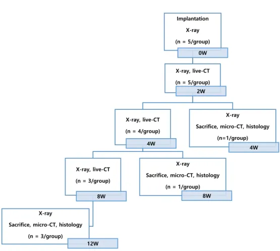
Radiographical evaluation
X-ray scanning and analysis
Live-CT scanning and analysis
Micro-CT scanning and analysis
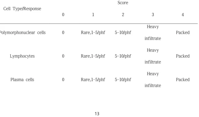
Histopathological evaluation
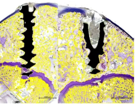
For the screw implanted in bone, a slide that was stained with Villanueva Osteochrome bone stain (BioLead Inc.) was observed to check the condition of the screw and surrounding bone tissue. For analysis, each article was evaluated by continuously enlarging all areas at a high magnification of X400 based on the interface of the test substance.
Bone evaluation method
Histomorphometric evaluation
Radiographical evaluation
Figures 6 and 7 present cross-sectional 2D micro-CT images at 4, 8, and 12 weeks after implant insertion at the point of the femoral condyle and prevertebral muscle. In the analysis, the Mg alloy screw and the bone have similar average molecular weights, so there were no contrast differences in the micro-CT images, and so the boundary between the bone and the Mg screw was manually drawn once in 3 to 5 sections. For all animals analyzed with micro-CT post-adhesion data, gas (blank space; black) generated by the screw at the implantation site was less than the previously reported trend 22), implying that the effect of reducing bone density due to gas accumulation was less.
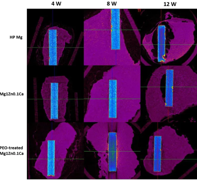
When comparing the amount of hydrogen generated by high-purity Mg, Mg1Zn0.1Ca and PEO-treated Mg1Zn0.1Ca, the largest amount was from high-purity Mg and the least amount was from PEO-treated. 3D modeling was performed to confirm the degree of degradation of the implanted materials and BIC (Figure 9). From the modeling results, it is clear that the helical shape of the Mg1Zn0.1Ca and PEO-treated Mg1Zn0.1Ca alloys was relatively intact compared to high-purity Mg.
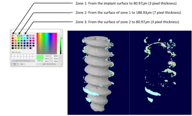
From the results, the degradation rate of the Mg1Zn0.1Ca implants was slower than that of the control high-purity Mg implants, while the PEO-treated Mg1Zn0.1Ca implants degraded even more slowly. Based on the distance from the screw surface, zones 1 to 3 (Figure 3) were divided and the bone occupancy ratio was calculated. It can be observed that the graft material is well present in the surgical site over time.
Micro-CT scans were taken 4, 8 and 12 weeks after surgery and used to measure implant volume, void volume and degenerated bone volume. The largest amount of hydrogen was produced by the high purity Mg implant and the smallest by the plasma electrolytic oxidation (PEO) treated Mg1ZnO.1Ca. BIC (Bone Implant Contact) 3D modeling of screws placed in femoral condyle notches of rabbits at 12 weeks.

Histopathological evaluation
Histopathologic results from un-decalcified bone slides
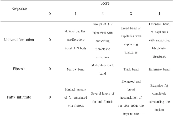
12-week samples decreased the infiltration of inflammatory cells in all groups, which was more pronounced in the test groups than the control group. In particular, the PEO-treated Mg1Zn0.1Ca group showed almost no inflammatory response (Figure 16). At week 12 (when bone union was complete), the PEO-treated Mg1Zn0.1Ca group showed little inflammatory response compared to the control group, indicating that the PEO-coated Mg1Zn0.1Ca implants were the least irritating (Table 6).
Histopathologic results for the muscle tissue
Histomorphometric test results
In the 12th week samples, the PEO-treated Mg1Zn0.1Ca group showed a smaller defect area than that of the control group and the Mg1Zn0.1Ca group. An increase in bone area in the 8th week for the Mg1Zn0.1Ca group was confirmed, but no significant differences were observed between the groups in the 4th and 8th weeks. In the 12th week samples, the bone area was smallest in the PEO-treated Mg1Zn0.1Ca group.
The soft tissue area of the PEO-treated Mg1Zn0.1Ca group at the 4th and 8th weeks was greater than the control and Mg1Zn0.1Ca groups. The void area of 4th week samples showed similar levels, and the 8th week sample showed higher control and Mg1Zn0.1Ca group than the PEO-treated Mg1Zn0.1Ca group. Also at 12 weeks, control group and Mg1Zn0.1Ca group were higher than the PEO-treated Mg1Zn0.1Ca group (Figure 20).
The aim of this study was to compare the biocompatibility of three biodegradable materials: high-purity Mg, Mg1Zn0.1Ca and PEO-treated Mg1Zn0.1Ca. Bar and bolt corrosion rates were calculated using the residual volume values from Figures 10 and 11, respectively; the corrosion rate of Mg1Zn0.1Ca was slower than that of pure Mg, and the PEO-treated Mg1Zn0.1Ca was even slower. The BIC values were in the order of PEO-treated Mg1Zn0.1Ca > Mg1Zn0.1Ca > high-purity Mg.
In particular, the inflammatory response was very low in the PEO-treated Mg1Zn0.1Ca group. At week 12 (when bone union was complete), this group showed little inflammatory response compared to the control group, so the PEO-treated Mg1Zn0.1Ca implants were the least irritating. Therefore, the Mg1Zn0.1Ca and PEO-treated Mg1Zn0.1Ca implants caused less irritation to muscle tissue than the control group.
In this study, the Mg1Zn0.1Ca and PEO-treated Mg1Zn0.1Ca groups showed similar levels of inflammation in the bone tissue observations compared to the control group at weeks 4 and 8. Therefore, the Mg1Zn0.1Ca and PEO-treated Mg1Zn0.1Ca implants were less irritating to the muscle tissue than the high-purity Mg implants. The bone tissue morphological results for the groups were similar at week 4, but the voids at week 8 and 12 were significantly reduced in the PEO-treated Mg1Zn0.1Ca group compared to the others (Figure 20).
Thus, it was confirmed that PEO-treated Mg1Zn0.1Ca implants produced less gas at the interface between the device and the tissue. It was confirmed that Mg1Zn0.1Ca had higher corrosion resistance than high-purity Mg and degraded safely over time without causing side effects (foreign body reaction, inflammatory reaction, etc.) in vivo.

BIC (bone implant contact) measurements of screws placed in rabbit femur