저작자표시 2.0 대한민국 이용자는 아래의 조건을 따르는 경우에 한하여 자유롭게
l 이 저작물을 복제, 배포, 전송, 전시, 공연 및 방송할 수 있습니다. l 이차적 저작물을 작성할 수 있습니다.
l 이 저작물을 영리 목적으로 이용할 수 있습니다. 다음과 같은 조건을 따라야 합니다:
l 귀하는, 이 저작물의 재이용이나 배포의 경우, 이 저작물에 적용된 이용허락조건 을 명확하게 나타내어야 합니다.
l 저작권자로부터 별도의 허가를 받으면 이러한 조건들은 적용되지 않습니다.
저작권법에 따른 이용자의 권리는 위의 내용에 의하여 영향을 받지 않습니다. 이것은 이용허락규약(Legal Code)을 이해하기 쉽게 요약한 것입니다.
Disclaimer
저작자표시. 귀하는 원저작자를 표시하여야 합니다.
약학석사학위논문
TM4SF5-mediated Invasion Cell Behaviors in 3D Gel Systems Consisting of Collagen I and/or
Matrigel
삼차원 세포외기질 환경에서 TM4SF5 발현 세포의 침윤능력 연구
2015 년 2 월
서울대학교 대학원 약학과 의약생명과학전공
이 규 호
Abstract
TM4SF5-mediated Invasive Cell Behaviors in 3D Gel Systems Consisting of Collagen I and/or
Matrigel
Gyuho Lee Pharmaceutical Bioscience College of pharmacy The Graduate School Seoul National University
Unlike 2 dimensional (2D) culture system, extracellular matrix (ECM)-surrounded 3 dimensional (3D) conditions differentially modulate cellular behaviors. Transmembrane 4 L six family member5 (TM4SF5), a branch of the tetraspanin superfamily, is a transmembrane glycoprotein. TM4SF5 is highly expressed in hepatocarcinoma. Previous studies showed that TM4SF5 is involved in epithelial-mesenchymal transition (EMT), cell migration and invasion. Here, we explored the TM4SF5-mediated behaviors of HCC cells embedded in 3D ECM gels,
with regards to invasive properties. To growing cells in 3D ECM Gel, I used 3D Matrigel (basement membrane) and/or Collagen Type I gel systems. In 3D Matrigel contidion, TM4SF5-positive cells formed aggressively invasive foci depending on PI3K or JNKs activity, and actin cytoskeletal organization, whereas TM4SF5-null cells did not. No invasive foci formation of TM4SF5-null cells in 3D Matrigel were recovered by ROCK inhibition itself, or by adding collagen I into Matrigel and additional EGF treatment in collagen I plus Matrigel condition. This TM4SF5-mediated invasive foci formation was inhibited by suppression of either p27kip1, TM4SF5 or pharmacological inhibition of either JNK or AKT and also RhoA activation inhibited the invasive foci formation. However, ERK or RacI inhibition didn't affected this invasvie foci formation. Further, when cell spheroids were embedded in 3D collagen I gels, TM4SF5-positive spheroids showed more protrusive morphology, compared with TM4SF5-null spheroids.
Also, TM4SF5-positive spheroids showed dramatic formation of invadopodia enriched with F-actin and cortactin at the tips of protrusive edges. Moreover, when TM4SF5-positive or TM4SF5-null cells were co-cultured in 3D Matrigel, even TM4SF5-null cells exhibited invasive foci formation surrounded by TM4SF5-positive cells.
All these observations suggest that TM4SF5 can play crucial roles in regulation of invasive properties in 3D ECM environment.
Keywords : 3D cell culture, Extracellular matrix, TM4SF5, Cell invasion, Tumor microenvironment
Student number : 2013-21601
CONTENTS
ABSTRACT ··· 1
LIST OF FIGURES ··· 4
INTRODUCTION ··· 5
MATERIALS AND METHODS ··· 8
RESULTS ··· 11
1. TM4SF5-expressing cells form invasive foci in 3D Matrigel system. ··· 11
2. TM4SF5-mediated ECM deformation during invasive foci formation might not be related to p130cas phosphorylation. ·· 17
3. Invasive foci formation in 3D Matrigel requires PI3K, JNK activity and RhoA inactivation. ··· 20
4. TM4SF5-mediated invasive outgrowth in 3D collagen I or Matrigel. ··· 24
5. Upon addition of collagen I to Matrigel, non-invasive cells showed invasive phenotype. ··· 27
DISCUSSION ··· 31
REFERENCES ··· 35
국문초록 ··· 40
LIST OF FIGURES
Figure 1. TM4SF5-positive cells formed invasive foci in a fully-embedded 3D Matrigel system.
Figure 2. TM4SF5-mediated invasive foci formation in 3D Matrigel On-Top system.
Figure 3. TM4SF5-mediated ECM deformation during invasive foci formation might not be related to p130cas phosphorylation.
Figure 4. TM4SF5-mediated invasive foci formation was inhibited by Pi3K, JNK pathway, p27Kip1 suppression, or RhoA activation.
Figure 5. TM4SF5-mediated invasive outgrowth in 3D collagen I or Matrigel.
Figure 6. Upon addition of collagen I to matrigel, non-invasive cells showed invasive phenotype.
Figure 7. The working model.
INTRODUCTION
During metastasis of cancer from a primary site to a distal site, cancer cells should communicate with their extracellular environmental cues including ECM, mechanical stimuli, cytokines, growth factors and neighboring cells containing leukocyte, endothelial cells and fibroblasts (1). Multiple mutations and genomic instabilities may allow cancer cells to disseminate from a primary tumor by abnormal alteration of cell-cell contact and cell-ECM interactions (2). In normal epithelial cells, cells maintain cell polarity which is supported by cell-cell adhesions and cell-ECM interactions. However, during cancer progression, some carcinoma cells lose the cell polarity and invade through their basement membrane. This phenomenon is referred to as epithelial-mesenchymal transition (EMT) and thought to be a crucial event of cancer progression (3-5). Also, In mammalian tissues, cells are not only interact each other, but also supported by extracellular matrix (ECM). Extracellular matrix consists of several proteins, such as collagen, laminin, elastin (9). However, in tumor mircroenvironment, there is an abnormal ECM remodeling near tumor mass. especially during the tumor progression, ECM scaffold undergoes structural changes including increased collagen I, III and IV, and fibronectin (10).
During metastasis, migration and invasion of tumor cell are regulated by signaling pathways that respond to the extracellular matrix or
soluble factors of tumor microenvironment (11).
Tissues and organs in vivo are made by three-dimensional constructs of organized assembly of different cell types that contribute to the architecture for functional differentiation (12). However, cells grown in vitro on flat 2D tissue culture substrates can differ considerably in their morphology, cell-cell and cell-ECM interactions, and differentiation from those growing in more physiological three-dimensional environments (13). 3D cell culture condition can recapitulating normal and pathological tissue architectures, hence providing physiologically relevant models to study normal development and disease (13). In 3D cell culture systems, cells attach and proliferate to one another and form natural cell-cell and cell-ECM interactions. Thus, by using 3D ECM cell culture system, it can mimic the tumor microenvironment in vivo. To growing cells in 3D ECM gel, I used 3D BME/Matrigel and/or collagen type I gel systems. Matrigel (basement membrane) is an important extracellular matrix that is found in all epithelial and endothelial tissues. Type I collagen is the major ECM component of fibrous connective tissue. Tumor cells in 3D extracellular matrix-surrounded gels can have dynamic behaviors including invasive protrusion, migration, foci formation, all of which can recapitulate tumor cell behaviors traveling through the stromal area enriched with ECM (14).
six family, and highly expressed in many types of cancers. TM4SF5 expression decreases expressions of E-cadherin and ZO-1, and increases expression of α-smooth muscle actin leading to a loss of cell-cell contacts, depending on cytosolic p27kip1-mediated RhoA inactivation and morphological changes (6). Also, TM4SF5 collaborates with integrins or growth factor receptors for cellular function (6-9).
However these TM4SF5 studies were mostly conducted in conventional 2D cell culture systems. Thus, we here explored TM4SF5-mediated cell behaviors for tumorigenic roles in 3D gel system. In this study, we investigated whether and how the invasive behaviors of TM4SF5-expressing cells embedded in 3D ECM gel could be regulated.
Material and Methods
1. 2D Cell culture
Control (SNU449Cp, SNU761-Mock), TM4SF5 WT (SNU449T7, SNU761-WT), or N-glycosylation mutant (N138A/N155Q) -expressing human hepatocellular carcinoma cells and HCC827 (mock, TM4SF5 WT1-9) human lung carcinoma cells have been described previously (7,18). SNU449T7 cells stably transfected with shRNA against TM4SF5 or scramble with tGFP (Origene Inc.) were selected by Puromycin 7µg/ml. every cells were cultured in RPMI-1640 (Welgene Inc.) containing 10% FBS and 1% penicillin/streptomycin (GenDEPOT Inc.) at 37⁰C in 5% CO2.
2. 3D Cell lysate preparation and Western blots.
Cells embedded into 3D Matrigel and cultured for 24 hrs were transferred into ice-cold 1.5ml tubes and centrifuged 5000 RPM for 1 min to remove residual medium. Cell pellet within Matrigel masses were washed with ice cold PBS and then homogenized in a modified RIPA buffer (50mM Tris-HCl, pH 7.4, 150 mM NaCl, 1% NP-40, and 0.25% sodium deoxycholate) with a protease inhibitor cocktail (GenDEPOT, USA), The extract were centrifuged for 30 min at 13000 RPM and 4⁰C. The samples were normalized for equal protein loading in standard immunoblots via α-tubulin levels.
3. Antibodies and reagents
Antibodies and reagents used in this study were listed below.
α-tubulin (Sigma-Aldrich), pS10p27kip1, pS473AKT (SantaCruz Biotechnology), pT473Y473ERK, pS63c-jun (Cell signaling), ERK, p27kip1 (BD Transduction Laboratory), RhoA (Transduction Laboratory), Matrigel, Rat-tail collagen type I (BD biosciences), Diaminophenylindole and Rhodamine phalloidin (Invitrogen), SP600125, U-0126, LY294002, Y27632 (LC-LABs), NSC23766 (SantaCruz Biotechnology)
4. Cell culture in three-dimensional Matrigel/Collagen I gels.
3D Collagen type I gels for cell culture were prepared using Rat tail Collagen I (BD biosciences). Collagen I mixtures (2.5 mg/ml) were made by adding the appropriate volumes of 10x reconstitution buffer (260 mM sodium bicarbonate and 200 mM HEPES), 10x RPMI (Sigma Aldrich) to the collagen I solution. To adjust the pH of the collagen I solution mixture, I used the ice-cold solution of 2 N NaOH. Collagen I mixtures with the cells were incubated in 37⁰C for 30 min. In case of 3D Matrigel, cells were mixed with Matrigel solution in 4⁰C and incubated in 37⁰C for 30 min.
5. Time-lapse imaging
Time-lapse cell images in 3D Matrigel were captured with
IX81-ZDC (Olympus) for 20 ~ 42 hrs, and the imaging system was driven by MetaMorph software (Universal imaging). For Time-lapse microscopes cells were plated onto 3D Matrigel in a Lab-TekTM Chamber Slide (Thermo Scientific). All microscopes were equipped with a Chamlide Incubator systems (LCI live cell instrument, Korea), and an environmental chamber mounted on the microscope maintained constantly at 37⁰C, 5% CO2 and 95% humidity.
6. 3D immunofluorescent analysis
Cells were cultured within polydimethylsiloxane prepolymer (PDMS) glass coverslip and fixed directly with 4% formaldehyde for 30 min at room temperature (RT), and subsequently tretead with 100mM glycine to quench residual aldehyde groups. After PBS washing, cells were permeabilized for 30 min with 0.5% Triton X-100 at RT and blocked for 2 hrs with PBS in 3% BSA. Cells were stained with either fluorescent-labeled phalloidin (Molecular probes, 1:250) or Alexa Fluor®
488-labeled anti-cortactin (Millipore, 1:200) at 4⁰C overnight. Cells were then washed with washing buffer (PBS in 130mM NaCl, 13mM Na10HPO4, 3.5mM NaH2PO4, pH 7.4). Nuclei were counterstained with DAPI (Molecular probes). Confocal images were captured using confocal microscope with a Nikon Plan-Apochromat and analyzed using the NIK software.
RESULT
1. TM4SF5-expressing cells form invasive foci in 3D Matrigel system.
SNU449Cp (TM4SF5-negative) or SNU449T7 (TM4SF5-positive) cell lines were fully embedded in 3D Matrigel and live imaged for more than 28 hrs. Time-lapse imaging showed that TM4SF5-positive cells interact each other and aggregated to form invasive foci. Whereas TM4SF5-negative cells are just stay around as single cells (Fig. 1A).
When more cells embedded into Matrigel, TM4SF5-positive cells formed invasive foci or branched cell network more obviously, whereas control cells did not (Fig. 1B). This TM4SF5-mediated phenotypes were inhibited by treatment of JNK inhibitor (SP600125) or anti-TM4SF5 chemical compund (TSAHC), although ERK inhibitor (U-0126) didn't affect it (Fig. 2C). Although immunoblot analysis showed elevated levels of pS10p27kip1, pS63c-jun, pS473AKT in control DMSO sample, JNK inhibitor and anti-TM4SF5 chemical compund not only block the invasive foci formation, but also downregulate the pS10p27kip1 and pS473AKT levels in TM4SF5-positive samples (Fig. 1D).
These results indicate that pS10p27kip1 and pS473AKT were involved in the TM4SF5-mediated invasive foci formation.
For experiments that needed to be done in a shorter term, or to get more clear image, cells were embedded in 3D On-Top system (15).
When cells were embedded in 3D Matrigel On-Top system similar to the fully-embedded condition, TM4SF5-positive cells gradually gathered together and form invasive foci, whereas TM4SF5-negative cells did not (Fig. 2A). To confirm this invasive foci formation is TM4SF5-dependent, TM4SF5 N-glycosylation mutant cells or TM4SF5-suppressed cells were embedded in 3D Matrigel On-Top. The results showed that TM4SF5-glycosylation mutant or -suppressed cells didn't show invasive foci formation (Fig. 2B). Also quantification of cell velocity showed that TM4SF5-positive cells exhibited more directional movements and higher movement speeds, compared with the TM4SF5-negative cells. These results indicates that TM4SF5-expressing cells gathered together and form invasive foci in 3D Matrigel.
Figure 1. TM4SF5-positive cells formed invasive foci in a fully-embedded 3D Matrigel system.
(A) SNU449 stable cell lines (SNU449Cp: control pooled clone, SNU449T7: stably TM4SF5-overexpressing clone) were embedded in full-3D Matrigel. After 18 hrs, cells were live imaged for 24 hrs. The lower parts showed endpoint snapshot 42 h after embedding. (B) SNU449T7 cells were embedded in full-3D Matrigel with inhibitors against JNK (SP600125, 50 μM), ERK1/2 (U0126, 50 μM), or anti-TM4SF5 TSAHC (50 μM). Images showed endpoint snapshots 42 h after the embedding. (C) SNU449Cp or T7 cells embedded in full-3D Matrigel with each inhibitor for 42 hrs were harvested, before
Figure 2. TM4SF5-mediated invasive foci formation in 3D Matrigel On-Top system
(A) SNU449Cp or T7 stable cell lines were embedded and live imaged in 3D Matrigel On Top for 20 hrs. Snapshot images were shown at each time point. (B) SNU449 stable cell lines (Cp, T7, TM4SF5 glycosylation mutant, or TM4SF5-suppressed) were embedded in 3D Matrigel On Top for 20 hrs. Endpoint snapshot images were shown.
(C) Quantification of the velocity of SNU449Cp or T7 cell lines in 3D Matrigel On Top. Right panels depict movement tracks of cells that
2. TM4SF5-mediated ECM deformation during invasive foci formation might not be related to p130Cas phosphorylation.
To determine whether this invasive foci formation was accompanied by extracellular matrix deformation, I was tracking the movement of microbeads that were seeded in the Matrigel together with the cells.
Microbead around the TM4SF5-negative cells showed only a slight movement, while microbead around the TM4SF5-positive cells showed more dynamic movements (Fig. 3A). Total movement of microbeads were quantified (Fig. 3B). Furthermore, based on p130Cas scaffold protein was known as a mechanosensor (16,17), I postulated that this invasive foci formation and ECM deformation would be regulated by p130Cas activation. Thus, TM4SF5-negative or TM4SF5-positive cells were transduced with GFP-tagged retrovirus-p130Cas witdtype (WT) or its phosphorylation mutant form (15F/mPR). The results showed that most cells were overexpressed with p130Cas WT or its phosphorylation-defective mutant form. However, there were no phenotype changes in both TM4SF5-positive or TM4SF5-negative cells, following the introduction of p130Cas WT or nonphosphorylatable form (Fig. 3C). These observation indicates that TM4SF5-mediated ECM deformation during invasive foci formation was not related to p130Cas phosphorylation.
Figure 3. TM4SF5-mediated ECM deformation during invasive foci formation might not be related to p130Cas phosphorylation.
(A-B) Movement of the microbead around cells that were embedded in 3D Matrigel On Top for 15 hrs. Microbead with 3 mm diameter were mixed with cells during the embedding. (A) Each graph showed total distance that the microbeads (n = 35) around each cell line. (B) The tracks depict microbead trajectories of the cells of each cell line. (C) Retroviral induction of p130Cas (WT) or mutant (15 Tyr to Phe, 15F/mPR) didn’t change the phenotype to form invasive foci in 3D Matrigel On Top.
3. Invasive foci formation in 3D Matrigel requires PI3K, JNK activity and RhoA inactivation.
To further gain the mechanistic insights into the formation of invasive foci, inhibitor treatments were carried out. Since the previous immunoblot analysis (Fig. 1D) showed elevated level of pS10p27kip1, pS63c-jun and pS473AKT, I treated JNK inhibitor (SP600125), AKT inhibitor (LY294002) and anti-TM4SF5 chemical compound (TSAHC).
These inhibitors effectively inhibit the TM4SF5-mediated invasive foci formation, while TM4SF5-negative cells didn't show phenotype changes. Also ERK inhibitor (U-0126), Rac1 inhibitor (NSC-23766) were treated to both TM4SF5-negative or TM4SF5-positive cell line.
However these inhibitors didn't affect the both cell lines, supporting for the fact that TM4SF5 expression didn't affect the ERK activation level. Interestingly, ROCK inhibitor (Y-27632) affected both TM4SF5-negative and TM4SF5-positive cells. TM4SF5-negative cells showed cellular aggregation, just like to TM4SF5-expression alone, consistent with the previous report showed that TM4SF5-expressing cells have downregulated RhoA activity. When I further suppressed RhoA activity in TM4SF5-positive cells, it showed another phenotype, cellular network formation (Fig. 4A). This observation was consistent with the previous report (6,19) showed that the TM4SF5-expressing
p27 (pS10p27kip1). Thus I postulated that p27kip1 knockdown or RhoA activation might inhibit the TM4SF5-mediated foci formation.
Therefore TM4SF5-expressing cells were transduced with adenovirus for p27kip1 knockdown or transduced with retrovirus for RhoA activation. Their result showed that both p27kip1 knockdown or RhoA activation (RhoA Q63L) inhibited the invasive foci formation, compared with no effects by control transductions. These results indicate that TM4SF5-mediated invasive foci formation needed JNK, AKT activation and RhoA inactivation.
Figure 4. TM4SF5-mediated invasive foci formation was inhibited by PI3K, JNK pathway, p27Kip1 suppression, or RhoA activation.
(A) SNU449 stable cell lines were embedded in 3D Matrigel On Top system with treatment of either DMSO, SP600125, TSAHC, LY294002, U-0126, NSC23766 (a specific Rac1 inhibitor), or Y-27632 for 20 hrs at the indicated concentrations. Live imagings were performed for 20 hrs, and the endpoint snapshots were shown for each condition. (B) TM4SF5-expressing SNU449T7 cell line was transduced with adenovirus for p27kip1 suppression (si-p27kip1) or a control scrambled sequence, embedded and live imaged in 3D Matrigel On TOP for 20 hrs. Upper panel showed representative endpoint (20 hrs) snapshots for each condition. Lower panel showed p27kip1 expression levels by an immunoblot. (C) TM4SF5-expressing SNU449T7 cell line was transduced with a control retrovirus or retrovirus for RhoA activation (Q63L:constitutively active), embedded and live imaged in 3D Matrigel On Top for 20 hrs. Upper panels showed representative endpoint (20 hrs) snapshots. Lower panel showed RhoA expression levels by an immunoblot.
4. TM4SF5-mediated invasive outgrowth in 3D collagen I or Matrigel.
Since TM4SF5-negative or TM4SF5-positive cells showed differential phenotype in 3D Matrigel, I have tried to co-culture both TM4SF5-negative and positive cell lines. Interestingly, the result showed that TM4SF5-negative cells formed cellular aggregates surrounded by TM4SF5-positive cell line (Fig. 5A). Further, TM4SF5-positive cells showed invasive cellular protrusion. Such invasive protrusion phenotype by TM4SF5-positive cells were recapitulated by embedding the cell spheroids in 3D collagen I gel.
Time-lapse imaging showed that TM4SF5-positive spheroids exhibited more outgrowth and dissemination of certain cells compared with control spheroids (Fig. 5B-C). Also, immunofluorescent analysis revealed that singnals for F-actin and cortactin, which is an indicator of invadopodia (20), were co-localized in leading edges of the TM4SF5-positive spheroids (Fig. 5D). while TM4SF5-negative spheroids didn't show such cellular outgrowth and co-staining. These observation revealed that TM4SF5-mediated cellular outgrowth involves more effective invadopodia formation.
Figure 5. TM4SF5-mediated invasive outgrowth in 3D collagen I or Matrigel.
(A) SNU449Cp, SNU449T7 cell lines were labeled with cell tracker fluorescent probe, and then cells were mixed at 50:50 (%/%) ratio for Cp and T7 cells before being embedded in 3D Matrigel On-Top for 20 hrs. SNU449Cp cells (green) were gathered together, but SNU449T7 cells (red) were located outside of the cell mass. Panel showed the endpoint fluorescent snapshots. (B) Spheroids of hepatic cancer SNU761 stable cell lines (SNU761-MOCK or SNU761-TM4SF5 WT) were fully-embedded in 3D collagen type I gels together with EGF 50 ng/ml for 26 hrs. Snapshots at the indicated time points were shown.
(C) Spheroids of lung cancer HCC827 stable cell lines (HCC827-MOCK or HCC827-TM4SF51-9) were fully embedded in 3D collagen type I gels for 22 hrs. Snapshots at the indicated time points were shown.
(D) Spheroids of HCC827 or SNU449 stable cell lines were fully-embedded in 3D collagen type I gels for 24 hrs and immunostained for cortactin (green) and stained with phalloidin for actin (red) and DAPI (blue). White arrows indicate co-localization
5. Upon addition of collagen I to Matrigel, non-invasive TM4SF5-null cells showed invasive phenotype.
Furthermore, by culturing cells in collagen I plus Matrigel condition, even TM4SF5-negative cells showed invasive foci formation. This phenotype is similar to RhoA inhibition in TM4SF5-negative cells.
Meanwhile, by adding collagen I to the Matrigel, TM4SF5-positive cells showed cellular network formation (Fig. 6A). This phenotype change became more invasive by additional EGF treatment into TM4SF5-negative cells (Fig. 6B). To sum up, In 3D Matrigel, hepatic epithelial cells became invasive and form invasive foci by TM4SF5 expression. This Invasive foci formation was inhibited by suppression of either p27kip1, TM4SF5 or pharmacological inhibition of either JNK or AKT and also RhoA activation inhibited the invasive foci formation.
TM4SF5-mediated invasive phenotype was more invasive by ROCK inhibition or adding collagen. On the other hands. TM4SF5-negative cells showed rounded morphologies and stayed in single cells in 3D matrigel. By treatment of ROCK inhibitor or adding collagen I, cells showed invasive foci formation. By further treatment of EGF in these cells, they showed invasive network formation (Fig. 7).
Figure 6. Upon addition of collagen I to Matrigel, non-invasive (i.e., TM4SF5-null) cells showed malignantly invasive phenotype.
(A) SNU449 stable cell lines were embedded in 3D ECM On-Top system (as indicated) for 20 hrs. Non-invasive SNU449Cp cells became to form invasive foci, upon addition of collagen I, like SNU449T7 cells in Matrigel alone (Upper panels). SNU449T7 cells became more invasive to form network-like structure in 3D Matrigel and collagen I (bottom panels). (B) SNU449 stable cell lines were embedded in 3D ECM (Matrigel plus collagen I) On-Top system for 20 hrs. SNU449Cp cells became to form invasive network, upon stimulation with EGF (100 ng/ml), like SNU449T7 cells without EGF (Upper panels).
SNU449T7 cells became more invasive to form network-like structure in 3D Matrigel and collagen I after EGF treatment (bottom panels).
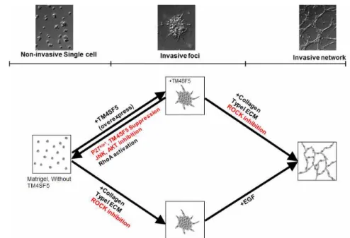
Figure 7. The working model
(A) In 3D Matrigel, non-invasive TM4SF5-null cells exhibited rounded morphologies and styed still in single cells. However, TM4SF5-expressing cells showed invasive protrusions and aggregated together to form invasive foci. Invasive foci was inhibited by suppression of either p27kip1 or TM4SF5 or pharmacological inhibition of either JNK or AKT. This invasive foci was observed in TM4SF5-null cells additionally by ROCK inhibition or by adding collagen type I ECM to the Matrigel. The TM4SF5-null cells showed invasive network in Matrigel/collagen I ECM further with EGF stimulation. Meanwhile, TM4SF5-expressing cells showed more aggressive foci formation even without collagen I addition or ROCK inhibition, and further invasive network formation without any stimulation.
Discussion
Recently tumor microenvironment has received growing attention from the biologist over the last decade. tumor microenvironment consists of various factors such as immune cells, fibroblast, soluble factors, and extracellular matrix. The tumor and the surrounding microenvironment are closely related and interact constantly. Also ECM is a key regulator of tumor cell behaviors (21). These crucial microenvironment may be partially restored by using 3D cultures of laminin-rich matrigel or collagen. Thus, The 3D cell culture represents an important bridge for linking 2D cell culture to the organ or even animal (22). In the basement membrane (Matrigel) non-cancerous cells are maintained by adhesions between integrins on cell surface and extracellular matrix proteins and by cell-cell adhesion (23). But cancer cells undergo morphological change and disturbance of epithelial cell-cell adhesions leading to EMT that occurs for more efficient migration and invasion (24). In previous study, TM4SF5 have been shown to be up-regulated in multiple tumors, have roles in EMT and involved in cacner invasion and metastasis (25,26). In this study, the use of hepatic cancer cells that overexpressing TM4SF5 or empty vector in 3D Matrigel or collagen I gel might demonstrates that formation of tumor mass in vivo-like conditions and dissemination from tumor mass. This study revealed that the TM4SF5-expressing cells showed elevated migration and invasion ability and formed
invasive foci in 3D Matrigel condition, whereas with TM4SF5-null cells showed lower migration ability in 3D Matrigel. When cells embedded in 3D Matrigel, TM4SF5-overexpressing cells interact each other and aggregated to form invasive foci (Fig . 1A, 2A). Immunoblot analysis revealed that this TM4SF-mediated phenotype needed JNK activation and cytosolic p27. Also, This foci formation phenotype was recapitulated by treatment of ROCK inhibitor (Y-27632) or adding collagen I into Matrigel in TM4SF5-null cells. Further, upon additional treatment of EGF into TM4SF5-null cells in collagen I plus Matrigel condition, TM4SF5-null cells form cellular network formation. In case of TM4SF5-expressing cells, it showed cellular network formation just by treatment of ROCK inhibitor or adding collagen I into Matirgel (Fig 5). These results indicates that in 3D Matrigel (basememt membrane), TM4SF5-expression might plays crucial roles in cancer cell migration or invasion from primary site to a distal site. Furthermore, by tracking microbeads that embedded with the TM4SF5-negative or -positive cells in 3D Matrigel, invasive foci formation accompanied by ECM deformation (Fig. 3A). Based on this result (Fig. 3A) and previous reports that cell-matrix adhesion and local ECM distribution is associated with cell functions and mechanotransduction (27), thus we hypothesized that this invasive foci formation and ECM deformation might be related to p130Cas, the mechanosensor scaffold protein (17).
suppression (Fig. 3C). In addition, when TM4SF5-negative of -positive cells were co-cultured in 3D Matrigel, TM4SF5-negative cells exhibited invasive foci formation surrounded by TM4SF5-positive cells (Fig 5A). This observation indicates that TM4SF5-expressing cells secretes some paracrine factors (i.e., TGF-β or MMP-2) that could affect the nearby non-cancerous cells. MMP-dependent EMT has been reported in a variety of epithelial cell types. (28-31) consistent with these reports, pharmacological inhibition of MMP2 or antibody blocking of TGF-βRII inhibited TM4SF5-mediated invasive foci formation (data not shown). With these results we hypothesized that tumor-associated stroma cells that are known as secretes various paracrine factors may play similar roles like TM4SF5-expressing cells. (32,33). Therefore, TM4SF5-negative cells were co-cultured with Carcinoma-associated fibroblast (CAF) or Mesenchymal stem cell-like cell in 3D Matrigel.
The result showed similar phenotype to the TM4SF5-positive cells, as shown in Fig. 5A. This observation might indicate that TM4SF5-overexpressing cells have similar roles such as carcinoma-associated stroma cells. Furthermore, we checked another ECM protein, collagen type I. collagen constitutes the scaffold of tumor microenvironment and affects tumor microenvironment such that it regulates ECM remodeling by collagen degradation and re-deposition, and promotes tumor infiltration, angiogenesis, invasion and migration (34). TM4SF5-positive or -negative cell spheroids were embedded into
3D collagen I gels. Time-lapse observation showed that TM4Sf5-positive spheroids showed more outgrowth and dissemination of certain cells (Fig. 5B-C). Also, Immunofluorescence analysis revealed that signals for F-actin and Cortactin, which is an indicator of invadopodia (20), were co-localized in leading edges of the TM4SF5-expressing cells, while TM4SF5-negative cells did not showed such cellular outgrowth and co-staining (Fig. 5D). These observasion indicates that TM4SF5-mediated outgrowth involves more effective invadopodia formation. Furthermore, in 3D ECM gel, hepatic cancer cells exhibited foci formation or cell network formation by adding collagen I into Matrigel or additional treatment of EGF (Fig. 6).
These observastions showed TM4SF5-expressing cells exhibited one-level more malignant cell phenotypes, compared with TM4SF5-negative cells. These results indicate that TM4SF5 expression itself could be enough to cause invasive phenotypes to the hepatic cancer cells even with a minimal environmental cues.
Moreover, these three-dimensional culture allow phenotypic discrimination between non-maliganant and malignant especially in breast cancer cell (36,36). This study revealed not only the TM4SF5-mediated invasive cell behoviors in 3D ECM conditions, but also represent the hepatic cancer cell model (i.e., invasive foci formation or cell network formation ability) in 3D cell culture system.
References
1. Whiteside, T. L. "The tumor microenvironment and its role in promoting tumor growth." Oncogene 27.45 (2008): 5904-5912.
2. Stamenkovic, Ivan. "Extracellular matrix remodelling: the role of matrix metalloproteinases." The Journal of pathology 200.4 (2003):
448-464.
3. Thiery, Jean Paul. "Epithelial–mesenchymal transitions in tumour progression." Nature Reviews Cancer 2.6 (2002): 442-454.
4. Christiansen, Jason J., and Ayyappan K. Rajasekaran. "Reassessing epithelial to mesenchymal transition as a prerequisite for carcinoma invasion and metastasis." Cancer research 66.17 (2006): 8319-8326.
5. Grünert, Stefan, Martin Jechlinger, and Hartmut Beug. "Diverse cellular and molecular mechanisms contribute to epithelial plasticity and metastasis." Nature Reviews Molecular Cell Biology 4.8 (2003):
657-665.
6. Lee, Sin-Ae, et al. "Tetraspanin TM4SF5 mediates loss of contact inhibition through epithelial-mesenchymal transition in human hepatocarcinoma." The Journal of clinical investigation 118.4 (2008):
1354-1366.
7. Lee, Sin, et al. "Blockade of four‐transmembrane L6 family member 5 (TM4SF5)‐mediated tumorigenicity in hepatocytes by a synthetic chalcone derivative." Hepatology 49.4 (2009): 1316-1325.
8. Lee, Sin-Ae, Ki Hun Park, and Jung Weon Lee. "Modulation of signaling between TM4SF5 and integrins in tumor microenvironment." Front Biosci 16 (2011): 1752-1758.
9. Abbott, Alison. "Cell culture: biology's new dimension." Nature 424.6951 (2003): 870-872.
10. Mathot, Lucy, and Johan Stenninger. "Behavior of seeds and soil in the mechanism of metastasis: a deeper understanding." Cancer science 103.4 (2012): 626-631.
11. Friedl, Peter, and Katarina Wolf. "Proteolytic interstitial cell migration: a five-step process." Cancer and Metastasis Reviews 28.1-2 (2009): 129-135.
12. Griffith, Linda G., and Melody A. Swartz. "Capturing complex 3D tissue physiology in vitro." Nature Reviews Molecular Cell Biology 7.3 (2006): 211-224.
13. Nelson, Celeste M., and Mina J. Bissell. "Of extracellular matrix, scaffolds, and signaling: tissue architecture regulates development, homeostasis, and cancer." Annual review of cell and developmental biology 22 (2006): 287.
14. Cox, Thomas R., and Janine T. Erler. "Remodeling and homeostasis of the extracellular matrix: implications for fibrotic diseases and cancer." Disease models & mechanisms 4.2 (2011): 165-178.
15. Lee, Genee Y., et al. "Three-dimensional culture models of normal
359-365.
16. Goldmann, Wolfgang H. "Mechanotransduction and focal adhesions."
Cell biology international 36.7 (2012): 649-652.
17. Meenderink, Leslie M., et al. "P130Cas Src-binding and substrate domains have distinct roles in sustaining focal adhesion disassembly and promoting cell migration." PLoS One 5.10 (2010):
e13412.
18. Lee, Mi-Sook, et al. "Gefitinib resistance of cancer cells correlated with TM4SF5-mediated epithelial–mesenchymal transition."
Biochimica et Biophysica Acta (BBA)-Molecular Cell Research 1823.2 (2012): 514-523.
19. Besson, Arnaud, et al. "p27Kip1 modulates cell migration through the regulation of RhoA activation." Genes & development 18.8 (2004): 862-876.
20. Albiges-Rizo, Corinne, et al. "Actin machinery and mechanosensitivity in invadopodia, podosomes and focal adhesions."
Journal of cell science 122.17 (2009): 3037-3049.
21. Koontongkaew, Sittichai. "The tumor microenvironment contribution to development, growth, invasion and metastasis of head and neck squamous cell carcinomas." Journal of Cancer 4.1 (2013): 66.
22. Page, Henry, Peter Flood, and Emmanuel G. Reynaud.
"Three-dimensional tissue cultures: current trends and beyond."
Cell and tissue research 352.1 (2013): 123-131.
23. Harris, Tony JC, and Ulrich Tepass. "Adherens junctions: from molecules to morphogenesis." Nature Reviews Molecular Cell Biology 11.7 (2010): 502-514.
24. Guarino, Marcello, Barbara Rubino, and Gianmario Ballabio. "The role of epithelial-mesenchymal transition in cancer pathology."
Pathology 39.3 (2007): 305-318.
25. Kao, Yu-Rong, et al. "Tumor-associated antigen L6 and the invasion of human lung cancer cells." Clinical cancer research 9.7 (2003): 2807-2816.
26. Muschel, Ruth J., and Annamaria Gal. "Tetraspanin in oncogenic epithelial-mesenchymal transition." Journal of Clinical Investigation 118.4 (2008): 1347-1350.
27. Vogel, Viola, and Michael Sheetz. "Local force and geometry sensing regulate cell functions." Nature Reviews Molecular Cell Biology 7.4 (2006): 265-275.
28. Cheng, Sunfa, and David H. Lovett. "Gelatinase A (MMP-2) is necessary and sufficient for renal tubular cell epithelial-mesenchymal transformation." The American journal of pathology 162.6 (2003): 1937-1949.
29. Tan, Thian Kui, et al. "Macrophage Matrix Metalloproteinase-9 Mediates Epithelial-Mesenchymal Transition< i> in Vitro</i> in Murine Renal Tubular Cells." The American journal of pathology
30. Zheng, Guoping, et al. "Disruption of E-cadherin by matrix metalloproteinase directly mediates epithelial-mesenchymal transition downstream of transforming growth factor-β1 in renal tubular epithelial cells." The American journal of pathology 175.2 (2009): 580-591.
31. Illman, Sara A., et al. "Epilysin (MMP-28) induces TGF-β mediated epithelial to mesenchymal transition in lung carcinoma cells." Journal of cell science 119.18 (2006): 3856-3865.
32. Yu, Y., et al. "Cancer-associated fibroblasts induce epithelial–
mesenchymal transition of breast cancer cells through paracrine TGF-β signalling." British journal of cancer (2013).
33. Hanahan, Douglas, and Robert A. Weinberg. "Hallmarks of cancer:
the next generation." Cell 144.5 (2011): 646-674.
34. Fang, Min, et al. "Collagen as a double-edged sword in tumor progression." Tumor Biology 35.4 (2014): 2871-2882.
35. Nelson, Celeste M., and Mina J. Bissell. "Modeling dynamic reciprocity: engineering three-dimensional culture models of breast architecture, function, and neoplastic transformation." Seminars in cancer biology. Vol. 15. No. 5. Academic Press, 2005.
36. Ghajar, Cyrus M., and Mina J. Bissell. "Extracellular matrix control of mammary gland morphogenesis and tumorigenesis: insights from imaging." Histochemistry and cell biology 130.6 (2008): 1105-1118.
국문 초록
세포외 미세환경은 이웃하는 다른 세포, 세포성장인자들, cytokine, chemokine, 및 세포외기질들로 구성되며, 이들은 세포의 기능 조절에 큰 영향을 미치는 것으로 잘 알려져 있다. 한편, 세포외기질(ECM)로 둘러쌓 여 있는 삼차원 배양조건에서는, 기존의 이차원 배양조건과는 다르게 세 포의 행동이 조절될 것으로 이해되고 있다. TM4SF5는 간암을 포함하여 여러 암종에서 높게 발현되며, EMT(Epithelial-mesenchymal transition, 상피-중배엽 세포 전이), 세포의 이동, 침윤을 촉진하는 역할을 하고 있음 이 알려져 있다. 본 연구에서, 나는 삼차원적으로 세포외기질로 둘러싸인 환경 속에서 분석하였을 경우, TM4SF5를 발현하는 세포는, TM4SF5가 발현되지 않는 세포에 비하여 향상된 세포의 이동/침윤 능력을 확인하였 다. 삼차원적 Matrigel 속에서는 TM4SF5를 발현하는 세포들은 PI3K, JNK 활성 및 액틴세포골격 형성 능력, 그리고 p27Kip1 발현에 의존하는 invasive foci (암세포가 모여들어 foci를 형성)를 형성하였고, TM4SF5를 발현하지 않는 세포들은 invasive foci를 형성하지 않았다. 삼차원적 Matrigel 속에서 invasive foci를 형성하지 않았던 TM4SF5가 발현되지 않는 세포들은 ROCK를 억제하거나, Matrigel에 Collagen I 을 추가로 넣 어주게 되면 TM4SF5를 발현하는 세포들처럼 invasive foci를 형성하는 것을 확인하였다. TM4SF5를 발현하는 세포와 발현하지 않는 세포를 삼 차원적 Matrigel 속에서 공동배양 하였을 때에는 TM4SF5의 유무에 상관
foci의 바깥 쪽에 위치하는 것을 확인하였다. 반면에 삼차원적 Collagen I 젤 속에서 TM4SF5를 발현하는 세포들은 TM4SF5가 발현되지 않는 세 포들에 비해서 더 침윤적으로 돌출된 형태를 나타내는 것을 확인하였으 며, 또한 이러한 세포의 말단 돌출부위에서는 invadopodia가 많이 형성되 어 F-actin과 cortactin이 같이 존재하는 것을 확인하였다. 이러한 결과들 은 TM4SF5가 삼차원적으로 ECM에 둘러싸인 배양환경에서 세포의 침윤 능력을 중요히 조절할 수 있다는 것을 제시한다.
주요어 : 삼차원 세포배양, 세포외기질, TM4SF5, 세포침윤, 종양미세환경 학 번 : 2013-21601