저작자표시-비영리-변경금지 2.0 대한민국 이용자는 아래의 조건을 따르는 경우에 한하여 자유롭게
l 이 저작물을 복제, 배포, 전송, 전시, 공연 및 방송할 수 있습니다. 다음과 같은 조건을 따라야 합니다:
l 귀하는, 이 저작물의 재이용이나 배포의 경우, 이 저작물에 적용된 이용허락조건 을 명확하게 나타내어야 합니다.
l 저작권자로부터 별도의 허가를 받으면 이러한 조건들은 적용되지 않습니다.
저작권법에 따른 이용자의 권리는 위의 내용에 의하여 영향을 받지 않습니다. 이것은 이용허락규약(Legal Code)을 이해하기 쉽게 요약한 것입니다.
Disclaimer
저작자표시. 귀하는 원저작자를 표시하여야 합니다.
비영리. 귀하는 이 저작물을 영리 목적으로 이용할 수 없습니다.
변경금지. 귀하는 이 저작물을 개작, 변형 또는 가공할 수 없습니다.
2 0 13 年 2 月 博士學位申請論文
Effects of Oxidative Stress on Endothelial Modulation of Contractions in Aorta
from Renal Hypertensive Rats
朝鮮大學校 大學院
醫 學 科
신 혜 랑
[UCI]I804:24011-200000263703
Effects of Oxidative Stress on Endothelial Modulation of Contractions in Aorta
from Renal Hypertensive Rats
신성 고혈압쥐에서 내피의존 혈관수축반응에 미치는 산화적 스트레스 영향
2013 年 2 月 日
朝鮮大學校 大學院
醫 學 科
신 혜 랑
Endothelium-Dependent Vasodilation by Ferulic Acid in Aorta from Chronic Renal
Hypertensive Rats 指導敎授 金 相 勳
이 論文을 醫學博士學位 申請論文으로 提出함
2012 年 10 月 日
朝鮮大學校 大學院
醫 學 科
신 혜 랑
신 혜 랑의 博士學位論文을 認准함
委員長 朝鮮大學校 敎授 박 상 학 印 委 員 朝鮮大學校 敎授 염 철 호 印 委 員 朝鮮大學校 敎授 전 제 열 印 委 員 全南大學校 敎授 김 재 민 印 委 員 朝鮮大學校 敎授 김 상 훈 印
2012 年 12 月 日
朝鮮大學校 大學院
CONTENTS
Korean Abstract --- 5
Ⅰ. INTRODUCTION --- 7
Ⅱ. METHODS --- 9
1. Induction of 2K1C Hypertension --- 9
2. Tissue Preparation --- 10
3. Protocol --- 12
4. Drugs --- 13
5. Statistics --- 13
Ⅲ. RESULTS --- 15
Ⅳ. DISCUSSION --- -- 30
Ⅴ. SUMMARY --- 36
REFERENCES --- 38
CONTENTS OF FIGURES
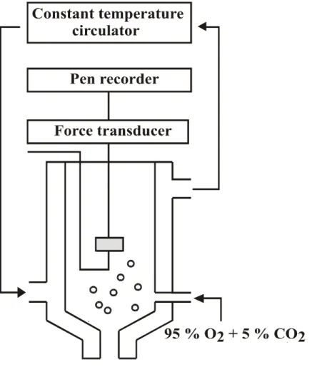
Fig. 1. A schematic representation of the recording system for isometric contraction with 15 mL tissue bath. --- 11
Fig. 2. Systolic blood pressure in 2K1C hypertensive and sham-operated control rats. --- 16
Fig. 3. Concentration-response curves to norepinephrine in aortic rings with and without endothelium from sham rats. --- 17
Fig. 4. Concentration-response curves to norepinephrine in aortic rings with and without endothelium from sham rats. 2K1C rats --- 18
Fig. 5. Effects of Vitamin C on concentration-response curves to norepinephrine in aortic rings with endothelium from sham rats. --- 19
Fig. 6. Effects of Vitamin C on concentration-response curves to norepinephrine in aortic rings with endothelium from 2K1C rats. --- 20 Fig. 7. Effects of diphenyleneiodonium on
concentration-response curves to norepinephrine in
aortic rings with endothelium from sham rats. ---- 22
Fig. 8. Effects of diphenyleneiodonium on concentration-response curves to norepinephrine in aortic rings with endothelium from 2K1C rats. ---- 23
Fig. 9. Effects of apocynin on concentration-response curves to norepinephrine in aortic rings with endothelium from sham rats. --- 24
Fig. 10. Effects of apocynin on concentration-response curves to norepinephrine in aortic rings with endothelium from 2K1C rats. --- 25
Fig. 11. Effects of allopurinol on the contractile response to norepinephrine in aortic rings with endothelium from sham rats. --- 26
Fig. 12. Effects of allopurinol on the contractile response to norepinephrine in aortic rings with endothelium from 2K1C rats. --- 27
Fig. 13. Effects of hypoxanthine plus xanthine oxidase and hypoxanthine plus xanthine oxidase plus vitamin C on concentration-response curves to norepinephrine in aortic rings with endothelium from sham
rats.--- 28
Fig. 14. Effects of hypoxanthine plus xanthine oxidase and hypoxanthine plus xanthine oxidase plus vitamin C on concentration-response curves to norepinephrine in aortic rings with endothelium from 2K1C rats.--- 29
국문초록
신성 고혈압쥐에서 내피의존 혈관수축반응에 미치는 산화적 스트레스 영향
신 혜 랑
지도교수: 김 상 훈 (金 相 勳) 조선대학교 대학원 의학과
혈관내피세포는 평활근층 긴장을 조절함으로써 혈압조절에 관여 하며 고혈압상태에서는 혈관내피세포 기능 변화가 일어남이 알려 져 있다. 저자는 신혈관성 고혈압상태에서 산화적 스트레스가 내 피의존 혈관수축반응에 미치는 영향이 차이가 있는지 밝히고자 본 연구를 시행하였다.
체중 160~180 g 흰쥐 (Sprague-Dawley, 수컷)의 일측 신동맥에 직경 0.2 mm 의 clip을 장치하고 10 주 동안 사육하여 2-kidney, 1 clip (2K1C) 고혈압을 유발시켰다. 대조군은 개복한 후 바로 봉합 하였다. 실험당일 흉부 대동맥을 채취하여 수조에 현수하고 그 등 장성 장력변화를 생리기록기에 기록하였다.
적출 흉부 대동맥 표본은 norepinephrine에 의해 용량의존 수축
반응을 보였으며 그 반응은 고혈압군에서 항진되었다.
Norepinephrine에 의한 수축반응은 혈관 내피층을 제거하거나 Nω -nitro-L-arginine methyl ester 처치시 증가되었으며 그 크기는
고혈압군에서 대조군에 비해 약화되었다. Vitamin C 는
norepinephrine의 수축반응을 고혈압군에서 억제시켰으나 대조군
은 영향받지 않았으며 diphenyleneiodonium 이나 apocynin 전처치 시에도 유사한 양상을 보였다. Hypoxanthine과 Xanthine oxidase 병용처치시 대조군은norepinephrine의 수축반응이 항진되었으나 고 혈압군은 영향이 없었으며 수축반응은 Vitamin C에 의해 양군 모 두 억제되었다.
이상의 실험결과로 보아 혈관내피세포는 nitric oxide를 유리시킴 으로써 수축반응을 조절하며 2K1C 신성고혈압상태에서 혈관내피 층 기능의 변이는 부분적으로 NADH/NADPH oxidase에 의한 superoxide anion 생성의 증가와 관련될 것으로 사료된다.
Ⅰ. INTRODUCTION
Initially regarded as an inert between blood and blood vessels, the vascular endothelium plays a pivotal role in maintaining the vascular tone by generating various vasoactive substances1). Under basal conditions, the endothelium releases relaxing and contracting factors, of which balance contributing to the local regulation of vascular tone2). Hypertension is associated with altered endothelial function in large conduit and small resistance arteries3-5), which is characterized by impaired endothelium-dependent relaxation in response to vasodilators such as acetylcholine. The endothelium-dependent vasodilation is indeed impaired in a number of experimental models, including two-kidney, one clip (2K1C) renal hypertension6-8). A decreased production of endothelial relaxing factors or an increased production of endothelial contractile substances may be responsible for the depressed endothelium-dependent relaxations in hypertension9).
Besides the direct relaxing or contracting effects of the endothelium, it also can modulate the effects of vasoconstrictor substances. It is widely recognized that the vascular endothelium plays an important role in the response of isolated vascular segments to several vasoconstrictors, including norepinephrine7, 10). The constrictors apparently interact with the endothelium to cause the release of relaxing factors, which then have an inhibitory effect on vascular smooth muscle tone7, 11). We have reported previously
that the endothelium negatively modulates contractions of rat aorta by releasing nitric oxide (NO)12). The inhibitory effect of the endothelium against contractions induced by norepinephrine was markedly deteriorated in 2K1C hypertensive rats compared to sham-clipped control rats12). However, the mechanisms that underlie the impaired endothelial modulation have not been extensively elucidated.
It is well known that the development of endothelial dysfunction is linked to an exaggerated production of superoxide anion. The evidence has accumulated that increased oxidative inactivation of NO by an excess of superoxide anions may account for the decrease in available NO and endothelial dysfunction observed in hypertension13, 14). Miyagawa et al15) have shown that vascular oxidative stress may contribute to the altered circulation by impairing endothelial modulation of vascular contractions in spontaneously hypertensive rats. In addition, an increase in oxidative stress systemically plays a major role in maintaining high arterial blood pressure and sympathetic drive in renal hypertension16), and we also observed that hydrogen peroxide may partly contribute to an altered vascular relaxation in 2K1C hypertensive rats17).
Therefore the purpose of resent study was designed to examine the possible role of oxidative stress in the impaired endothelium dependent modulation of vascular contractions in 2K1C renal hypertensive and sham-clipped normotensive rats isolated aortic ring preparations.
Ⅱ. METHODS
1. Induction of 2K1C renal hypertension
Male Sprague-Dawley rats (160~180 g) were anesthetized with sodium thiopental (40 mg/kg, IP). The left posterior side of the animal was shaved and sterilized with 70 % ethanol. An incision about 2 cm long was made through the skin and muscles just below the ribs. The left kidney was then exposed and retracted to expose the renal artery. A silver clip with an internal diameter of 0.2 mm was applied on the exposed renal artery. The clip was then turned so that the slit opening faces the abdomen. The contralateral kidney was left intact. The muscles and skin were sutured and the rats were allowed to recover from anaesthesia. Control rats received a sham treatment: they were operated as in 2K1C rats except for that no clip was made. All rats were maintained on standard chow with free access to drinking water. They were used at 10 weeks after clipping, since the endothelial dysfunction is associated with a duration of hypertension18). Hypertensive rats were selected on the basis of the systolic blood pressure measured obtained in conscious rats by use of the tail-cuff method with a piezoelectric pneumotaxic pulse transducer.
2. Tissue preparation
The thoracic aorta between the aortic arch and diaphragm was rapidly removed and placed into a physiological salt solution (PSS) of the following composition (mM): NaCl 118.3, KCl 4.7, NaHCO3 25, MgCl2 1.2, KH2PO4 1.2, CaCl2 2.5 and glucose 11.1. Vessels were cleaned of adhering tissue and cut into rings (2 mm in width) under a dissection microscope. Care was taken not to stretch the artery or damage the luminal surface. In some preparations, the endothelium was removed by gentle rubbing of the intimal surface with a moistened cotton swab. Successful removal of endothelial cells from aortic rings was confirmed by the inability of acetylcholine to induce relaxation.
The rings were suspended by means of two triangle-shaped stainless steel holders in the vessel lumen in organ chambers containing 15 ml of PSS maintained at 37 ℃, aerated with a mixture of 95 % O2 and 5 % CO2. One of the holders was fixed at the bottom of the chambers and the other was connected to a force displacement transducer (Grass FTO3) for measurement of isometric tension development (Fig. 1). Before initiating specific experimental protocols, aortae were equilibrated under a resting tension of 2 g for at least 90 min. This tension was found to be optimal for contraction of the preparation as assessed by repeated exposure to a 60 mM KCl solution (obtained by equimolar replacement of NaCl by KCl in the
Fig. 1. A schematic representation of the recording system for isometric contraction with 15 mL tissue bath.
physiological solution) under various resting tensions. After the equilibration period, the rings were maximally contracted by the 60 mM KCl solution two times at 45 min intervals.
3. Protocol
Concentration-response curves to norepinephrine (10-10 to 10-5 M) were obtained in rings with or without endothelium. To obtain α-adrenoceptor mediated responses to norepinephrine, aortic rings were pretreated with β-adrenoceptor antagonist timolol (3×10⁻⁷ M). The contribution of NO to the endothelium-dependent modulation of vascular contractions was assessed by analyzing the effect of Nω-nitro-L-arginine methyl ester (L-NAME 10-4 M), which inhibits the endogenous production of NO from L-arginine, on the contractile response to norepinephrine. To examine a possible role of oxidative stress and the involvement of nicotinamide adenine dinucleotide/nicotinamide adenine dinucleotide phosphate (NADH/NADPH) oxidase in the impaired modulation of contractions by the endothelium in 2K1C rats, we obtained concentration-response curves to norepinephrine of aortic rings with endothelium in the absence and presence of vitamin C (10-4 M) or inhibitors of NADH/NADPH oxidase, diphenyleneiodonium (DPI, 10-5 M) or apocynin (3×10⁻⁵ M)15). The potential role of other enzymatic sources of superoxide was assessed by
examining the effects of allopurinol (3×10⁻⁴ M), a xanthine oxidase inhibitor, on concentration–response curves to norepinephrine. The effects of superoxide on the contractile response to norepinephrine were examined using the hypoxanthine–xanthine oxidase system, which generates superoxide anion and hydrogen peroxide19). Concentration–
response curves for norepinephrine were constructed in the absence and presence of hypoxanthine (2×10⁻⁴ M)/xanthine oxidase (20 U/L) plus catalase (1000 U/mL).
4. Drugs
Drugs used were acetylcholine chloride, apocynin (Calbiochem, California, USA), catalase, diphenyleneiodonium, hypoxanthine, L-norepinephrine bitartrate, L-NAME, timolol maleate, vitamin C (L-ascorbic acid) and xanthine oxidase. They were purchased from Sigma Chemical Co (St. Louis, MO, USA) unless otherwise stated. Drug concentrations are expressed as the final molar concentration within the organ chamber and all drugs were administered in volumes not exceeding 0.5 % of the bath volume.
5. Statistics
Contractions were expressed as a percentage of the contraction developed by the 60 mM KCl. Data are presented as means and standard error of the mean for the number of aortic rings indicated in parentheses. Statistical comparisons were performed by means of unpaired t test or analysis of variance with repeated measurements and Fischer post-hoc test.
Differences were considered to be statistically significant when P value was less than 0.05.
Ⅲ. RESULTS
Ten weeks after the operative intervention, the systolic blood pressure were 136±3 mmHg (n=38) and 190±4 mmHg (n=40) in sham-clipped control and 2K1C hypertensive rats, respectively (P<0.05, Fig. 2). The magnitude of KCl (60 mM)-induced isometric tension development was comparable between the two groups (1.41±0.10 g in control; 1.49±0.13 g in 2K1C).
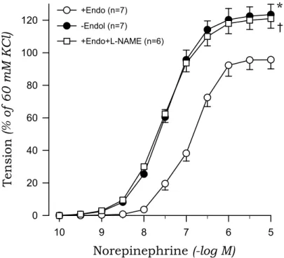
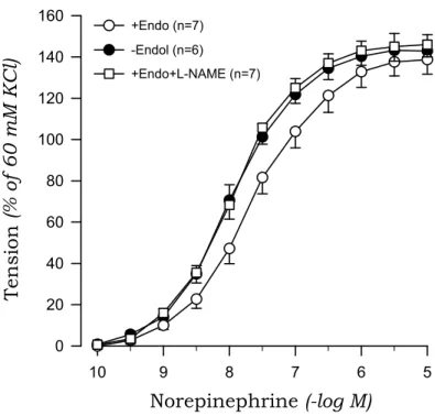
Norepinephrine evoked a concentration-dependent contraction of aortic rings from sham-clipped and 2K1C rats. The contractile response to norepinephrine of aortic rings with endothelium was greater in 2K1C than in those from sham rats.
Removal of the endothelium augmented the norepinephrine-induced contraction, and this effect was more pronounced for aortic rings from sham rats compared to 2K1C rats. Treatment with L-NAME mimicked the effects of endothelium removal on norepinephrine-induced contraction (Fig.
3, Fig. 4).
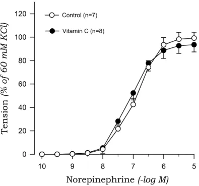
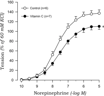
To investigate whether oxidative stress is involved in the impaired endothelial inhibition of norepinephrine-induced contraction in 2K1C hypertensive rats, we examined the effects of vitamin C on norepinephrine-induced contraction in aortic rings with endothelium. Vitamin C reduced the contractile response to norepinephrine in 2K1C rats but not sham rats (Fig.
5, Fig. 6).
0 50 100 150
200
*
Sham (n=38)
2K1C (n=40)
S B P ( m m H g )
Fig. 2. Systolic blood pressure (SBP) in 2K1C hypertensive and sham-operated control rats. The values were obtained at 10 weeks after inducing the hypertension. * P<0.05, compared with the sham value.
Norepinephrine (-log M)
5 6
7 8
9 10
Tension (% of 60 mM KCl)
0 20 40 60 80 100 120
Sham
*
+Endo+L-NAME (n=6) +Endo (n=7)
-Endol (n=7) †
Fig. 3. Concentration-response curves to norepinephrine in aortic rings with (+) and without (-) endothelium (Endo) from sham rats. The results obtained in rings with endothelium in the presence of L-NAME (10-4 M) are also shown. Points represent means±SE for number(n) of experiments in parentheses. *, ✝ P〈0.05, compared with the +Endo value, respectively.
Norepinephrine (-log M)
5 6
7 8
9 10
Tension (% of 60 mM KCl)
0 20 40 60 80 100 120 140 160
2K1C
+Endo+L-NAME (n=7) +Endo (n=7)
-Endol (n=6)
Fig. 4. Concentration-response curves to norepinephrine in aortic rings with (+) and without (-) endothelium (Endo) from 2K1C rats. The results obtained in rings with endothelium in the presence of L-NAME (10-4 M) are also shown.
Norepinephrine (-log M)
5 6
7 8
9 10
Tension (% of 60 mM KCl)
0 20 40 60 80 100 120
Sham
Control (n=7) Vitamin C (n=8)
Fig. 5. Effects of Vitamin C on concentration-response curves to norepinephrine in aortic rings with endothelium from sham rats.
Norepinephrine (-log M)
5 6
7 8
9 10
Tension (% of 60 mM KCl)
0 20 40 60 80 100 120 140 160
2K1C
Control (n=6) Vitamin C (n=7)
*
Fig. 6. Effects of Vitamin C on concentration-response curves to norepinephrine in aortic rings with endothelium from 2K1C rats. * P<0.05, compared with the control value.
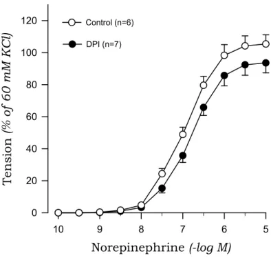
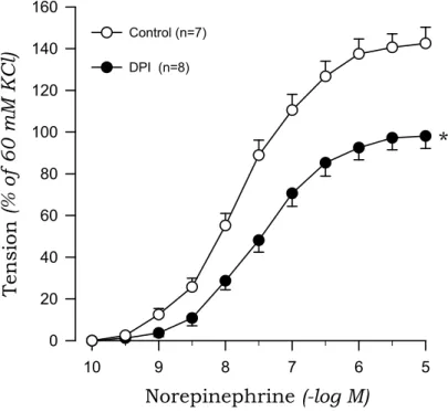
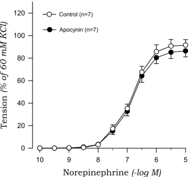
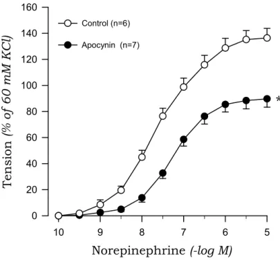
To investigate the origin of oxidative stress, we examined the effects of DPI and apocynin on norepinephrine-induced contraction in aortic rings from 2K1C and sham rats. DPI markedly suppressed the contractile response to norepinephrine of aortic rings with endothelium from 2K1C rats but not sham rats (Fig. 7, Fig. 8). Similarly, apocynin inhibited the norepinephrine-induced contraction in aortic rings with endothelium obtained from 2K1C rats but not sham rats (Fig. 9, Fig. 10).
A xanthine oxidase inhibitor, allopurinol affected the contraction to norepinephrine neither in 2K1C nor in sham rats (Fig. 11, Fig. 12).
In aortic rings with endothelium from sham rats, xanthine oxidase in the presence of hypoxanthine enhanced norepinephrine-induced contraction. This enhancement disappeared in the presence of vitamin C (Fig. 13). In contrast, the hypoxanthine–xanthine oxidase system did not augment the contractile response to norepinephrine in 2K1C rats (Fig. 14).
Norepinephrine (-log M)
5 6
7 8
9 10
Tension (% of 60 mM KCl)
0 20 40 60 80 100 120
Sham
Control (n=6) DPI (n=7)
Fig. 7. Effects of diphenyleneiodonium (DPI) on concentration-response curves to norepinephrine in aortic rings with endothelium from sham rats.
Norepinephrine (-log M)
5 6
7 8
9 10
Tension (% of 60 mM KCl)
0 20 40 60 80 100 120 140 160
2K1C
Control (n=7) DPI (n=8)
*
Fig. 8. Effects of diphenyleneiodonium (DPI) on concentration-response curves to norepinephrine in aortic rings with endothelium from 2K1C rats. * P<0.05, compared with the control value.
Norepinephrine (-log M)
5 6
7 8
9 10
Tension (% of 60 mM KCl)
0 20 40 60 80 100 120
Sham
Control (n=7) Apocynin (n=7)
Fig. 9. Effects of apocynin on concentration-response curves to norepinephrine in aortic rings with endothelium from sham rats.
Norepinephrine (-log M)
5 6
7 8
9 10
Tension (% of 60 mM KCl)
0 20 40 60 80 100 120 140 160
2K1C
Control (n=6) Apocynin (n=7)
*
Fig. 10. Effects of apocynin on concentration-response curves to norepinephrine in aortic rings with endothelium from 2K1C rats. * P<0.05, compared with the control value.
0 20 40 60 80 100 120
Norepinephrine
(10 uM)T e nsi on (% of 60m M KCl )
Control (n=6) Allopurinol (n=7)
Sham
Fig. 11. Effects of allopurinol on the contractile response to norepinephrine in aortic rings with endothelium from sham rats.
0 20 40 60 80 100 120 140 160
Norepinephrine
(10 uM)T e nsi on (% of 60m M KCl )
Control (n=7) Allopurinol (n=8)
2K1C
Fig. 12. Effects of allopurinol on the contractile response to norepinephrine in aortic rings with endothelium from 2K1C rats.
Norepinephrine (-log M)
5 6
7 8
9 10
Tension (% of 60 mM KCl)
0 20 40 60 80 100 120
Sham
HX+XO+Vit C (n=7)
*
Control (n=6) HX+XO (n=7)
†
Fig. 13. Effects of hypoxanthine plus xanthine oxidase (HX+XO) and hypoxanthine plus xanthine oxidase plus vitamin C (HX+XO+Vit. C) in the presence of catalase on concentration-response curves to norepinephrine in aortic rings with endothelium from sham rats. * P<0.05, compared with the control value. ✝ P〈0.05, compared with HX+XO values.
Norepinephrine (-log M)
5 6
7 8
9 10
Tension (% of 60 mM KCl)
0 20 40 60 80 100 120 140 160
2K1C
HX+XO+Vit. C (n=8) Control (n=7) HX+XO (n=8)
†
Fig. 14. Effects of hypoxanthine plus xanthine oxidase (HX+XO) and hypoxanthine plus xanthine oxidase plus vitamin C (HX+XO+Vit. C) in the presence of catalase on concentration-response curves to norepinephrine in aortic rings with endothelium from 2K1C rats. ✝ P
〈0.05, compared with HX+XO values.
Ⅳ. DISCUSSION
The vascular responsiveness induced by norepinephrine was enhanced in 2K1C hypertensive rats compared to sham-clipped control rats, as has been shown in our previous findings11,12). Augmented contractile responses in hypertension may be attributed to an augmented phosphoinositide hydrolysis, a greater release of intracellular Ca2+ from a cellular pool, an increased activation of protein kinase C, or alterations in number of affinity of inositol trisphosphate receptors on the endoplasmic reticulum of vascular smooth muscle20). Many of these processes may also be responsible for the increased sensitivity of vascular response to norepinephrine in 2K1C hypertension. Our observation that L-NAME mimicked the effects of endothelium removal on norepinephrine-induced contraction indicates that NO plays a pivotal role in the endothelial modulation of norepinephrine-induced contraction. The inhibitory role of the endothelium was markedly impaired in 2K1C rats, confirming our previous data12). This clearly demonstrates that ‘endothelial dysfunction’ indicates not only impaired relaxations but also exaggerated contractile responses in hypertension. The impaired endothelial modulation of vascular contractions induced by norepinephrine in 2K1C hypertensive rats could be due to a decrease in the production of NO, an increase in the inactivation of NO, and/or altered NO-cGMP signaling in the hypertensive
vascular tissue. However, an altered NO-cGMP signaling is not likely to be involved in the impaired endothelial modulation in 2K1C rats, because in our previous study12), relaxations induced by sodium nitroprusside were not different for aortic rings obtained from 2K1C and sham rats. On the other hand, Miyagawa et al15) have demonstrated that NO levels are not altered after contractions induced by norepinephrine, although the inhibitory role of the endothelium is impaired in hypertensive rats compared to normotensive rats. In addition, we have also found that plasma concentrations of NO is comparable between 2K1C and sham rats21). These observations imply that the production of NO is preserved in hypertensive endothelium, and it may be speculated that the inactivation of NO is more pronounced in aortic rings from 2K1C hypertensive rats. In line with this view, contractile responses to norepinephrine in aortic rings with endothelium obtained from 2K1C rats were significantly depressed after the treatment with vitamin C in this study. Vitamin C is an antioxidant that improves impaired endothelium-dependent relaxation induced by acetylcholine in hypertension; this effect is attributed to the scavenging of free radicals by vitamin C22). Therefore, production of reactive oxygen species is likely involved in the impaired endothelial modulation of arterial contractions in 2K1C hypertensive rats, although vitamin C may have increased endothelial NO synthase (eNOS) activity or reduced the production of superoxide by inhibiting NADH/NADPH oxidase23). An enhanced production of
super oxide anion may result in inactivation of NO and generation of peroxynitrite16). The resulting decrease in NO availability might be involved in the impaired endothelial modulation of arterial contractions.
Exaggerated production of superoxide anions by the vascular wall has been observed in different animal models of hypertension, including 2K1C rats15,16,24). There are several possible sources of superoxide anion, including NADH/NADPH oxidase, eNOS, and xanthine oxidase. However, NADH/NADPH oxidase accounts for most of the superoxide generated within the vessel wall15) and was reported to be involved in superoxide production in the arteries obtained from 2K1C hypertensive rats25). In the present study, the NADH/NADPH oxidase inhibitor DPI reduced norepinephrine-induced contraction in aortic rings with endothelium obtained from 2K1C rats, but had no such effect on rings from sham rats. The results suggest that hypertensive vascular tissue produces superoxide anions via a NADH/NADPH oxidase-dependent mechanism during the contractions induced by norepinephrine. Although DPI may also inhibit eNOS, NADH/NADPH oxidase is the most likely candidate for the source of excessive superoxide production in the present study because the other specific inhibitor of NADH/NADPH oxidase, apocynin, also reversed the impaired endothelial modulation in 2K1C rats. On the other hand, allopurinol, a xanthine oxidase inhibitor, did not affect the norepinephrine-induced contraction either in sham or in 2K1C
rats, as has been shown previously in genetically hypertensive rats15). Therefore, it is unlikely that xanthine oxidase is involved for the source of superoxide production under the experimental conditions in this study.
Renovascular hypertension caused by renal artery stenosis leads to the stimulation of the renin-angiotensin system, thereby elevating circulating angiotensin II (Ang II) levels. However, despite the fact that the 2K1C model has been extensively investigated, the exact mechanisms responsible for the maintenance of this type of hypertension remain unclear. One hypothesis is that an increase in Ang II can activate the NADH/NADPH oxidase that will produce superoxides, modifying the bioavailavility of NO and increasing vasoconstriction, in addition to generating an increase in the sympathetic activity16). In association of this notion, Ang II has been shown to stimulate the generation of superoxide anion radical in cultured vascular smooth muscle cells26) and in intact aortae of rats made hypertensive by chronic Ang II infusion27). Taken together, it can not be ruled out the possibility that Ang II may affect on the production of superoxide anion in 2K1C hypertensive rats, although we did not confirm the concentration of Ang II in this study.
To further verify that superoxide is responsible for the impaired endothelial modulation of arterial contractions in 2K1C rats, we conducted experiments using a superoxide generation system. Oxidant stress conditions can be created in isolated
aortic rings by the generation of superoxide anions via the hypoxanthine-xanthine oxidase system28). However, these conditions are complicated by the fact that superoxide anion generating systems also given rise to other reactive oxygen species such as hydrogen peroxide, hydroxyl radical and peroxynitrite which can damage the endothelium and thereby impair endothelial modulation29). We wished to examine specifically the interaction between NO and superoxide anion and, consequently, catalase was included15) in our experiment.
Hypoxanthine-xanthine oxidase in the presence of catalase enhanced the norepinephrine-induced contraction of aortic rings with endothelium from sham-clipped control rats; this effect was reversed by the application of vitamin C, which imply that increased production of superoxide anions can impair the endothelial modulation of arterial contractions. Effects of hypoxanthine-xanthine oxidase was negligible in 2K1C rats, possibly because most of the NO released is inactivated by endogenous superoxide derived from NADH/NADPH oxidase and, thus, exogenous superoxide generated by hypoxanthine-xanthine oxidase system does not have additional inhibitory effects against NO.
In summary, modulation of vascular contractions is one of the most important regulatory roles of the endothelium in the circulation, and this feature is impaired in the aorta of 2K1C hypertensive rats. The impairment is related to increased production of superoxide anions by NADH/NADPH oxidase in
response to norepinephrine. Vascular oxidative stress may contribute to the altered circulation in renal hypertension by impairing the regulation of contraction by the endothelium.
Ⅴ. SUMMARY
The endothelium modulates vascular contractions. This study was conducted to examine the effects of oxidative stress on endothelial modulation of contractions in chronic two-kidney, one clip (2K1C) renal hypertensive rats. 2K1C hypertension was made by clipping the left renal artery and age-matched rats received a sham treatment served as control. Thoracic aortae were mounted in tissue baths for measurement of isometric tension. The contractile response to norepinephrine of aortic rings with endothelium was greater in 2K1C rats than in sham rats. Endothelium removal augmented the norepinephrine-induced contraction and the augmentation was more pronounced in sham rats than in 2K1C rats. Nω-nitro-L-arginine methyl ester, which inhibits the production of nitric oxide (NO), mimicked the effect of endothelium removal. Vitamin C suppressed the contraction of aortae with endothelium from 2K1C rats but not from sham rats. Diphenyleneiodonium and apocynin, inhibitors of nicotinamide adenine dinucleotide/nicotinamide adenine dinucleotide phosphate (NADH/NADPH) oxidase, attenuated the contraction of aortae with endothelium from 2K1C rats but not sham rats. Superoxide generated by xanthine oxidase/hypoxanthine enhanced the norepinephrine-induced contraction of aortae with endothelium from sham rats, while it did not affect on 2K1C rats. Enhanced contractile responses to
norepinephrine by xanthine oxidase/hypoxanthine in sham rats were reversed by vitamin C. These results suggest that the endothelium modulates vascular contraction by releasing NO and the endothelial modulation is impaired in 2K1C renal hypertension. The impairment is, at least in part, related to increased production of superoxide anions by NADH/NADPH oxidase.
REFERENCES
1) Triggle CR, Ding H.A: Review of endothelial dysfunction in diabetes: a focus on the contribution of a dysfunctional eNOS. J Am Soc Hypertens 4:102-115, 2010
2) McIntyre M, Bohr DF, Dominiczak AF: Endothelial function in hypertension: the role of superoxide anion. Hypertension 34:539-545, 1999
3) Taddei S, Virdis A, Ghiadoni L, Magagna A, Favilla S, Pompella A, Salvetti A: Restoration of nitric oxide availability after calcium antagonist treatment in essential hypertension.
Hypertension 37:943-948, 2001
4) Brunner H, Cockcroft JR, Deanfield J, Donald A, Ferrannini E, Halcox J, Kiowski W, Lüscher TF, Mancia G, Natali A, Oliver JJ, Pessina AC, Rizzoni D, Rossi GP, Salvetti A, Spieker LE, Taddei S, Webb DJ: Working Group on Endothelins and Endothelial Factors of the European Society of Hypertension. Endothelial function and dysfunction. Part II:
Association with cardiovascular risk factors and diseases. A statement by the Working Group on Endothelins and Endothelial Factors of the European Society of Hypertension.
J Hypertens 23:233-246, 2005
5) Zhong MF, Shen WL, Wang J, Yang J, Yuan WJ, He J, Wu PP, Wang Y, Zhang L, Higashino H, Chen H: Paradoxical effects of streptozotocin-induced diabetes on endothelial
dysfunction in stroke-prone spontaneously hypertensive rats.
J Physiol 589: 5153-5165, 2011
6) Luscher TF, Raij L, Vanhoutte PM: Endothelium-dependent responses in normotensive and hypertensive Dahl rats.
Hypertension 9:157-163, 1987
7) Dohi Y, Kojima M, Sato K: Endothelial modulation of contractile responses in arteries from hypertensive rats.
Hypertension 28:732-737, 1996
8) Callera GE, Varanda WA, Bendhack LM: Impaired relaxation to acetylcholine in 2K-1C hypertensive rat aortas involves an abnormal contribution of endothelial factors. Gen Pharmacol 34:379-389, 2000
9) Vanhoutte PM, Boulanger CM: Endothelium-dependent responses in hypertension. Hypertens Res 18:87-98, 1995 10) Cocks TM, Angus JA: Endothelium-dependent relaxation of
coronary arteries by noradrenaline and serotonin. Nature 305:627-630, 1983
11) Choi S, Jun KB, Jun JY, Yoon PJ, Kim JS, Kim MY, Mun HS, Yeum CH: Effect of aminoguanidine on norepinephrine-Induced vascular contraction in renovascular hypertensive rats. Kor J Nephrol 23:703-713, 2004
12) Jun JY, Yeum CH, Moon SH, Cho CH, Jun KB, Chung JH, Yoon PJ: Altered endothelial modulation of vasoconstriction in chronic two-kidney, one clip hypertensive rats. Kor J Nephrol 20:381-392, 2001
13) Cuzzocrea S, Mazzon E, Dugo L, Di Paola R, Caputi AP,
Salvemini D: Superoxide: a key player in hypertension.
FASEB J 18:94-101, 2004
14) Takiguchi S, Ayaori M, Uto-Kondo H, Iizuka M, Sasaki M, Komatsu T, Takase B, Adachi T, Ohsuzu F, Ikewaki K:
Olmesartan improves endothelial function in hypertensive patients: link with extracellular superoxide dismutase.
Hypertens Res 34: 686-692, 2011
15) Miyagawa K, Ohashi M, Yamashita S, Kojima M, Sato K, Ueda R, Dohi Y: Increased oxidative stress impairs endothelial modulation of contractions in arteries from spontaneously hypertensive rats. J Hypertens 25:415-421, 2007
16) Oliveira-Sales EB, Dugaich AP, Carillo BA, Abreu NP, Boim MA, Martins PJ, D'Almeida V, Dolnikoff MS, Bergamaschi CT, Campos RR: Oxidative stress contributes to renovascular hypertension. Am J Hypertens 21:98-104, 2008 17) Choi S, Yoo IJ, Whi HW, Jun JY, Kim HI, Shin HR, Oh HJ,
Chung JH, Yeum CH: The role of oxygen-derived free radicals in vascular relaxations to pinacidil in renal hypertensive rats. Kor J Nephrol 29: 695-701, 2010
18) Sigmon DH, Beierwaltes WH: Influence of nitric oxide in the chronic phase of two-kidney one clip renovascular hypertension. Hypertension 31:649-656, 1998
19) Lawler JM, Hu Z: Interaction of nitric oxide and reactive oxygen species on rat diaphragm contractility. Acta Physiol Scand 169:229-236, 2000
20) Turla MB, Webb RC: Augmented phosphoinositide metabolism in aortas from genetically hypertensive rats. Am J Physiol 258:H173-H178, 1990
21) Yeum CH, Choi KC, Lee JU, Chung JH, Jun JY, Yoon PJ, Yeum CH: Altered resting nitric oxide vasodilator tone in two-kidney, one clip rats. Korean J Nephrol 20:955-963, 2001
22) Taddei S, Virdis A, Ghiadoni L, Magagna A, Salvetti A:
Vitamin C improves endothelium-dependent vasodilation by restoring nitric oxide activity in essential hypertension.
Circulation 97:2222-2229, 1998
23) Ulker S, McKeown PP, Bayraktutan U: Vitamins reverse endothelial dysfunction through regulation of eNOS and NAD(P)H oxidase activities. Hypertension 41:534-539, 2003 24) Wu R, Millette E, Wu L, de Champlain J: Enhanced
superoxide anion formation in vascular tissues from spontaneously hypertensive and deoxycorticosterone acetate-salt hypertensive rats. J Hypertens 19:741-748, 2001
25) Costa CA, Amaral TA, Carvalho LC, Ognibene DT, da Silva AF, Moss MB, Valença SS, de Moura RS, Resende AC:
Antioxidant treatment with tempol and apocynin prevents endothelial dysfunction and development of renovascular hypertension. Am J Hypertens 22:1242-1249, 2009
26) Griendling KK, Minieri CA, Ollerenshaw JD, Alexander RW:
Angiotensin II stimulates NADH and NADPH oxidase
activity in cultured vascular smooth muscle cells. Circ Res 74:1141-1148, 1994
27) Rajagopalan S, Kurz S, Munzel T, Tarpey M, Freeman BA, Griendling KK, Harrison DG: Angiotensin II-mediated hypertension in the rat increases vascular superoxide production via membrane NADH/NADPH oxidase activation.
Contribution to alterations of vasomotor tone. J Clin Invest 97:1916-1923, 1996
28) MacKenzie A, Martin W: Loss of endothelium-derived nitric oxide in rabbit aorta by oxidant stress: restoration by superoxide dismutase mimetics. Br J Pharmacol 124:719-728, 1998
29) Dowell FJ, Hamilton CA, McMurray J, Reid JL: Effects of a xanthine oxidase/hypoxanthine free radical and reactive oxygen species generating system on endothelial function in New Zealand white rabbit aortic rings. J Cardiovasc Pharmacol 22:792-797, 1993