저작자표시-비영리-변경금지 2.0 대한민국 이용자는 아래의 조건을 따르는 경우에 한하여 자유롭게
l 이 저작물을 복제, 배포, 전송, 전시, 공연 및 방송할 수 있습니다. 다음과 같은 조건을 따라야 합니다:
l 귀하는, 이 저작물의 재이용이나 배포의 경우, 이 저작물에 적용된 이용허락조건 을 명확하게 나타내어야 합니다.
l 저작권자로부터 별도의 허가를 받으면 이러한 조건들은 적용되지 않습니다.
저작권법에 따른 이용자의 권리는 위의 내용에 의하여 영향을 받지 않습니다. 이것은 이용허락규약(Legal Code)을 이해하기 쉽게 요약한 것입니다.
Disclaimer
저작자표시. 귀하는 원저작자를 표시하여야 합니다.
비영리. 귀하는 이 저작물을 영리 목적으로 이용할 수 없습니다.
변경금지. 귀하는 이 저작물을 개작, 변형 또는 가공할 수 없습니다.
의학박사 학위논문
심폐소생술 중 적절한 호흡량 제공을 위한 실시간 호흡량 감시 장치의 개발
Development of a Real-time Tidal Volume Monitoring Device to Guide Optimal Ventilation
during Cardiopulmonary Resuscitation
2017년 2월
심폐소생술 중 적절한 호흡량 제공을 위한 실시간 호흡량 감시 장치의 개발
지도교수 이 정 찬
이 논문을 의학 박사 학위논문으로 제출함 2016년 10월
서울대학교 대학원 의학과 의공학 전공
유 경 민
유경민의 박사학위논문을 인준함 2017년 1월
위 원 장 김 희 찬 (인)
부 위 원 장 이 정 찬 (인) 위 원 서 길 준 (인)
위 원 권 운 용 (인)
위 원 조 익 준 (인)
Development of a Real-time Tidal Volume Monitoring Device
to Guide Optimal Ventilation
during Cardiopulmonary Resuscitation
by
Kyoung Min You
A thesis submitted to the Department of Biomedical Engineering in partial fulfillment of the requirements for
the Degree of Doctor of Philosophy in Medicine at Seoul National University College of Medicine
January 2017
요약 (국문초록)
연구 목적: 본 연구에서는 실시간 호흡량 감시 장치(real-time tidal volume montoring device, TVD)를 개발하고 이를 이용하여 심폐소생술을 시행하였을 때 적정 호흡을 늘리고, 과호흡을 줄일 수 있는지 평가하고자 하였다.
연구 방법: 자석 유량계의 원리를 이용하여 실시간으로 일회 호흡량을 측정할 수 있는 감시 장치를 개발 후 실제 사용 중인 인공호흡기 및 인공 폐에 장치를 연결, 인공호흡기를 volume control mode로 설정하여 기계적으로 설정되어 제공되는 호 흡량과 실시간 호흡량 감시 장치에서 측정되는 호흡량의 일치도를 다양한 호흡량에 서 비교, 평가하였다.
이후 마네킹을 이용한 무작위 교차(randomized, cross-over) 심폐소생술 시뮬레 이션을 통하여 기기의 유효성을 평가하였다. 총 14명의 실험 참가자를 각 군당 7명 씩 무작위로 대조군 및 실험군으로 배정한 후, 대조군은 실시간 호흡량 감시 장치 없이 기존의 방법대로, 실험군은 실시간 호흡량 감시 장치를 이용하여 마네킹 시뮬 레이션 심폐소생술을 진행하였다. 적정 호흡은 70 kg 성인을 기준으로 일회 호흡 량 420-490 ml로 정의하였다. 첫 실험 후 2주간의 wash-out 기간을 가진 뒤, 각 군을 서로 교차(cross-over)하여 다시 시뮬레이션을 반복하여 진행하였다.
Primary outcome은 대조군과 실험군 간의 각 실험 참가자 당 적정 호흡의 비율 차이(the difference in the proportion of optimal ventilation per person between the control and TVD groups)로 설정하였다.
결과: 인공호흡기와 실시간 호흡량 감시 장치의 일치도 평가에서 인공호흡기와 실
시간 호흡량 감시 장치의 일회 호흡량 간의 difference-ratio는 전체 측정값의 97.6%, 100%에서 각각 ± 1.5%, ± 2.5% 이내로 측정되었다. 마네킹 시뮬레이션 심폐소생술에서 실시간 호흡량 감시 장치를 이용하였을 때 실험 참가자 당 제공하 는 적정 호흡의 비율이 증가하였다. 대조군과 실험군의 실험 참가자 당 적정 호흡 비율의 중간값(범위)(median value(range))은 각각 37.5%(0.0–65.0), 87.5%(65.0–100.0)으로 측정되었다 (p < 0.001). 또한 실시간 호흡량 감시 장치 를 이용하였을 때 과호흡도 감소함을 확인할 수 있었다. 대조군과 실험군에서 과호 흡의 비율은 각각 25.0%와 8.9%로 측정되었다 (p < 0.001).
결론: 마네킹 시뮬레이션 심폐소생술에서 실시간 호흡량 감시 장치를 통하여 호흡 량에 대한 피드백을 제공하였을 때 적정 호흡은 증가하고 과호흡은 감소하였다.
………
주요어 : 심폐소생술; 일회 호흡량; 유량계; 적정 호흡; 과호흡 학 번 : 2014 – 21980
본 학위논문의 연구 결과를 포함한 내용은 2016년 10월 11일 American Journal of Emergency Medicine에 accept되어 게재 예정임을 밝힘 (Available online 4 November 2016) (1).
목차
초록 ‥‥‥‥‥‥‥‥‥‥‥‥‥‥‥‥‥‥‥‥‥‥‥‥‥‥·i
목차 ‥‥‥‥‥‥‥‥‥‥‥‥‥‥‥‥‥‥‥‥‥‥‥‥‥‥ iv
List of Tables ‥‥‥‥‥‥‥‥‥‥‥‥‥‥‥‥‥‥‥‥‥ vi
List of Figures ‥‥‥‥‥‥‥‥‥‥‥‥‥‥‥‥‥‥‥‥‥vii
List of abbreviations and symbols ‥‥‥‥‥‥‥‥‥‥‥‥x
서론 ‥‥‥‥‥‥‥‥‥‥‥‥‥‥‥‥‥‥‥‥‥‥‥‥‥‥·1
1. 연구 배경 ‥‥‥‥‥‥‥‥‥‥‥‥‥‥‥‥‥‥‥‥‥‥‥‥‥‥‥‥‥ 1
2. 연구 가설 ‥‥‥‥‥‥‥‥‥‥‥‥‥‥‥‥‥‥‥‥‥‥‥‥‥‥‥‥‥ 4
3. 연구 목적 ‥‥‥‥‥‥‥‥‥‥‥‥‥‥‥‥‥‥‥‥‥‥‥‥‥‥‥‥‥ 4
연구 방법 ‥‥‥‥‥‥‥‥‥‥‥‥‥‥‥‥‥‥‥‥‥‥‥‥ 5
3. 장치의 기능 평가 (Device validation) ‥‥‥‥‥‥‥‥‥‥‥‥‥‥‥19
4. 마네킹 시뮬레이션 (Manikin simulation)·‥‥‥‥‥‥‥‥‥‥‥‥‥19
5. 결과 분석 ‥‥‥‥‥‥‥‥‥‥‥‥‥‥‥‥‥‥‥‥‥‥‥‥‥‥‥‥ 24
연구 결과 ‥‥‥‥‥‥‥‥‥‥‥‥‥‥‥‥‥‥‥‥‥‥‥‥25
1. 실시간 호흡량 감시 장치의 유효성 확인 (TVD validation) ‥‥‥‥‥ 25
2. 적정 호흡의 비율 (Proportion of optimal ventilation per person)
‥‥‥‥‥‥‥‥‥‥‥‥‥‥‥‥‥‥‥‥‥‥‥‥‥‥‥‥‥‥‥‥‥‥ 28
3. 측정 호흡량의 분포 (Distribution of hypoventilation, optimal ventilation,
and hyperventilation) ‥‥‥‥‥‥‥‥‥‥‥‥‥‥‥‥‥‥‥‥‥‥‥35
4. 과호흡의 비율 (Proportion of hyperventilation)‥‥‥‥‥‥‥‥‥‥42
고찰 ‥‥‥‥‥‥‥‥‥‥‥‥‥‥‥‥‥‥‥‥‥‥‥‥‥‥ 48
결론 ‥‥‥‥‥‥‥‥‥‥‥‥‥‥‥‥‥‥‥‥‥‥‥‥‥‥ 52
참고 문헌 ‥‥‥‥‥‥‥‥‥‥‥‥‥‥‥‥‥‥‥‥‥‥‥‥53
Supplemental Data ‥‥‥‥‥‥‥‥‥‥‥‥‥‥‥‥‥‥
‥59
초록 (영문) ‥‥‥‥‥‥‥‥‥‥‥‥‥‥‥‥‥‥‥‥‥‥‥62
List of Tables
Table 1. Randomized participants’characteristics ‥‥‥‥‥‥‥‥ 29
Table 2. Results of manikin simulation. Compression rate, compression depth, and ventilation interval between groups during manikin simulation ‥‥‥‥‥‥‥‥‥‥‥‥‥‥‥‥‥‥‥‥‥‥‥ 30
Table 3. Results of manikin simulation. Proportion of hypoventilation, optimal ventilation, and hyperventilation per person ‥‥‥‥‥‥‥ 31
List of Figures
Fig. 1. System overview of the real-time tidal volume monitoring device (TVD) ‥‥‥‥‥‥‥‥‥‥‥‥‥‥‥‥‥‥‥‥‥‥‥‥‥‥‥6
Fig. 2. Components of the real-time tidal volume monitoring device (TVD) ‥‥‥‥‥‥‥‥‥‥‥‥‥‥‥‥‥‥‥‥‥‥‥‥‥‥‥‥‥‥‥7
Fig. 3. Circuit of the controller part in the real-time tidal volume monitoring device (TVD) ‥‥‥‥‥‥‥‥‥‥‥‥‥‥‥‥‥‥‥‥‥ 8
Fig. 4. The position of the round magnet in the flowmeter part
‥‥‥‥‥‥‥‥‥‥‥‥‥‥‥‥‥‥‥‥‥‥‥‥‥‥‥‥‥‥‥‥‥‥10
Fig. 5. The real-time tidal volume monitoring device (TVD) prototype ‥‥‥‥‥‥‥‥‥‥‥‥‥‥‥‥‥‥‥‥‥‥‥‥‥‥‥‥ 12
Fig. 6. Calibration equation of tidal volumes of the Hall-effect sensor 3 (H3)‥‥‥‥‥‥‥‥‥‥‥‥‥‥‥‥‥‥‥‥‥‥‥‥‥‥ 15
Fig. 7. Calibration equation of tidal volumes of the Hall-effect sensor 4 (H4)‥‥‥‥‥‥‥‥‥‥‥‥‥‥‥‥‥‥‥‥‥‥‥‥‥‥ 16
Fig. 8. Calibration equation of tidal volumes of the Hall-effect sensor 5 (H5)‥‥‥‥‥‥‥‥‥‥‥‥‥‥‥‥‥‥‥‥‥‥‥‥‥‥ 17
Fig. 9. Calibration equation of tidal volumes of the Hall-effect sensor 6 (H6)‥‥‥‥‥‥‥‥‥‥‥‥‥‥‥‥‥‥‥‥‥‥‥‥‥‥ 18
Fig. 10. Randomized, cross-over, manikin-simulation study
‥‥‥‥‥‥‥‥‥‥‥‥‥‥‥‥‥‥‥‥‥‥‥‥‥‥‥‥‥‥‥‥‥‥22
Fig. 11. Protocol of randomized, cross-over, manikin-simulation study ‥‥‥‥‥‥‥‥‥‥‥‥‥‥‥‥‥‥‥‥‥‥‥‥‥‥‥‥‥‥ 23
Fig. 12. Values of tidal volumes of the real-time tidal volume monitoring device (TVD) and a mechanical ventilator (MV)
‥‥‥‥‥‥‥‥‥‥‥‥‥‥‥‥‥‥‥‥‥‥‥‥‥‥‥‥‥‥‥‥‥‥26
Fig. 13. Difference-ratios in tidal volumes between the real-time tidal volume monitoring device (TVD) and a mechanical ventilator (MV) ‥‥‥‥‥‥‥‥‥‥‥‥‥‥‥‥‥‥‥‥‥‥‥‥‥‥‥‥‥‥‥27
Fig. 14. Proportion of optimal ventilation per person in the control group and the real-time tidal volume monitoring device (TVD) group in the first trial ‥‥‥‥‥‥‥‥‥‥‥‥‥‥‥‥‥‥‥‥‥‥‥‥‥ 32
Fig. 15. Proportion of optimal ventilation per person in the control group and the real-time tidal volume monitoring device (TVD) group in the second trial ‥‥‥‥‥‥‥‥‥‥‥‥‥‥‥‥‥‥‥‥‥‥‥‥33
Fig. 16. Proportion of optimal ventilation per person in the control group and the real-time tidal volume monitoring device (TVD) group in the first and second trials ‥‥‥‥‥‥‥‥‥‥‥‥‥‥‥‥‥‥‥34
Fig. 17. Distribution of hypoventilation, optimal ventilation, and hyperventilation in the control group and the real-time tidal volume
monitoring device (TVD) group in the first and second trials
‥‥‥‥‥‥‥‥‥‥‥‥‥‥‥‥‥‥‥‥‥‥‥‥‥‥‥‥‥‥‥‥‥‥38
Fig. 20. Distribution of hypoventilation, optimal ventilation, and hyperventilation. Comparison between the first trial and the second trial in the control groups ‥‥‥‥‥‥‥‥‥‥‥‥‥‥‥‥‥‥‥‥ 40
Fig. 21. Distribution of hypoventilation, optimal ventilation, and hyperventilation. Comparison between the first trial and the second trial in the real-time tidal volume monitoring device (TVD) group
‥‥‥‥‥‥‥‥‥‥‥‥‥‥‥‥‥‥‥‥‥‥‥‥‥‥‥‥‥‥‥‥‥‥41
Fig. 22. Proportion of hyperventilation. Comparison between the control group and the real-time tidal volume monitoring device (TVD) group in the first trial ‥‥‥‥‥‥‥‥‥‥‥‥‥‥‥‥‥‥ 43
Fig. 23. Proportion of hyperventilation. Comparison between the control group and the real-time tidal volume monitoring device (TVD) group in the second trial ‥‥‥‥‥‥‥‥‥‥‥‥‥‥‥‥‥44
Fig. 24. Proportion of hyperventilation. Comparison between the control group and the real-time tidal volume monitoring device (TVD) group in the first and second trials ‥‥‥‥‥‥‥‥‥‥‥‥45
Fig. 25. Proportion of hyperventilation. Comparison between the first trial and the second trial in the control groups ‥‥‥‥‥‥‥‥‥ 46
Fig. 26. Proportion of hyperventilation. Comparison between the first trial and the second trial in the real-time tidal volume monitoring device (TVD) groups ‥‥‥‥‥‥‥‥‥‥‥‥‥‥‥‥‥‥‥‥‥‥‥47
List of abbreviations and symbols
TVD real-time tidal volume monitoring device OHCA out-of-hospital cardiac arrest
CPR cardiopulmonary resuscitation ROSC return of spontaneous circulation AHA American Heart Association ECC emergency cardiovascular care IHCA in-hospital cardiac arrest ITD impedance threshold device VDDs Voltage Drain, power supply pin GNDs GrouND pin
CAD computer-aided design 3D 3 dimensional
UV ultraviolet
EP emergency physician
EMT emergency medical technician
서론
1. 연구 배경
심정지는 전 세계적으로 중요한 의료 문제 중의 하나이다. 매년 병원 밖 심정지 (out-of-hospital cardiac arrest, OHCA) 환자의 수가 증가하고 있으며, 2011년 미국에서 발생한 병원 밖 심정지 환자는 약 350,000명으로 추산되고 있다 (2). 그 동안 소생 의학의 발전과 심폐소생술(cardiopulmonary resuscitation, CPR)의 질 을 향상시키기 위한 많은 노력들이 있었지만, 심정지 환자의 생존율은 여전히 낮은 수준이다 (3). 특히 국내 심정지 환자의 생존율은 3-4%에 불과하며 이는 선진국 의 15-18%에 비하여 현저히 낮은 수준이다 (4, 5).
심폐소생술에서 심정지 환자의 자발순환 회복(return of spontaneous circulation, ROSC)에는 심박출량 및 관상동맥 관류압이 관련 있으며 (6-8), 심폐 소생술 중 적절한 인공호흡(ventilation)을 통한 충분한 호흡량의 제공 역시 자발순 환 회복에 중요한 요소로 작용한다 (9-11). 반면 과호흡(hyperventilation)은 기 존의 임상 연구 및 실험을 통하여 심정지 환자의 예후를 악화시키는 요인 중 하나 로 알려져 있다. 과호흡은 흉강 내 압력을 증가시켜 우심장으로 들어오는 혈류량을 감소시키고, 이로 인하여 심폐소생술에 의해 발생하는 심박출량이 감소되어 심폐소 생술의 효과가 떨어지게 된다 (12-15). 또한 과호흡은 심폐소생술 중 흉부 압박을 방해함으로써 관상동맥 관류압 감소를 유발, 심정지 환자의 신체 각 장기로 가는 혈류량을 떨어뜨린다 (16). Aufderheide 등은 2004년 병원 밖 심정지 환자들을 대상으로 한 심폐소생술 및 돼지 심정지 모델 연구에서 과호흡으로 인한 흉강 내 압력 증가와 관상동맥 관류압 감소가 심정지 생존율 감소에 유의한 영향을 미침을
보고하였다 (12). 뿐만 아니라 과호흡은 혈중 이산화탄소를 감소시켜 호흡성 알칼 리증을 유발하게 되고, 이로 인한 대뇌 자율신경계 장애 및 뇌내 혈관 축소는 신경 계의 허혈성 손상을 초래하여 심정지 후 생존환자의 신경학적 예후에 불량한 영향 을 미친다 (17).
이러한 이유로 현재 심폐소생술 가이드라인(2015 American Heart Association (AHA) guidelines update for CPR and emergency cardiovascular care (ECC))에서는 심폐소생술 중의 심정지 환자에게 과호흡의 제공을 피하도록 권고하 고 있으며, 심폐소생술 시 제공하는 분당 호흡수(ventilation rate) 및 일회 호흡량 (tidal volume)의 기준을 각각 분당 10회, kg당 6-7 ml로 제시하고 있다 (10, 11). 그러나 이러한 가이드라인에도 불구하고, 실제 심폐소생술 현장에서는 여전히 과호흡이 빈번하게 시행되고 있다 (18, 19). Abella 등은 2005년 병원 내 심정지 (in-hospital cardiac arrest, IHCA) 환자를 대상으로 심폐소생술의 질을 조사한 연구에서 60% 이상의 병원 내 심정지 환자에게 심폐소생술 초반 5분 동안 분당 호흡수 20회 이상의 과호흡이 제공되고 있음을 보고하였다 (18).
기존에 진행된 여러 연구들에서 심폐소생술 중 제공되는 인공호흡의 분당 호흡수 또는 일회 호흡량을 줄임으로써 과호흡을 방지하고자 하는 노력이 있었다. 심폐소 생술 중 메트로놈(metronome)의 사용은 기관 삽관 전후로 제공되는 인공호흡 시 적절한 호흡수를 제시하는 데 효과적임이 보고되었다 (20). Nehm 등은 2009년 마네킹 심폐소생술 시뮬레이션에서 기관 삽관 후 각각 1600 ml 백과 1000 ml 백
Wenzel 등은 1999년 연구에서 심폐소생술 중 기관 삽관을 시행하지 않은 환자들 을 두 군으로 나누어 각각 성인용 백(1000 ml)과 소아용 백(500 ml)을 사용하여 인공호흡을 시행하였을 때, 성인용 백을 사용한 환자군에 비해 소아용 백을 사용한 환자군에서 일회 호흡량 감소 및 적절한 산소 공급과 이산화탄소의 배출, 최고 기 도 압력의 감소, 위장 팽창의 감소 등이 관찰됨을 보고하였다 (22). 그러나 이러한 연구 결과들에도 불구하고, 실제 심폐소생술 현장에서 일회 호홉량을 효율적으로 감시하고 조절할 수 있는 방법은 아직 전무한 실정이다 (23, 24).
최근에는 심정지 환자에서의 impedance threshold device (ITD) 사용에 대한 임상 연구 및 실험들이 보고되었다 (25-27). ITD는 심폐소생술 시 흉부압박 중, 흉벽 반동(chest wall recoil)시기에 흉강 내로 들어가는 인공호흡량을 감소시켜 흉강 내 압력을 낮추어 우심장으로 들어가는 정맥 혈류량을 늘리기 위해 개발된 장 치이다. 2002년 Lurie 등은 돼지 심실 세동 심정지 모델에서 심폐소생술 중 ITD 의 사용이 24시간 생존율 및 신경학적 회복을 향상시킴을 보고하였다 (25). 또한 병원 밖 심정지 환자에서 심폐소생술 중 ITD의 사용은 대조군에 비해 평균 수축기 혈압의 상승 및 단기 생존율 향상에 영향을 줌이 보고되었다 (26, 27). 그러나 이 후 연구들에서 ITD의 사용이 심폐소생술 중 관상동맥 관류압 증가 및 자발순환 회 복 향상에 유의한 효과를 주는지에 대한 의문들이 제기되고 있으며 (28, 29), Aufderheide 등은 2011년 병원 밖 심정지 환자들을 대상으로 한 대규모 무작위 대조 연구(randomized controlled trial)에서 심폐소생술 중 ITD의 사용이 심정지 장기 생존율 향상에 도움을 주지 못함을 보고하였다 (30). 해당 연구에서 저자들은 자발순환 회복 후의 지속적인 ITD 사용은 폐 간질 내액과 폐혈관 압력을 증가시킴 으로써 심부전을 야기할 수 있음을 제시하였고, 이는 결국 정맥압을 증가시켜 결과 적으로 뇌 관류압을 감소시킬 수 있음을 보고하였다 (30). 또한 ITD는 실제 일회 호흡량에 대한 임상적인 정보를 제시하지 못할 뿐 아니라 심정지 환자의 체중 및
다양한 임상적 상황에 맞추어 인공호흡을 제공할 수 없다는 한계가 있다.
2. 연구 가설
심폐소생술 중 제공되는 백-밸브 인공호흡 중 실시간으로 일회 호흡량을 감시할 수 있는 장치를 개발하여 이를 심폐소생술에 적용한다면, 심폐소생술 중 적정 호흡 을 제공하고 과호흡을 효과적으로 예방할 수 있을 것이다.
3. 연구 목적
본 연구에서는 실시간 호흡량 감시 장치(real-time tidal volume monitoring device, TVD)를 개발하고 이 장치를 마네킹 시뮬레이션 심폐소생술 중 적용하여 호흡량에 대한 피드백을 제공함으로써 적정 호흡을 증가시키고 과호흡을 감소시킬 수 있는지 확인하고자 한다.
연구 방법
1. 실시간 호흡량 감시 장치의 개발 (Development of a real-time tidal volume monitoring device, TVD)
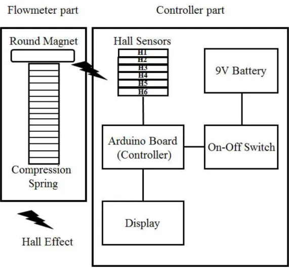
본 연구에서 개발한 실시간 호흡량 감시 장치의 전체적인 시스템 구성도는 Fig.
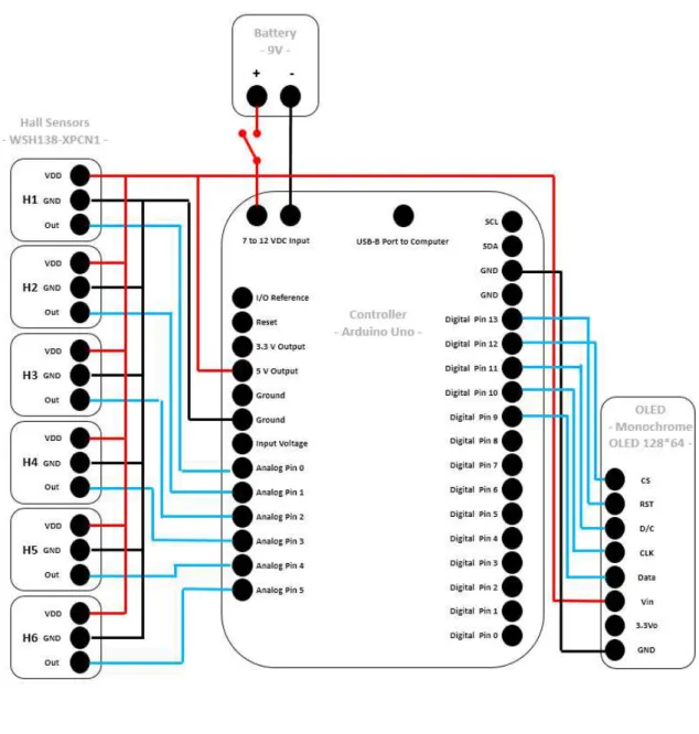
1, 2와 같다. 전체 시스템을 조절하는 controller는 Arduino Board(UNO, Arduino, Italy)를 사용하였고, On-off 스위치와 9 V 배터리(9 V battery holder with switch, Adafruit, New York, NY, USA)가 power source로 사용되었다.
여섯 개의 highly sensitive sensor(H1-H6, WSH138-XPCN1, Winson Semiconductor Corp., Hsinchu, Taiwan)에서 Hall-effect를 감지하여 그 analogue output이 Arduino Board의 analogue input pin으로 전달되게 연결하였 고, 각 Hall-effect sensor(H1-H6)의 VDDs(Voltage Drain, power supply pin), GNDs(GrouND pin), signal pin을 Arduino Board에 적절하게 배치하여 부착하였다 (Fig. 3). Arduino Board의 source code는 supplemental data에 제 시하였다. Hall-effect data는 specific numerical equation을 통해 실제 일회 호 흡량으로 수치화되어 장치의 display part(Monochrome OLED 128*64, Adafruit)에 표시되게 하였다. Flowmeter part와 Hall-effect sensor, Arduino Board, 배터리, on-off 스위치 및 display part를 싸고 있는 케이스는 computer-aided design(CAD) software(SolidWorks, Dassault Systemes, Paris, France)를 이용하여 디자인하였고, 3 dimensional(3D) printer(ProJet 3500, 3D systems, Rock Hill, SC, USA)와 ultraviolet(UV) curing plastic (VisiJet M3 Crystal, 3D systems)을 이용하여 제작하였다.
Fig. 1. System overview of the real-time tidal volume monitoring device (TVD)(1)
Fig. 2. Components of the real-time tidal volume monitoring device (TVD) (1)
Fig. 3. Circuit of the controller part in the real-time tidal volume monitoring device (TVD)
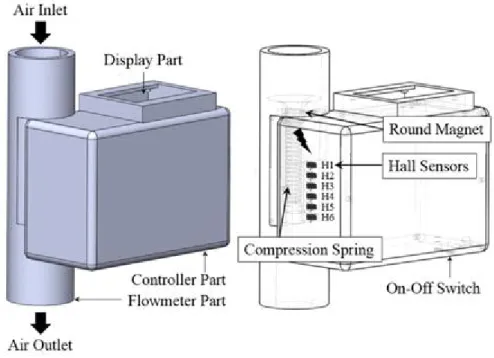
Flowmeter part는 본체(main body)와 원형 자석(round magnet, HXNN-8-3, diameter: 8 mm; length: 3 mm; material: neodymium;
MISUMI, Tokyo, Japan), 스프링(customized spring, outer diameter: 8 mm;
free length: 35 mm; wire diameter: 0.3 mm; material: stainless steel) 및 본 체 커버(main body cover)로 구성되었다 (Fig. 4).
Fig. 4. The position of the round magnet in the flowmeter part (1)
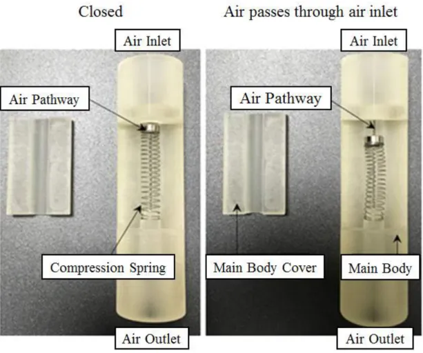
공기가 air inlet을 통하여 flowmeter part의 본체로 들어오면 원형 자석이 파이 프 형태의 flowmeter part 내부에서 움직이면서 air pathway가 열리게 되고, air outlet을 통하여 공기가 기기 외부로 빠져나가게 된다. 공기가 흐르지 않을 때는 스 프링의 restoring force 때문에 원형 자석이 원래의 위치로 돌아가고 air inlet을 완전히 막게 된다. 본체 및 그 커버는 epoxy glue(Locktite Epoxy Instant Mix 5 minutes, Locktite, Thief River Falls, MN, USA)로 밀봉하였다. 여섯 개의 Hall-effect sensor는 flowmeter와 controller part 사이에 일정한 간격으로 배치 하였다 (Fig. 2). Air trapping 및 환자로부터 역류하는 air 또는 secretion의 유입 을 막기 위하여 air outlet의 원위부에 one-way check valve를 추가적으로 설치 하였다 (Fig. 5).
A
B
본 장치에서는 Hall-effect sensor에 의해 감지되는 원형 자석의 위치와 air pathway의 total open time에 대한 정보를 이용하여 실제 일회 호흡량을 계산하 였으며, Arduino Board의 universal serial bus port와 laptop computer를 연결 하여 Hall-effect sensor의 측정값을 기록 후, calibration equation을 추론하였다.
또한 Hall-effect sensor에 의해 감지되는 자기장의 세기(magnetization intensity) 및 관련 hardware wiring status에 대한 기록을 위하여 Arduino program, version 1.7. 및 관련 library(Adafruit SSD1306 and Adafruit GFX, Adafruit)를 사용하였다.
2. 장치의 보정 (Device calibration)
실시간 호흡량 감시 장치의 보정(calibration)은 다음과 같은 단계로 시행하였다.
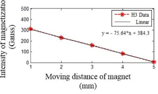
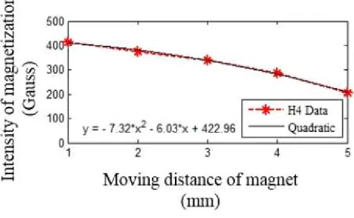
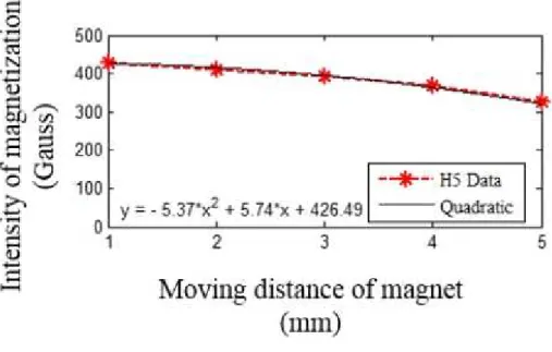
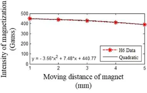
먼저, non-conducting plastic stick을 이용하여 원형 자석을 1 mm에서 5 mm까 지 1 mm 간격으로 translational하게 움직이도록 하였다. 이 과정을 다섯 차례 반 복 후, 각 Hall-effect sensor(H1-H6)에서 감지된 원형 자석의 자기장의 세기의 평균값을 계산하였다. 장치에 사용된 원형 자석 양면의 극성 때문에 H1와 H2에서 의 측정값이 모든 높이에서 saturate되어 이 두 Hall-effect sensor에서의 값은 제외하였고, 나머지 H3, H4, H5, H6에서의 측정값을 calibration에 사용하였다.
다음으로 air pathway의 total open time을 실시간으로 기록하였다. 이 과정에는 6개의 Hall-effect sensor에서의 측정값 중 가장 linear한 변화를 보인 H3의 데이 터를 사용하였다 (Fig. 6). 장치 내부로 유입되는 air flow volume에 대한 calibration equation은 H3의 linear interpolation equation을 air pathway의 total open time으로 적분하여 구하였다 (Fig. 6). 이러한 과정을 통하여 얻어진
calibration equation을 실제 air flow의 volume과 비교하여(장치의 기능 평가, device validation 부분 참조) 최종적으로 conversion equation을 구하였다 (Fig.
6-9).
Fig. 6. Calibration equations of tidal volumes of the real-time tidal volume monitoring device (TVD). The calibration equation of tidal volumes of the Hall-effect sensor 3 (H3) (1).
Fig. 7. Calibration equations of tidal volumes of the real-time tidal volume monitoring device (TVD). The calibration equation of tidal volumes of the Hall-effect sensor 4 (H4) (1).
Fig. 8. Calibration equations of tidal volumes of the real-time tidal volume monitoring device (TVD). The calibration equation of tidal volumes of the Hall-effect sensor 5 (H5) (1).
Fig. 9. Calibration equations of tidal volumes of the real-time tidal volume monitoring device (TVD). The calibration equation of tidal volumes of the Hall-effect sensor 6 (H6) (1).
3. 장치의 기능 평가 (Device validation)
본 연구에서 개발된 실시간 호흡량 측정 장치를 인공호흡기(GE Datex-Ohmeda S/5 Aespire Anesthesia Machine, GE Healthcare, Buckinghamshire, UK)에 연결, 인공호흡기를 volume-controlled mode(frequency of 10 minute-1, I:E ratio of 1:5)로 설정 후 인공호흡기에서 설정되어 제공되는 일회 호흡량과 실시간 호흡량 감시 장치에서 측정되는 일회 호흡량을 비교하여 일치도를 평가하였다. 일 회호흡량은 200, 300, 400, 500, and 600 ml로 설정, 각 일회호흡량 당 25회씩,
총 125회 측정하여 실시간 호흡량 감시 장치와 인공호흡기 간의
difference-ratios in tidal volumes, 즉 (tidal volume of TVD - tidal volume of mechanical ventilator) / tidal volume of mechanical ventilator 값을 계산하 였다.
4. 마네킹 시뮬레이션 (Manikin simulation)
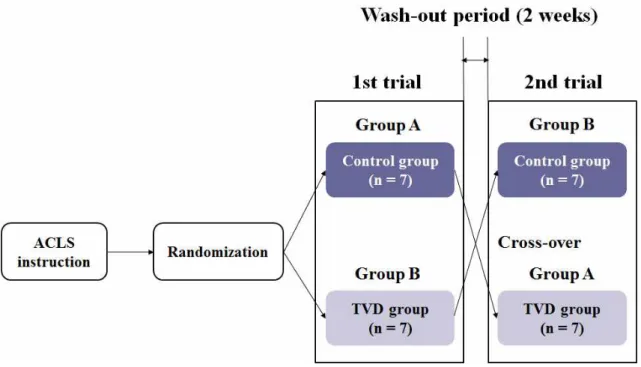
심폐소생술 시뮬레이션 중 실시간 호흡량 감시 장치의 유효성을 평가하기 위하여 무작위 교차 마네킹 시뮬레이션(randomized, cross-over, manikin-simulation study)을 시행하였다. 본 연구는 헬싱키 선언(Fortaleza, Brazil, 2013)에 의거하 여 시행하였고, 서울대학교의과대학/서울대학교병원 의학연구윤리심의위원회를 통 한 IRB 승인(IRB number, H-1605-085-762) 이후에 시행하였다. 연구에 참가 하는 모든 참가자들에게 written informed consent를 구하였다. Primary outcome은 대조군과 실험군 간의 각 실험 참가자 당 적정 호흡의 비율 차이(the
difference in the proportion of optimal ventilation per person between the control and TVD groups)이다. 적정 호흡(optimal ventilation)은 일회 호흡량 6-7 ml/kg을 70 kg 성인 마네킹을 기준으로 적용할 때 420-490 ml로 정의하였 다. 기존 연구들에 의하면 심폐소생술 중 적정 호흡량의 비율은 25%를 넘지 않는 것으로 보고되어 있으며 (21, 31, 32), 실시간 호흡량 감시 장치를 통하여 적정 호 흡의 비율을 95%까지 향상시킬 수 있을 것으로 기대하였을 때, 필요한 sample size는 각 군당 7명으로 계산되었다 (power of 0.8, significance level of 0.05).
Secondary outcome은 대조군과 실험군 간의 저호흡, 적정 호흡, 과호흡의 횟수 차이(the differences in the numbers of hypoventilation, optimal ventilation, and hyperventilation events between the control and TVD groups)로 설정하 였다.
4명의 응급의학과 전문의 및 10명의 응급구조사를 포함한 총 14명의 참가자가 마네킹 시뮬레이션 심폐소생술에 참여하였다. 1차 실험에서 참가자들을 무작위로 대조군(n = 7, 2명의 응급의학과 전문의와 5명의 응급구조사)과 실험군(n = 7, 2 명의 응급의학과 전문의와 5명의 응급구조사)으로 배정, 대조군은 실시간 호흡량 감시 장치의 피드백 없이 마네킹에게 백-밸브 인공호흡을 실시하도록 하였고, 실험 군은 실시간 호흡량 감시 장치의 피드백 하에 백-밸브 인공호흡을 실시하도록 하 였다 (Fig. 10). 무작위 배정(randomization)은 서울대학교병원 의학연구협력센터 (Medical Research Collaborating Center of Seoul National University
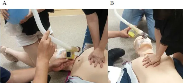
cardiovascular life support, ACLS) 교육을 시행하였다. 교육 내용에는 현재 CPR and ECC guidelines에서 제시하는 적절한 인공호흡에 대한 내용을 포함하였 다. 이후, 마네킹(Resusci Anne® QCPR-D 176-01250, Leardal Medical, Stavanger, Norway)에 기관 삽관을 시행하였고, 실시간 호흡량 감시 장치를 1500 ml 용량의 백-밸브(Ambu® Silicone Resuscitator, Ambu, Ballerup, Denmark)와 기관 내관(endotracheal tube) 사이에 연결한 후 (Fig. 5), 마네킹 시뮬레이션 심폐소생술을 시행하였다. 한 참가자가 백-밸브 인공호흡을 실시하는 동안 다른 참가자는 메트로놈 가이드 하에 마네킹에 깊이 5 cm, 분당 속도 100회 로 흉부 압박을 실시하였다. 각 참가자 당 2분 동안 분당 10회의 호흡수로 총 20 회의 인공호흡을 시행하였다. 실험 참가자의 흉부 압박 속도(compression rate), 흉부 압박 깊이(depth) 및 인공호흡 간격(ventilation interval)은 SimPad SkillReporter (Laerdal Medical)를 이용하여 기록하였다.
Fig. 10. Randomized, cross-over, manikin-simulation study comparing the control group, in which participants provided bag-valve ventilation to a manikin without tidal volume feedback (The participants could not see the display part of the real-time tidal volume monitoring device.)(A), and the TVD group, in which participants provided bag-valve ventilation to a manikin under real-time tidal volume feedback with the TVD (The participants could see the display part of TVD.) (B).
Fig. 11. Protocol of randomized, cross-over, manikin-simulation study comparing the control group and the real-time tidal volume monitoring device (TVD) group (1).
5. 결과 분석
각 군의 결과 변수의 비교에는 Mann-Whitney U test, Χ2 test 및 Fisher’s exact test를 사용하였다. P 값이 0.05 미만인 경우 통계적으로 유의한 의미가 있 는 것으로 판단하였으며, 유의수준 설정에는 양측검정을 사용하였다. 통계 분석에는 SPSS version 22.0 for Windows (SPSS, Chicago, IL, USA)를 사용하였다.
연구 결과
1. 실시간 호흡량 감시 장치의 유효성 확인 (TVD validation)
실시간 호흡량 감시 장치에서 측정한 일회 호흡량과 인공호흡기의 일회 호홉량의 일치도는 Fig. 12와 같다. 실시간 호흡량 감시 장치와 인공호흡기 간의 일회 호흡 량 차이의 평균(the mean value of the differences in tidal volumes between the TVD and mechanical ventilator)은 1.02 ± 0.15 ml로 측정되었다. 총 125 개의 측정값(the difference-ratios in tidal volumes between the TVD and mechanical ventilator) 중, 122개(97.6%)가 오차 범위 ± 1.5% 이내, 125개 (100%)가 오차 범위 ± 2.5%, 이내로 측정되었다 (Fig. 13).
Fig. 12. Validation data. Values of tidal volumes of the real-time tidal volume monitoring device (TVD) and a mechanical ventilator (MV) (1).
Fig. 13. Validation data. The difference-ratios in tidal volumes, defined as (tidal volume of TVD - tidal volume of MV) / tidal volume of MV, between the TVD and MV (1).
2. 적정 호흡의 비율 (Proportion of optimal ventilation per person)
대조군과 실험군 간의 참가자의 성별 및 연령에는 유의한 차이가 없었고 (Table 1), 1차 실험, 2차 실험 모두 흉부 압박 속도, 흉부 압박 깊이 및 인공호흡 간격에 양 군 간의 차이는 관찰되지 않았다 (Table 2).
1차 실험과 2차 실험 각각에서 참가자 당 적정 호흡의 비율은 실험군에서 대조 군에 비해 높게 측정되었다 (p = 0.001 and 0.001, respectively) (Table 3, Fig. 14 and 15). 1차 및 2차 실험을 종합했을 때의 참가자 당 총 적정 호흡의 비 율 역시 실험군에서 대조군에 비해 높게 측정되었다 (p < 0.001) (Table 3, Fig.
16).
각 참가자 당 저호흡의 비율은 2차 실험에서 실험군에서 대조군에 비해 낮게 측 정되었다 (p = 0.004) (Table 3).
Table 1. Randomized participants’characteristics (1)
Group A (n = 7) Group B (n = 7) Pa
EP (n = 2) EMT (n = 5) Total (n = 7) EP (n = 2) EMT (n = 5) Total (n = 7)
Male gender N (%) 2 (100.0) 3 (60.0) 5 (71.4) 1 (50.0) 3 (60.0) 4 (57.1) 0.500
Age (years) Mean ± SD 33.0 ± 1.4 29.2 ± 5.5 30.3 ± 4.9 35.0 ± 2.8 26.8 ± 3.6 29.1 ± 5.1 0.710b Median
(range)
33.0
(32.0-34.0)
27.0
(24.0-36.0)
32.0
(24.0-36.0)
35.0
(33.0-37.0)
25.0
(24.0-33.0)
27.0
(24.0-37.0) Abbreviations: EP, Emergency Physician; EMT, Emergency Medical Technician; SD, standard deviation
aP-values comparing the total of group A and B
bP-values calculated by Mann-Whitney U test
Table 2. Results of manikin simulation (1)
Compression rate, compression depth, and ventilation interval between groups during manikin simulation 1st trial (n = 7/group) 2nd trial (n = 7/group) Total (n = 14)
Control TVD Control TVD Control TVD
Group A Group B Pa Group B Group A Pa Pa
Compression rate
(/minutes)
Mean ± SD 99.9 ± 0.4 99.9 ± 0.4 1.000 99.7 ± 0.5 99.7 ± 0.5 1.000 99.8 ± 0.4 99.8 ± 0.4 1.000 Median
(range)
100.0
(99.0-100.0)
100.0
(99.0-100.0)
100.0
(99.0-100.0)
100.0
(99.0-100.0)
100.0
(99.0-100.0)
100.0
(99.0-100.0) Compression
depth (cm)
Mean ± SD 5.2 ± 0.6 5.4 ± 0.8 0.620 5.2 ± 0.6 5.0 ± 0.6 1.000 5.2 ± 0.6 5.2 ± 0.7 0.769 Median
(range)
5.3 (4.5-6.1)
5.0 (4.3-6.3)
5.0 (4.6-6.1)
5.0 (4.0-5.6)
5.0 (4.5-6.1)
5.0 (4.0-6.3)
Ventilation Mean ± SD 6.0 ± 0.1 6.0 ± 0.0 0.620 6.2 ± 0.1 6.1 ± 0.1 0.209 6.1 ± 0.1 6.1 ± 0.1 0.285
Table 3. Results of manikin simulation (1)
Proportion of hypoventilation, optimal ventilation, and hyperventilation per person
1st trial (n = 7/group) 2nd trial (n = 7/group) Total (n = 14)
Control TVD Control TVD Control TVD
Group A Group B Pa Group B Group A Pa Pa
Proportion/person (%) Hypo-
ventilation
Mean ± SD 36.4 ± 44.6 8.6 ± 12.8 0.456 50.0 ± 31.8 5.0 ± 5.0 0.004b 43.2 ± 37.9 6.8 ± 9.5 0.014b Median
(range)
10.0
(0.0-100.0)
5.0
(0.0-35.0)
55.0 (5.0-95.0)
5.0
(0.0-15.0)
50.0
(0.0-100.0) 5.0
(0.0-35.0) Optimal-
ventilation
Mean ± SD 27.9 ± 25.3 78.6 ± 13.1 0.001b 35.7 ± 21.1 90.0 ± 8.2 0.001b 31.8 ± 22.8 84.3 ± 12.1 <0.001b Median
(range)
20.0 (0.0-65.0)
70.0
(65.0-100.0)
40.0 (5.0-60.0)
90.0
(75.0-100.0)
37.5 (0.0-65.0)
87.5
(65.0-100.0) Hyper-
ventilation
Mean ± SD 35.7 ± 39.4 12.9 ± 12.9 0.456 14.3 ± 13.7 5.0 ± 7.6 0.209 25.0 ± 30.4 8.9 ± 11.0 0.194 Median
(range)
35.0 (0.0-95.0)
10.0 (0.0-30.0)
10.0 (0.0-35.0)
0.0
(0.0-20.0)
12.5 (0.0-95.0)
5.0
(0.0-30.0) Abbreviations: TVD, real-time tidal volume monitoring device; SD, standard deviation
aP-values calculated by Mann-Whitney U test
bP-values < 0.05 between the control and TVD groups
Fig. 14. Proportion of optimal ventilation per person. Comparison between the control group (group A, n = 7 participants), in which participants provided bag-valve ventilation to a manikin without tidal volume feedback, and the real-time tidal volume monitoring device (TVD) group (group B, n
= 7 participants), in which participants provided bag-valve ventilation to a
Fig. 15. Proportion of optimal ventilation per person. Comparison between the control group (group B, n = 7) and the real-time tidal volume monitoring device (TVD) group (group A, n = 7) in the second trial (1).
The plots are presented as the median(quartiles, ranges).
Fig. 16. Proportion of optimal ventilation per person. Comparison between the control group (groups A and B, n = 14) and the real-time tidal volume monitoring device (TVD) group (groups B and A, n = 14) in the first and second trials (1).
The plots are presented as the median(quartiles, ranges).
3. 측정 호흡량의 분포 (Distribution of hypoventilation, optimal ventilation, and hyperventilation)
실시간 호흡량 감시 장치의 사용은 1차 실험에서 대조군 대비, 실험군의 적정 호 흡의 횟수를 증가시켰고, 저호흡 및 과호흡의 횟수를 감소시켰으며 (대조군 vs. 실 험군 = 39/140 vs. 110/140 of optimal ventilation, 51/140 vs. 12/140 of hypoventilation, and 50/140 vs. 18/140 of hyperventilation, respectively, p
< 0.001) (Fig. 17), 이러한 결과는 마찬가지로 2차 실험에서도 관찰되었다 (대조 군 vs. 실험군 = 50/140 vs. 126/140 of optimal ventilation, 70/140 vs.
7/140 of hypoventilation, and 20/140 vs. 7/140 of hyperventilation, respectively, p < 0.001) (Fig. 18). 또한 1차 및 2차 실험을 종합한 결과에서도 실시간 호흡량 감시 장치의 사용이 대조군 대비, 실험군의 적정 호흡의 횟수를 증 가시킴과 저호흡 및 과호흡의 횟수를 감소시킴을 알 수 있었다 (대조군 vs. 실험군
= 89/280 vs. 236/280 of optimal ventilation, 121/280 vs. 19/280 of hypoventilation, and 70/280 vs. 25/280 of hyperventilation, respectively, p
< 0.001) (Fig. 19).
Fig. 17. Distribution of hypoventilation, optimal ventilation, and hyperventilation. Comparison between the control group (group A, n = 140 ventilations) and the real-time tidal volume monitoring device (TVD) group (group B, n = 140 ventilations) in the first trial (1).
Fig. 18. Distribution of hypoventilation, optimal ventilation, and hyperventilation. Comparison between the control group (group B, n = 140) and the TVD group (group A, n = 140) in the second trial (1).
Fig. 19. Distribution of hypoventilation, optimal ventilation, and hyperventilation. Comparison between the control group (groups A and B, n = 280) and the real-time tidal volume monitoring device (TVD) group (groups B and A, n = 280) in the first and second trials (1).
대조군에서 적정 호흡과 저호흡의 횟수는 1차 실험에 비해 2차 실험에서 증가하 였고 (1차 실험 vs. 2차 실험 = 39/140 vs. 50/140 of optimal ventilation and 51/140 vs. 70/140 of hypoventilation, respectively), 과호흡의 횟수는 2차 실 험에서 1차 실험 대비, 감소하였다 (1차 실험 vs. 2차 실험 = 50/140 vs.
20/140) (p < 0.001) (Fig. 20). 실험군에서 적정 호흡의 횟수는 1차 실험에 비 해 2차 실험에서 증가하였고 (1차 실험 vs. 2차 실험 = 110/140 vs. 126/140), 저호흡 및 과호흡의 횟수는 2차 실험에서 1차 실험 대비, 감소하였다 (1차 실험 vs. 2차 실험 = 12/140 vs. 7/140 of hypoventilation and 18/140 vs. 7/140 of hyperventilation, respectively) (p = 0.027) (Fig. 21).
Fig. 20. Distribution of hypoventilation, optimal ventilation, and hyperventilation. Comparison between the first trial (group A, n = 140) and the second trial (group B, n = 140) in the control groups (1).
Fig. 21. Distribution of hypoventilation, optimal ventilation, and hyperventilation. Comparison between the first trial (group B, n = 140) and the second trial (group A, n = 140) in the real-time tidal volume monitoring device (TVD) groups (1).
4. 과호흡의 비율 (Proportion of hyperventilation)
실시간 호흡량 감시 장치의 사용은 1차 실험에서 대조군 대비, 실험군의 과호흡 의 비율을 감소시켰고 (대조군 vs. 실험군 = 35.7% vs. 12.9%, p < 0.001) (Fig. 22), 2차 실험에서도 대조군 대비, 실험군의 과호흡 비율을 감소시켰다 (대 조군 vs. 실험군 = 14.3% vs. 5.0%, p = 0.009) (Fig. 23). 또한 1차 실험과 2 차 실험을 종합한 결과에서도 실시간 호흡량 감시 장치의 사용이 대조군 대비, 실 험군에서의 과호흡의 비율을 감소시킴을 알 수 있었다 (대조군 vs. 실험군 = 25.0% vs. 8.9%, p < 0.001) (Fig. 24).
대조군과 실험군 모두에서 2차 실험에서의 과호흡의 비율이 1차 실험 대비, 감소 하였음을 확인하였다 (1차 실험 vs. 2차 실험 = 35.7% vs. 14.3%, p < 0.001 and 12.9% vs. 5.0%, p = 0.021, respectively) (Fig. 25 and 26).
Fig. 22. Proportion of hyperventilation. Comparison between the control group (group A, n = 140 ventilations) and the real-time tidal volume monitoring device (TVD) group (group B, n = 140 ventilations) in the first trial (1).
Fig. 23. Proportion of hyperventilation. Comparison between the control group (group B, n = 140) and the real-time tidal volume monitoring device (TVD) group (group A, n = 140) in the second trial (1).
Fig. 24. Proportion of hyperventilation. Comparison between the control group (groups A and B, n = 280) and the real-time tidal volume monitoring device (TVD) group (groups B and A, n = 280) in the first and second trials (1).
Fig. 25. Proportion of hyperventilation. Comparison between the first trial (group A, n = 140) and the second trial (group B, n = 140) in the control groups(1).
Fig. 26. Proportion of hyperventilation. Comparison between the first trial (group B, n = 140) and the second trial (group A, n = 140) in the real-time tidal volume monitoring device (TVD) groups (1).
고찰
본 연구에서는 실시간 호흡량 감시 장치를 개발하여 마네킹 시뮬레이션 심폐소생 술을 통하여 그 유효성을 확인하였다. 심폐소생술에서 호흡량을 감시하기 위한 장 치는 휴대 및 사용이 편리해야 하고, 정확한 실시간 호흡량 감시 및 피드백이 가능 하여야 하며, 심폐소생술 중 사용되는 모든 인공호흡 방법에 대해 적용할 수 있어 야 한다 (24). 또한 심폐소생술 중 장치를 쉽고 빠르게 이용 가능하여야 하며, 장 치의 사용이 심폐소생술 중 시행되는 술기 및 치료를 방해하지 않아야 한다 (24).
기존 연구들에서 심폐소생술 중 호흡량을 감시하기 위한 몇 가지 방법들이 제시 되어 왔다 (24). Thoracic impedance pneumography는 호흡 주기 동안의 폐의 전기적 저항 변화(trans-thoracic electrical impedance variation)를 이용하여 간 접적으로 폐 용적 변화와 일회호흡량을 측정하는 기기이다 (24, 33, 34). 그러나 thoracic impedance pneumography는 조직 특이성이 떨어지고 심폐소생술 중 흉 부 압박에 의해 환자의 몸이 흔들림에 따라 측정값의 정확도가 영향을 받게 된다는 한계가 있다 (24, 35). 또한 흉부 압박에 의한 impedance variance 및 심폐소생 술 중 흉강 내 혈류 변화에 대한 보정이 필요하기 때문에 실시간으로 호흡량을 감 시하기에는 어려움이 있다 (24, 36).
Pneumotachometer는 전자식 폐활량계로 환자의 들숨 및 날숨에 따른 일회호흡
pneumotachometer와 마찬가지로 hot wire anemometry 역시 기관 삽관이 되어 있는 환자 이외에서는 사용이 어렵다는 단점이 있다 (24).
Turbine flowmeter 역시 호흡량 감시 장치로 제안되어 왔고 (24), 본 연구에서 도 처음에는 turbine flowmeter의 원리를 이용한 장치 개발을 시도하였다. 그러나 pilot 실험에서 turbine flowmeter를 이용하여 일회 호흡량의 측정을 시도하였을 때 turbine flowmeter의 날개(impeller)가 무거울 경우 큰 관성 때문에 적은 양의 호흡량은 측정할 수가 없었고, 반대로 turbine flowmeter에 마찰이 적은 가벼운 날개를 이용하면 적은 양의 호흡량을 감지하는 데는 민감성이 높아지나 장치에 가 해지는 호흡량이 클 때는 한 번의 호흡량 제공에 날개의 회전이 6–10초 가량 지 속되어 연속적인 일회 호흡량의 측정이 불가능하다는 한계가 있었다. 따라서 본 연 구에서는 variable area flowmeter의 원리와 contactless magnetic measurement mechanism을 이용한 새로운 장치를 고안하게 되었다. 본 장치는 어떠한 물리적 접촉 없이 air flow에 의한 원형 자석의 constant area change와 Hall-effect sensor를 이용하여 자석의 위치 변화를 감지하며, 일회 호흡량의 연속 적 반복 측정이 가능하다.
본 연구에서 개발된 실시간 호흡량 감시 장치는 일회 호흡량을 구체적인 수치로 보여줌으로써 인공호흡 시행자에게 호흡량에 대한 피드백을 줄 수 있다. 또한 기존 의 장치들과는 달리 기관 내관을 통하여 인공호흡을 제공받는 환자 뿐만 아니라 facial mask를 이용하는 환자들에게도 사용이 가능하다. 장치의 원위부에 one-way check valve를 부착하여 air trapping을 예방하고 환자로부터 기기로 들어오는 air의 backflow나 secretion을 막을 수 있다는 것도 본 장치의 장점이다.
본 연구에서는 마네킹 시뮬레이션 심폐소생술 중 실시간 호흡량 감시 장치를 이 용하여 백-밸브 인공호흡을 시행하였을 때 적정 호흡의 비율이 유의하게 증가하고 과호흡의 비율이 감소함을 확인하였다. 또한 실시간 호흡량 감시 장치의 사용은 저
호흡의 비율도 감소시킴을 알 수 있었다. 이전 연구들은 주로 심폐소생술 중 과호 흡을 방지하는 데 주력하고 있으나, 실제 본 연구에서는 마네킹을 이용한 심폐소생 술 시뮬레이션 중 과호흡 뿐 아니라 저호흡 또한 상당한 비율로 관찰되었다. 심폐 소생술 중 인공호흡은 체내 산소 공급과 이산화탄소 제거를 담당하고 있고, 심정지 시간이 길어질 경우 흉부 압박만으로는 충분한 폐내 가스 교환이 이루어지지 않게 된다. 따라서 심정지 시간이 길어질 경우 적절한 양의 인공호흡의 제공은 심정지 환자의 자발순환 회복과 관련이 있는 것으로 알려져 있다 (38, 39). 본 연구에서 개발된 장치는 일회 호흡량의 값을 실시간으로 직접적인 수치로 제시함으로써 과호 흡 뿐 아니라 저호흡을 방지하는 데에도 도움이 될 것으로 생각된다.
심폐소생술 중 과호흡에 대하여 연구한 기존 논문들에서는 과도한 일회 호흡량보 다는 주로 빠른 호흡수에 의한 과호흡에 대하여 보고하고 있다 (19, 40). 본 연구 에서는 마네킹 시뮬레이션 심폐소생술 시 제공하는 인공호흡 속도를 메트로놈을 이 용하여 분당 호흡수 10회로 고정하였고, 대조군 및 실험군 간의 인공호흡 속도에는 유의한 차이가 관찰되지 않았다. 만약 본 연구에서 개발된 실시한 호흡량 감시 장 치에 추가적으로 메트로놈과 같은 기능을 더한다면 일회 호흡량 뿐만 아니라 인공 호흡 속도에 대한 가이드도 가능하여 심폐소생술 중 적절한 인공호흡을 실시하는 데 더욱 더 유용하게 사용될 것으로 생각된다.
본 연구에는 다음과 같은 제한점이 있다. 첫째, 인공호흡기를 이용한 장치의 기능 평가(validation)에서 인공호흡기와 실시간 호흡량 감시 장치 간의 일회 호흡량의
이가 커졌을 것으로 추정된다. 본체 내부 벽을 좀 더 마찰력이 작은 소재로 제작한 다면 이 문제를 해결할 수 있을 것으로 생각된다.
둘째, 1차 실험과 2차 실험 사이에 2주간의 wash-out period를 두었으나, 대조 군과 실험군 모두에서 1차 실험 대비, 2차 실험에서의 적정 호흡의 비율이 증가하 였고, 과호흡의 비율은 감소하였다. 이러한 결과는 2주간의 wash-out period가 1 차 실험에서의 실시간 호흡량 감시 장치의 사용으로 인한 효과를 완전히 없애기에 는 충분하지 않았다는 점을 시사한다. 그러나 이러한 결과로 미루어 볼 때, 실시간 호흡량 감시 장치를 활용하여 심폐소생술 교육을 실시한다면 인공호흡의 질 평가 및 향상과 같은 교육적 효과 또한 기대할 수 있겠다. 최근 보고된 연구 결과들에 따르면 아직까지 심폐소생술 중 인공호흡의 질을 평가할 수 있는 정형화된 방법은 전무한 실정이다 (23).
셋째, 본 연구에서 실시간 호흡량 감시 장치의 임상적 유효성 평가는 마네킹 시 뮬레이션 심폐소생술을 통해서만 이루어졌다. 향후 돼지 심정지 모델을 이용한 추 가적인 연구를 통하여 (41), 실시간 호흡량 감시 장치를 통해 측정되는 일회 호흡 량과 동맥혈 가스, 호기말 이산화탄소, 흉강 압력 및 심박출량과 같은 physiologic data 간의 연관성을 분석하고 실시간 호흡량 감시 장치의 임상적 유효성에 대하여 확인할 수 있을 것으로 생각된다.
결론
본 연구에서 개발된 실시간 호흡량 감시 장치를 통하여 호흡량에 대한 피드백을 제공함으로써 마네킹 시뮬레이션 심폐소생술 중 인공호흡의 적정 호흡을 증가시키 고 과호흡을 감소시킬 수 있었다.
References
1. You KM, Lee C, Kwon WY, et al. Real-time tidal volume feedback guides optimal ventilation during simulated cardiopulmonary resuscitation.
Am J Emerg Med 2016 pii: S0735-6757(16)30822-1. doi:
10.1016/j.ajem.2016.10.085.
2. Mozaffarian D, Benjamin EJ, Go AS, et al. Heart disease and stroke statistics-2015 update: a report from the American Heart Association.
Circulation 2015;131:e29–322.
3. Mottram AR, Page RL. Advances in resuscitation. Circulation 2012;126:991–1002.
4. Shin SD, Suh GJ, Ahn KO, et al. Cardiopulmonary resuscitation outcome of out-of-hospital cardiac arrest in low-income versus high-volume emergency department: An observational study and propensity score matching analysis. Resuscitation 2011;82:32-9.
5. Weisfeldt ML, Sitlani CM, Ornato JP, et al. Survival after application of automatic external defibrillators before arrival of the emergency medical system: evaluation in the resuscitation outcomes consortium population of 21 million. J Am Coll Cardiol 2010;55:1713-20.
6. Wolfe JA, Maier GW, Newton JR Jr, et al. Physiologic determinants of coronary blood flow during external cardiac massage. J Thorac Cardiovasc Surg 1998;95:523-32.
7. Paradis NA, Martin GB, Rivers EP, et al. Coronary perfusion pressure
and the return of spontaneous circulation in human cardiopulmonary resuscitation. JAMA 1990;263:1106-13.
8. Kern KB, Ewy GA, Voorhees WD, et al. Myocardial perfusion pressure:
a predictor of 24 h survival during prolonged cardiac arrest in dogs.
Resuscitation 1988;16:241-50.
9. Neumar RW, Shuster M, Callaway CW, et al. Part 1: Executive summary: 2015 American Heart Association guidelines update for cardiopulmonary resuscitation and emergency cardiovascular care.
Circulation 2015;132:S315-67.
10. Kleinman ME, Brennan EE, Goldberger ZD, et al. Part 5: Adult basic life support and cardiopulmonary resuscitation quality: 2015 American Heart Association guidelines update for cardiopulmonary resuscitation and emergency cardiovascular care. Circulation 2015;132:S414-35.
11. Link MS, Berkow LC, Kudenchuk PJ, et al. Part 7: Adult advanced cardiovascular life support: 2015 American Heart Association guidelines update for cardiopulmonary resuscitation and emergency cardiovascular care. Circulation 2015;132:S444-64.
12. Aufderheide TP, Sigurdsson G, Pirrallo RG, et al.
Hyperventilation-induced hypotension during cardiopulmonary
14. Cheifetz IM, Craig DM, Quick G, et al. Increasing tidal volumes and pulmonary overdistention adversely affect pulmonary vascular mechanics and cardiac output in a pediatric swine model. Crit Care Med 1998;26:710-6.
15. Yannopoulos D, Tang W, Roussos C, Aufderheide TP, Idris AH, Lurie KG. Reducing ventilation frequency during cardiopulmonary resuscitation in a porcine model of cardiac arrest. Respir Care 2005;50:628-35.
16. Berg RA, Sanders AB, Kern KB, et al. Adverse hemodynamic effects of interrupting chest compressions for rescue breathing during cardiopulmonary resuscitation for ventricular fibrillation cardiac arrest.
Circulation 2001;104:2465-70.
17. Roberts BW, Kilgannon JH, Chansky ME, Mittal N, Wooden J, Trzeciak S. Association between postresuscitation partial pressure of arterial carbon dioxide and neurological outcome in patients with post cardiac arrest syndrome. Circulation 2013;127:2107-13.
18. Abella BS, Alvarado JP, Myklebust H, et al. Quality of cardiopulmonary resuscitation during in-hospital cardiac arrest. JAMA 2005;293:305-10.
19. Talikowska M, Tohira H, Finn J. Cardiopulmonary resuscitation quality and patient survival outcome in cardiac arrest: a systematic review and meta-analysis. Resuscitation 2015;96:66-77.
20. Kern KB, Stickney RE, Gallison L, Smith RE. Metronome improves compression and ventilation rates during CPR on a manikin in a
randomized trial. Resuscitation 2010;81:206-10.
21. Nehme Z, Boyle MJ. Smaller self-inflating bags produce greater guideline consistent ventilation in simulated cardiopulmonary resuscitation. BMC Emerg Med 2009;9:4.
22. Wenzel V, Keller C, Idris AH, et al. Effects of smaller tidal volumes during basic life support in patients with respiratory arrest: good ventilation, less risk? Resuscitation 1999;43:25-9.
23. Khoury A, De Luca A, Sall FS, Pazart L, Capellier G. Performance of manual ventilation: how to define its efficiency in bench studies? A review of the literature. Anaesthesia 2015;70:985-92.
24. Terndrup TE, Rhee J. Available ventilation monitoring methods during pre-hospital cardiopulmonary resuscitation. Resuscitation 2006;71:10-8.
25. Lurie KG, Zielinski T, McKnite S, Aufderheide T, Voelckel W. Use of an inspiratory impedance threshold valve improves neurologically intact survival in a porcine model of ventricular fibrillation. Circulation 2002;105:124-9.
26. Pirrallo RG, Aufderheide TP, Provo TA, Lurie KG. Effect of an inspiratory impedance threshold device on hemodynamics during
28. Mader TJ, Kellogg AR, Smith J, et al. A blinded, randomized controlled evaluation of an impedance threshold device during cardiopulmonary resuscitation in swine. Resuscitation 2008;77:387–94.
29. Menegazzi JJ, Salcido DD, Menegazzi MT, et al. Effects of an impedance threshold device on hemodynamics and restoration of spontaneous circulation in prolonged porcine ventricular fibrillation.
Prehosp Emerg Care 2007;11:179–85.
30. Aufderheide TP, Nichol G, Rea TD, et al. A trial of an impedance threshold device in out-of-hospital cardiac arrest. N Engl J Med 2011;365:798-806.
31. Losert H, Sterz F, Kohler K, et al. Quality of cardiopulmonary resuscitation among highly trained staff in emergency department setting. Arch Intern Med 2006;166:2375-80.
32. Park SO, Shin DH, Baek KJ, et al. A clinical observational study analysing the factors associated with hyperventilation during actual cardiopulmonary resuscitation in the emergency department.
Resuscitation 2013;84:298-303.
33. Frerichs I, Hahn G, Hellige G. Thoracic electrical impedance tomographic measurements during volume controlled ventilation-effects of tidal volume and positive end-expiratory pressure. IEEE Trans Med Imag 1999;18(9):764—73.
34. Cotter G, Moshkovitz Y, Kaluski E, et al. Accurate, noninvasive continuous monitoring of cardiac output by whole-body electrical bioimpedance. Chest 2004;125(4):1431—40.
35. Cohen KP, Ladd WM, Beams DM, et al. Comparison of impedance and inductance ventilation sensors on adults during breathing, motion, and simulated airway obstruction. IEEE Trans Biomed Eng 1997;44(7):555-66.
36. Malmivuo J, Plonsey R. Bioelectromagnetism-principles and applications of bioelectric and biomagnetic fields. New York: Oxford University Press, 1995: 461
37. Quanjer PH, Tammeling GJ, Cotes JE, Pedersen OF, Peslin R, Yernault JC. Lung volumes and forced ventilatory flow. Report Working Party Standardization of Lung Function Tests, European Community for Steel and Coal. Official Statement of the European Respiratory Society. Eur Respir J Suppl 1993;16:540.
38. Idris AH. Reassessing the need for ventilation during CPR. Ann Emerg Med 1996;27:569-75.
39. Idris AH, Banner MJ, Wenzel V, Fuerst RS, Becker LB, Melker RJ.
Ventilation caused by external chest compression is unable to sustain effective gas exchange during CPR: a comparison with mechanical ventilation. Resuscitation 1994;28:143-50.
40. O’Neill JF, Deakin CD. Do we hyperventilate cardiac arrest patients?
Supplemental data.
Source codes of the Arduino Board in the real-time tidal volume monitoring device (TVD)#include <SPI.h>
#include <Wire.h>
#include <Adafruit_GFX.h>
#include <Adafruit_SSD1306.h>
#define OLED_MOSI 9
#define OLED_CLK 10
#define OLED_DC 11
#define OLED_CS 12
#define OLED_RESET 13
Adafruit_SSD1306 display(OLED_MOSI, OLED_CLK, OLED_DC, OLED_RESET, OLED_CS);
#define NUMFLAKES 10
#define XPOS 0
#define YPOS 1
#define DELTAY 2
void setup() {
display.begin(SSD1306_DISPLAYON);
display.clearDisplay();
Serial.begin(9600);
}
const int analogInPin3 = A3;
int sensorValue3 = 0;
double a=0;
double b=0;
double c=0;
int flag=0;
double bValue;
void loop() {
int aValue =analogRead(analogInPin3)+10;
if (aValue<438){
if (flag>15) {
a=0;
flag=0;
}
c=979772652.1-2109520*(aValue);
bValue=(5*sqrt(10*c)-87305)/52738;
a=a+bValue;
} else{
flag=flag+1;
b=0;
}
if (a>5 || a==5){
b=0;
}
Serial.print(aValue);
Serial.print("₩t");
Serial.print(bValue);
Serial.print("₩t");
Serial.print(a);
Serial.print("₩t");
Serial.print(b);
Serial.print("₩t");
Serial.println();
display.setTextSize(2);
display.setTextColor(WHITE);
display.setCursor(30,40);
display.println(b);
display.display();
display.clearDisplay();
delay(2);
}
Abstract
Development of a Real-time Tidal Volume Monitoring Device to Guide Optimal Ventilation during
Cardiopulmonary Resuscitation
Kyoung Min You Dept. of Medicine, Biomedical Engineering Major The Graduate School Seoul National University
Purpose: The purpose of this study is to develop a real-time tidal volume monitoring device (TVD) and investigate whether the real-time tidal volume feedback using TVD increases optimal ventilation and decreases hyperventilation during manikin-simulated cardiopulmonary resuscitation (CPR).
Methods: In the present study, a new TVD was developed, which estimated tidal volume in real time using a magnetic flowmeter. The device
tidal volumes for 70 kg adult manikin. After 2 weeks of the wash-out period, the simulation was repeated via the cross-over of the participants.
The primary outcome was the difference in the proportion of optimal ventilation per person between the control and TVD groups.
Result: In the validation study, among 125 values of the difference-ratios in tidal volumes between the mechanical ventilator and TVD, 122(97.6%) and 125(100%) of the difference-ratios in tidal volumes between the two devices were within ± 1.5% and ± 2.5%, respectively. During manikin-simulated CPR, TVD use increased the proportion of optimal ventilation per person. Its median values(range) of the control group and the TVD group were 37.5%(0.0-65.0) and 87.5%(65.0-100.0), respectively, p < 0.001). TVD use also decreased hyperventilation. The proportions of hyperventilation in the control group and the TVD group were 25.0% vs. 8.9%, respectively (p < 0.001).
Conclusion: Real-time tidal volume feedback using the newly developed TVD guided rescuers to provide optimal ventilation and to avoid hyperventilation during manikin-simulated CPR.
………
Keywords : Cardiopulmonary resusci