https://doi.org/10.5468/ogs.2019.62.3.194 pISSN 2287-8572 · eISSN 2287-8580
Introduction
Ovarian Sertoli-Leydig cell tumor (SLCT) is a rare ovarian neo- plasm belonging to the subgroup of sex cord-stromal tumors, with a worldwide incidence rate of <0.5% [1], which can manifest as either benign or malignant. Although approxi- mately 75% of cases occur in female before the age of 40 years, the disease can still affect females of all ages. As the tumor comprises a testicular structure, it often results in overproduction of androgens; therefore, overt androgenic effects occur in approximately 30% of patients; clinical symptoms, such as hirsutism, thickening of voice, and male patterns of fat distribution may also occur [2]. SLCT may be suspected if the clinical manifestation is virilization or the plasma testosterone level is >6.5 nmol/L [1]. Mostly, con- ventional radiologic imaging studies can localize SLCT when the size of the tumor mass is significant. However, some tumors may be too small to localize and make an appropri- ate differential diagnosis before surgical exploration. When conventional imaging modalities commonly performed for ovarian tumors fail, the profiles of adjunctive imaging stud- ies are not well described for SLCT. Here, we report a patient with clinical symptoms and signs of SCLT; however, findings
of abdominal-pelvic computed tomography (CT) and pelvic magnetic resonance imaging (MRI) were inconclusive, where- as positron emission tomography-CT (PET-CT) findings aided in precisely localizing the tumor.
Case report
A 51-year-old female patient presented with hirsutism on her face, chest, abdomen, and legs together with facial swell-
Diagnosis of an indistinct Leydig cell tumor by positron emission tomography-computed tomography
Jinkyoung Kong 1 , Yoo Mee Park 1 , Young Sik Choi 2 , SiHyun Cho 1 , Byung Seok Lee 2 , Joo Hyun Park 1
Department of Obstetrics and Gynecology,
1Gangnam Severance Hospital,
2Severance Hospital, Yonsei University College of Medicine, Seoul, Korea
A 51-year-old perimenopausal female patient presented with hirsutism and voice thickening which was started approximately one and a half years ago. Her initial hormone assay revealed elevated plasma testosterone, 5a-dihydrotestosterone, and dehydroepiandrosterone (DHEA) levels and therefore androgen-secreting tumor was first suspected. However, the lesion was inconspicuous on transvaginal sonography, abdominal-pelvic computed tomography (CT) scan, and pelvic magnetic resonance (MRI) imaging. Consequently,
18F-fluorodeoxyglucose (FDG) positron emission tomography-CT was performed, which localized the lesion as a focal FDG uptake within the right adnexa. Total laparoscopic hysterectomy with bilateral salpingo-oophorectomy was performed, and although visible gross mass lesions were not observed intraoperatively, pure Leydig cell tumor was pathologically confirmed within the right ovary. Plasma testosterone, 5a-dihydrotestosterone, and DHEA levels were normalized postoperatively. Clinical signs of virilization were also significantly resolved after 3-months of follow-up.
Keywords: Sertoli-Leydig cell tumor; PET-CT; Diagnosis
Received: 2018.07.19. Revised: 2018.09.24. Accepted: 2018.10.24 Corresponding author: Joo Hyun Park
Department of Obstetrics and Gynecology, Gangnam Severance Hospital, Yonsei University College of Medicine, 211 Eonju-ro, Gangnam-gu, Seoul 06273, Korea
E-mail: [email protected]
https://orcid.org/0000-0001-6388-5975
Articles published in Obstet Gynecol Sci are open-access, distributed under the terms of the Creative Commons Attribution Non-Commercial License (http://creativecommons.
org/licenses/by-nc/3.0/) which permits unrestricted non-commercial use, distribution, and reproduction in any medium, provided the original work is properly cited.
Copyright © 2019 Korean Society of Obstetrics and Gynecology
ing and deepening of voice, which started approximately 18 months ago. The patient had an obstetric history of grav- ida 2, para 2, and had irregular menstrual bleeding. Her last menstrual period was about 6 weeks ago. She was under medication for hypertension and asthma, and had received bilateral thyroidectomy 4 years ago due to thyroid cancer.
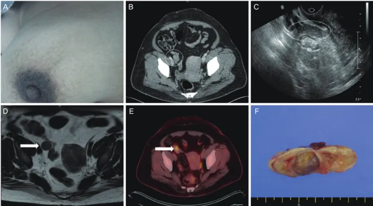
Physical examination revealed hirsutism involving the chin, philtrum, chest, abdomen, and extremities (Fig. 1A). Signs of moon face and central obesity were also manifested, with a body mass index (BMI) of 37.8 kg/m
2. No palpable abdomi- nal or pelvic lesions were observed.
The patient was on a fluticasone furoate-containing inhaler for 20 years due to asthma and was first admitted to the endocrinology department to rule out Cushing’s syndrome.
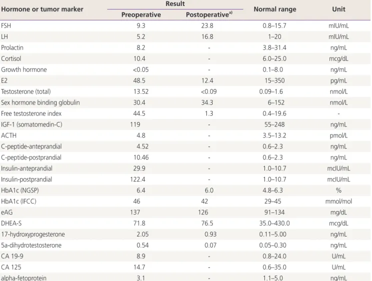
However, the result of the overnight dexamethasone sup- pression test was negative. The initial laboratory investiga- tions revealed normal complete blood count and normal hepatic and renal function. Tumor markers, including alpha-
fetoprotein, cancer antigen (CA) 125, and CA 19-9, were not elevated. The concomitant hormonal test results were as follows: follicle-stimulating hormone (FSH), 9.3 mIU/mL; lu- teinizing hormone (LH), 5.2 mIU/mL; estradiol (E2), 48.5 pg/
mL; cortisol, 10.4 mcg/dL; total testosterone, 13.52 nmol/L;
free testosterone, 44.5; 5a-dihydrotestosterone, 0.54 ng/mL;
and dehydroepiandrosterone (DHEA), 71.8 mcg/dL (Table 1).
With the highest likelihood of hormone-producing tumor as the differential diagnosis, abdominal-pelvis CT was per- formed, but no remarkable findings were noted (Fig. 1B).
The patient was then referred to our reproductive endocri- nology division for further evaluation, where she underwent transvaginal ultrasonography. The examination showed that the volume of the left ovary was 4.9 cm
3and of the right ovary was 9.7 cm
3, with neither ovary showing any abnormal echogenicity other than a slight discrepancy in size (Fig. 1C).
Identifiable lesions were not detected on pelvic MRI (Fig. 1D).
A
18F-fluorodeoxyglucose (FDG) PET-CT scan was then per-
Fig. 1. (A) Truncal hirsutism: male pattern hair distribution on the patient’s chest. (B) Lopromide contrast-enhanced abdominopelvic CT:
nonspecific findings of the right ovary (white circle). (C) Transvaginal ultrasonography: no abnormal echogenicity in the patient’s right ovary (white ellipse) and other pelvic structures. (D) T2-weighted MRI: ovoid-shaped homogeneous right ovary, and nonspecific findings of the right ovary (arrow). (E)
18F-FDG PET-CT: small focal FDG uptake in the right adnexa (arrow). (F) Macroscopic features of the pure Leydig cell tumor: yellow solid mass of 1.5 cm in size. CT, computed tomography; MRI, magnetic resonance imaging, FDG, fluorodeoxyglucose;
PET-CT, positron emission tomography computed tomography.
A B C
D E F
formed, revealing 0.7 cm focal FDG uptake within the right adnexa, with no abnormal uptake in other pelvic or distant organs (Fig. 1E).
Upon laparoscopic inspection of the abdominal cavity, the right ovary did not show any gross abnormalities on the cor- tical surface, and no other additional visible anomalies, such as nodules or ascites, were observed. After initially perform- ing right salpingo-oophorectomy, the specimen was sent out for frozen section pathologic analysis, and was identified as pure Leydig cell tumor. Since the patient had no desire to preserve fertility, additional total laparoscopic hysterectomy with left salpingo-oophorectomy was performed. Grossly, the
right ovary weighed 8.6 gm, and serial sections of the ovary revealed a yellow solid mass measuring approximately 1.5 cm (Fig. 1F). Microscopically, no capsular involvement was noted.
On immunohistochemical staining, the tumor was positive for inhibin-a, calretinin, androgen, and Melan-A, with Ki-67 positivity of 1%. The final pathology report concluded that the right ovarian mass was pure Leydig cell tumor.
The first assay of postoperative hormone levels, measured 3 weeks postoperatively, was as follows: testosterone of
<0.09 nmol/L, 5a-dihydrotestosterone of 0.07 ng/mL, LH of 16.8 mIU/mL, FSH of 23.8 mIU/mL, and E2 of 12.4 pg/mL.
These results indicated that the patient’s hormone levels had
Table 1. Laboratory hormone and tumor markers of the patient
Hormone or tumor marker Result
Normal range Unit Preoperative Postoperative
a)FSH 9.3 23.8 0.8–15.7 mIU/mL
LH 5.2 16.8 1–20 mIU/mL
Prolactin 8.2 - 3.8–31.4 ng/mL
Cortisol 10.4 - 6.0–25.0 mcg/dL
Growth hormone <0.05 - 0.1–8.0 ng/mL
E2 48.5 12.4 15–350 pg/mL
Testosterone (total) 13.52 <0.09 0.09–1.6 nmol/L
Sex hormone binding globulin 30.4 34.3 6–152 nmol/L
Free testosterone index 44.5 1.3 0.4–19.6 -
IGF-1 (somatomedin-C) 119 - 55–248 ng/mL
ACTH 4.8 - 3.5–13.2 pmol/L
C-peptide-anteprandial 4.52 - 0.6–2.3 ng/mL
C-peptide-postprandial 10.46 - 0.6–2.3 ng/mL
Insulin-anteprandial 29.9 - 1.0–10.7 mcIU/mL
Insulin-postprandial 122.4 - 1.0–10.7 mcIU/mL
HbA1c (NGSP) 6.4 6.0 4.8–6.3 %
HbA1c (IFCC) 46 42 29–45 mmol/mol
eAG 137 126 91–134 mg/dL
DHEA-S 71.8 76.5 35.0–430.0 mcg/dL
17-hydroxyprogesterone 2.05 0.93 0.11–5.00 ng/mL
5a-dihydrotestosterone 0.54 0.07 0.05–0.30 ng/mL
CA 19-9 8.9 - 0.8–24.0 U/mL
CA 125 14.7 - 0.6–35.0 U/mL
alpha-fetoprotein 3.1 - 1.1–5.0 ng/mL
FSH, follicle-stimulating hormone; LH, luteinizing hormone; E2, estradiol; IGF-1, insulin-like growth factor 1; ACTH, adrenocorticotropic hor- mone; NGSP, national glycohemoglobin standardization program; IFCC, International Federation of Clinical Chemistry; eAG, estimated average glucose; DHEA-S, dehydroepiandrosterone sulfate; CA, cancer antigen.
a)