Micro-computed tomography evaluation and pathological analyses of female rats with collagen-induced arthritis JVS
7
0
0
전체 글
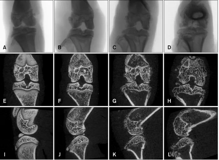
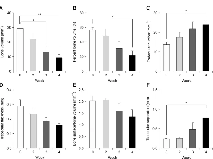
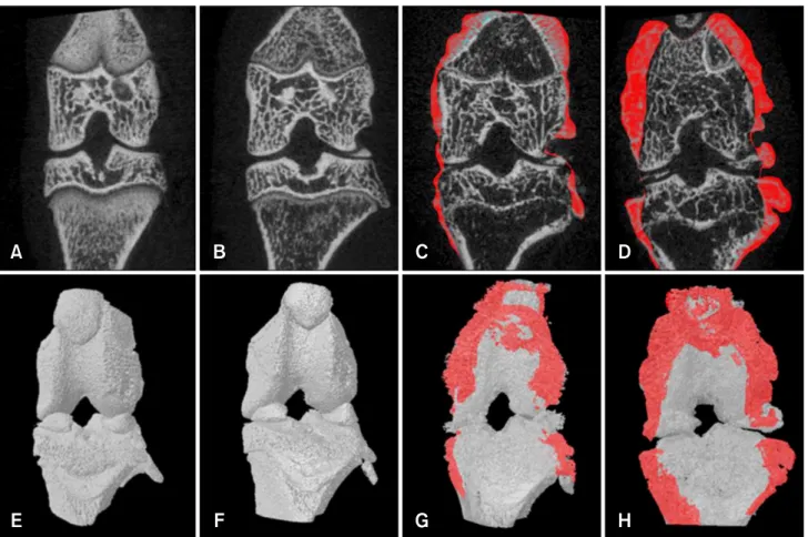
(2) 166 Young Hee Kim, Jin Seok Kang rats have not been assessed. Therefore, the purpose of the present study was to perform a comprehensive characterization of rat CIA-induced arthritis using micro-CT. The results were to compared to data from a conventional pathological examination.. Materials and Methods CIA model Twenty-four female Wistar rats (8 weeks old) were obtained from Samtaco Bio (Korea) and acclimated for 7 days before the study was initiated. The animals were kept in a temperature-controlled environment (22 ± 3oC), with 55 ± 5% relative humidity and a 12-h light/dark cycle. The rats were fed a gamma ray-irradiated rodent diet (Cargill Agri Purina, Korea) and filtered water ad libitum. The rats were randomly divided into four equal groups based on time points: week 0, 2, 3, and 4. Each rat was injected with an emulsion to induce arthritis except for the week 0 group. CIA was induced as follows. Bovine type II collagen (Chondrex, USA) was dissolved overnight in 0.01 M acetic acid at 4oC. This was emulsified in an equal volume of incomplete Freund's adjuvant (Chondrex). The rats were immunized intradermally at the base of the tail with 0.1 mL of emulsion containing 100 g of type II collagen. On day 7 after primary immunization, the rats were boosted using the same technique that was used for the first round of injections. The animals were sacrificed at week 0 as well as week 2, 3, and 4 after the initial collagen immunization. All hind knee joints were fixed in 10% neutral phosphate-buffered formalin. This study was approved by the animal ethics committee of Namseoul University (Cheonan) according to the Animal Protection Act. Micro-CT analysis Quantitative analysis of the hind knee joints was performed using a micro-CT system (SKYSCAN 1172; Bruker, Belgium). Specimens were scanned with an X-ray source of 40 kV/250 V, pixel size of 23 m, and a 0.5 mm aluminum filter. After scanning, cross-sectional slices were generated and each scan result was reconstructed using 0∼0.14 threshold values to distinguish bone from air. Three-dimensional analysis was performed using CTAn software (Bruker). The fraction of bone volume (BV), percent bone volume (BV/TV), trabecular number (Tb.N), trabecular thickness (Tb.Th), bone surface/bone volume (BS/BV), and trabecular separation (Tb.Sp) were evaluated at weeks 0, 2, 3, and 4 using the built-in software. In addition, osteophytes within each contiguous coronal image section were manually outlined and the volume was calculated using CTvol software (Bruker). Pathological assessment The hind knee joints were fixed in 10% phosphate-buffered Journal of Veterinary Science. formalin for 24 h and decalcified in 14% EDTA-glycerol for 14 days at room temperature. The specimens were routinely processed, embedded in paraffin, and cut into 4-m sections that were stained with hematoxylin and eosin (H&E). To examine the cartilage, safranin O-fast green staining of the hind knee joints was performed. Pathological analyses of the joints were carried out by monitoring infiltration of inflammatory cells, cartilage degradation, synovial proliferation, and chondrocyte hyperplasia. Lesion of severity was was classified according to four grades: 0, no detectable change; 1, slight; 2, moderate; 3, severe. Statistical analysis Statistical analyses were performed using GraphPad Prism 6 (GraphPad Software, USA). All data were analyzed using Student’s t-test and are expressed as the mean ± standard deviation (SD). P values < 0.05 were considered statistically significant.. Results CIA model X-ray, coronal, and sagittal micro-CT images showed that the joints had a normal appearance at week 0. Joint destruction was observed in the collagen-treated rats at week 2, 3, and 4. Additionally, destruction of bony surfaces was increased at weeks 2, 3, and 4 in a time-dependent manner (Fig. 1). Several bone parameters were altered during the development of arthritis according to the micro-CT findings (Fig. 2). BV was significantly decreased in the tibia of the rats at weeks 3 and 4 compared to week 0 (p < 0.05 and p < 0.01, respectively). BV/TV was significantly reduced in the tibia at week 4 compared to week 0 (p < 0.05). In contrast, BS/BV and Tb.Sp were significantly increased in the tibia at week 4 compared to week 0 (p < 0.05). Tb.N and Tb.Th, both tended to decrease in a time-dependent manner. Using CTvol software, osteophytes in the hind knee joint of collagen-treated rats were visualized. Osteophyte development appeared as red coloring surrounding the irregular bone surface at weeks 3 and 4 (Fig. 3). Quantitative assessment of osteophytes over the knee was performed and calculated osteophyte volumes in the knee joints were 0 at week 0, 0 at week 2, 2.2 ± 3.8 at week 3, and 3.4 ± 5.8 at week 4. Pathological observation Time-dependent pathological alterations were observed in the rat knee joints. No inflammation or tissue destruction was found at week 0. However, knee joints of the collagen-treated rats showed severe joint destruction with inflammatory cell infiltration. This was accompanied by erosion of cartilage and bone along with the appearance of enlarged cavities filled with synovial fibroblasts and inflammatory cells in a time-dependent.
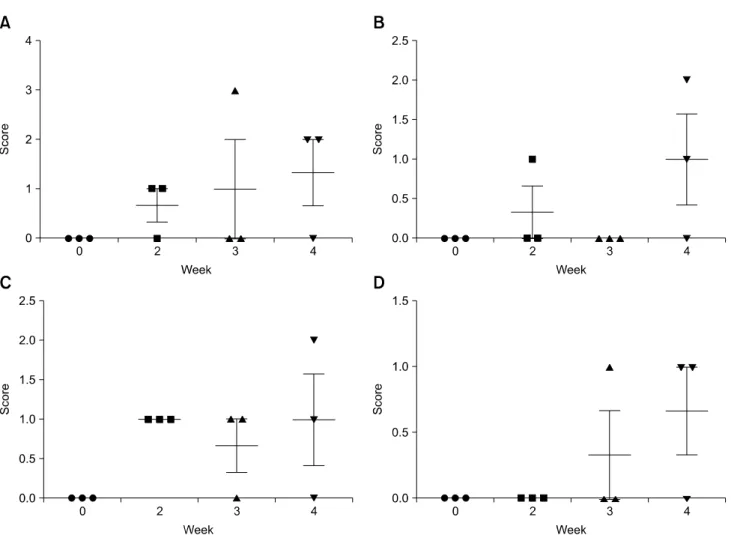
(3) Micro-CT and pathological analyses of female rat collagen-induced arthritis 167. Fig. 1. X-ray projection image along with coronal and sagittal images of the hind knee joint of collagen-treated rats generated by micro-CT. Note the X-ray projection images (A~D), coronal images (E∼H), and sagittal images (I∼L) at weeks 0, 2, 3, and 4. Joints appeared normal at week 0 while destruction of the bony surface occurred at weeks 2, 3, and 4 in a time-dependent manner.. manner (Fig. 4). Safranin O-fast green staining demonstrated that the joints of the rats had normal cartilage structures (indicated by red pigmentation) at week 0. However, significant cartilage destruction appeared in time-dependent manner in the joints of rats treated with collagen (Fig. 5). Scoring of the pathological findings, revealed that infiltration of inflammatory cells, cartilage degradation, synovial proliferation, and hyperplasia of chondrocytes, increased in a time-dependent manner (Fig. 6).. Discussion The current study showed that micro-CT analysis could be performed to evaluate several quantitative parameters of CIA, and these data generally correlated to microscopic finding. Several bone parameters were altered during arthritis development according to micro-CT analyses that showed. progressive bony structure changes at weeks 2, 3, and 4. BV was significantly decreased at weeks 3 and 4 while BV/TV was significantly reduced at week 4 compared to week 0. Both Tb.N and Tb.Th tended to decrease in a time-dependent manner. These data indicated that bone destruction was evident in rats treated with collagen at weeks 3 and 4. As BS/BV and Tb.Sp were significantly increased at week 4 compared to week 0, there could be the deposition of osteophytes around the surface and the evidence of bone loss. Micro-CT can measure osteophytes formation as well as trabecular bone structures [12]. Therefore, we quantified osteophytes volumes in the knee joints (0, 0, 2.2 ± 3.8, and 3.4 ± 5.8 at weeks 0, 2, 3, and 4, respectively). Even though osteophytes volumes increased, this characteristic varied widely among the animals. In the present study, cartilage destruction was observed. Safranin O-fast green staining showed that the joints of the rats represented normal cartilage structures at week 0. Significant. www.vetsci.org.
(4) 168 Young Hee Kim, Jin Seok Kang. Fig. 2. Micro-CT analysis parameters of the hind knee joint in collagen-treated rats at weeks 0, 2, 3, and 4. *, **Significantly different compared to week 0 (p < 0.05 and p < 0.01, respectively). Values are presented as the mean ± SD.. cartilage destruction appeared in the joints of the collagen-treated animals in a time-dependent manner. Since the ability of micro-CT analysis to detect cartilage is limited, other imaging techniques should be performed. High-resolution magnetic resonance imaging (MRI) has been found to be sensitive to changes in cartilage integrity in rats [9]. It was also reported that MRI assessment of cartilage degeneration correlated to macroscopic grading [1]. Additionally, imaging devices such as positron emission tomography and single photon emission computed tomography can be applied to assess arthritis [6,12]. Furthermore, it was reported that intravital microscopy is a powerful tool for measuring the functionality and dynamics of individual regulators of cell migration [7]. Since collagenases, matrix metalloproteinase, or gelatinases promote joint destruction in cases of arthritis [10], quantitative analysis of the breakdown products of matrix components in joints is important for evaluating the effects of inflammatory joint disease [15]. Using a matrix metalloproteinase 3-specific polymeric probe, an early diagnosis of arthritis and visualization of arthritis progression and this approach could be used for early diagnosis as well as Journal of Veterinary Science. monitoring the effects of drug and surgical therapies [16]. Further studies are warranted to monitor animals treated with pharmaceuticals using in situ imaging modalities such as MRI and intravital microscopy as well as micro-CT. In this investigation, HE staining showed that inflammatory cells invaded the synovium, sometimes forming a fibrovascular pannus eroding articular cartilage and subchondral bone in collagen-treated animals, in a time-dependent manner. Hypertrophy of the synovial lining is the first histological manifestation of early lesions associated with arthritis and mild inflammatory infiltrate into the synovium rapidly forms a fibrovascular pannus [11]. When monitoring cartilage degradation, highly varied pathological scores were obtained even at week 4. Immunization with type II collagen leads to the development of severe polyarticular arthritis in rodents that is mediated by an autoimmune response [14]. In cases of RA, macrophages are responsible for inducing inflammation, matrix destruction, and angiogenesis [13]. Further studies are needed to analyze the types of immune cells involved in the process of arthritis development in rats..
(5) Micro-CT and pathological analyses of female rat collagen-induced arthritis 169. Fig. 3. Visualization of osteophytes in micro-CT coronal (A~D) and 3D images (E~H) of the hind knee joint of collagen-treated rats at weeks 0, 2, 3, and 4. Osteophyte development appeared as red coloring surrounding the irregular bone surface at weeks 3 and 4.. Fig. 4. Pathological examination of the hind knee joint from rats treated with collagen. (A) Week 0. (B) Week 2. (C) Week 3. (D) Week 4. Note the normal microscopic structure of the joint of the control rat (A). Knee joints of the collagen-treated animals showed severe joint destruction with inflammatory cell infiltration. Cartilage and bone erosion along with enlarged cavities filled with synovial fibroblasts and inflammatory cells appeared in a time-dependent manner (B~D). H&E staining of paraffin-embedded sections from the hind joint of the rats. 200× magnification.. Fig. 5. Safranin O-fast green staining of the hind knee joint from rats treated with collagen. (A) Week 0. (B) Week 2. (C) Week 3. (D) week 4. Note the normal cartilage structure of the joint in the rats that appeared as red pigmentation at week 0 (A). However, remarkable cartilage destruction was observed in the joint of the collagen-treated rats in a time-dependent manner (B~D). 200× magnification.. www.vetsci.org.
(6) 170 Young Hee Kim, Jin Seok Kang. Fig. 6. Pathological scores for the hind knee joint of rats treated with collagen. (A) Infiltration of inflammatory cells. (B) Cartilage degradation. (C) Synovial proliferation. (D) Hyperplasia of chondrocytes. Values represent the mean ± SD.. For semi-quantitative analysis of the pathological findings, infiltration of inflammatory cells, cartilage degradation, synovial proliferation, and hyperplasia/hypertrophy were scored. Even though infiltration of inflammatory cells, cartilage degradation, synovial proliferation, and hyperplasia tended to increase over time, none of the changes observed at weeks 2, 3, and 4 were significant compared to week 0. This is probably due to animal variation. Even though the scoring paradigm is sufficiently sensitive to evaluate cases of arthritis [8], it seems to be difficult to show statistic difference of these lesions in this study. Based on our findings, we concluded that micro-CT is capable of characterizing degenerative changes, associated with arthritis that were detected by pathological examination. Development of an image-based model of RA using rats with CIA will be of great use to researches for examining the disease process. Taken together, analysis by micro-CT makes it possible to quantify CIA lesions in rats when performed with pathological examination.. Journal of Veterinary Science. Acknowledgments We would like to thank Ms. Kyung A Kwak (Namseoul University) for her technical assistance. Funding for this paper was provided by Namseoul University.. Conflict of Interest There is no conflict of interest.. References 1. Batiste DL, Kirkley A, Laverty S, Thain LMF, Spouge AR, Holdsworth DW. Ex vivo characterization of articular cartilage and bone lesions in a rabbit ACL transection model of osteoarthritis using MRI and micro-CT. Osteoarthritis Cartilage 2004, 12, 986-996. 2. Bendele A. Animal models of rheumatoid arthritis. J Musculoskelet Neuronal Interact 2001, 1, 377-385. 3. Bevaart L, Vervoordeldonk MJ, Tak PP. Evaluation of therapeutic targets in animal models of arthritis: how does it.
(7) Micro-CT and pathological analyses of female rat collagen-induced arthritis 171. 4. 5. 6. 7. 8.. 9.. 10.. relate to rheumatoid arthritis? Arthritis Rheum 2010, 62, 2192-2205. Brand DD, Latham KA, Rosloniec EF. Collagen-induced arthritis. Nat Protoc 2007, 2, 1269-1275. Carvalheiro H, da Silva JAP, Souto-Carneiro MM. Potential roles for CD8+ T cells in rheumatoid arthritis. Autoimmun Rev 2013, 12, 401-409. de Jong M, Essers J, van Weerden WM. Imaging preclinical tumour models: improving translational power. Nat Rev Cancer 2014, 14, 481-493. Ellenbroek SIJ, van Rheenen J. Imaging hallmarks of cancer in living mice. Nat Rev Cancer 2014, 14, 406-418. Gerwin N, Bendele AM, Glasson S, Carlson CS. The OARSI histopathology initiative - recommendations for histological assessments of osteoarthritis in the rat. Osteoarthritis Cartilage 2010, 18 (Suppl 3), S24-34. Goebel JC, Bolbos R, Pham M, Galois L, Rengle A, Loeuille D, Netter P, Gillet P, Beuf O, Watrin-Pinzano A. In vivo high-resolution MRI (7T) of femoro-tibial cartilage changes in the rat anterior cruciate ligament transection model of osteoarthritis: a cross-sectional study. Rheumatology (Oxford) 2010, 49, 1654-1664. Ishikawa T, Nishigaki F, Miyata S, Hirayama Y, Minoura K, Imanishi J, Neya M, Mizutani T, Imamura Y, Naritomi Y, Murai H, Ohkubo Y, Kagayama A, Mutoh S. Prevention of progressive joint destruction in collagen-induced arthritis in rats by a novel matrix metalloproteinase inhibitor, FR255031. Br J Pharmacol 2005, 144, 133-143.. 11. Knoerzer DB, Donovan MG, Schwartz BD, Mengle-Gaw LJ. Clinical and histological assessment of collagen-induced arthritis progression in the diabetes-resistant BB/Wor rat. Toxicol Pathol 1997, 25, 13-19. 12. Koba W, Jelicks LA, Fine EJ. MicroPET/SPECT/CT imaging of small animal models of disease. Am J Pathol 2013, 182, 319-324. 13. Maruotti N, Cantatore FP, Crivellato E, Vacca A, Ribatti D. Macrophages in rheumatoid arthritis. Histol Histopathol 2007, 22, 581-586. 14. Myers LK, Rosloniec EF, Cremer MA, Kang AH. Collagen-induced arthritis, an animal model of autoimmunity. Life Sci 1997, 61, 1861-1878. 15. Otero M, Goldring MB. Cells of the synovium in rheumatoid arthritis. Chondrocytes. Arthritis Res Ther 2007, 9, 220. 16. Ryu JH, Lee A, Chu JU, Koo H, Ko CY, Kim HS, Yoon SY, Kim BS, Choi K, Kwon IC, Kim K, Youn I. Early diagnosis of arthritis in mice with collagen-induced arthritis, using a fluorogenic matrix metalloproteinase 3-specific polymeric probe. Arthritis Rheum 2011, 63, 3824-3832. 17. Schambach SJ, Bag S, Schilling L, Groden C, Brockmann MA. Application of micro-CT in small animal imaging. Methods 2010, 50, 2-13. 18. Scott DL. Biologics-based therapy for the treatment of rheumatoid arthritis. Clin Pharmacol Ther 2012, 91, 30-43. 19. Scott DL, Wolfe F, Huizinga TWJ. Rheumatoid arthritis. Lancet 2010, 376, 1094-1108.. www.vetsci.org.
(8)
수치
관련 문서
After first field tests, we expect electric passenger drones or eVTOL aircraft (short for electric vertical take-off and landing) to start providing commercial mobility
1 John Owen, Justification by Faith Alone, in The Works of John Owen, ed. John Bolt, trans. Scott Clark, "Do This and Live: Christ's Active Obedience as the
In addition, the expression of REDD1 was increased when Huh7 cells were treated with PA, and overexpression of REDD1 affected cell survival and lipid
A and E, In control group, a small amount of new bone was observed at the margin of bone defect (40×); B and F, In experimental group 1, a large amount of new bone was formed
Quantitation of procollagen synthesis by Hs68 cells treated with fish collagen hydrolysates (FCHs), their <1 kDa fraction, and their >1 kDa fraction for 24 hrs.
Consequently, the present study was designed to investigate the effect of adding 60 mg of raloxifene hydrochloride daily to women treated with GnRH-a for severe endometriosis
Pro-allergic cytokines were important mediators of allergic inflammation, cell recruitment and allergenic response decided to further investigate the
Bone transplants, autogenous bones, allogenic bones, xenogenic bones, and synthetic bone substitutes have all been used as bone graft materials.. However,
