Hydroxyapatite coated magnesium alloy for peripheral nerve regeneration
9
0
0
전체 글
(2) Magnesium nerve conduit for peripheral nerve regeneration. like donor site morbidity (traumatic injury, infection & neuropathic pain), the need for specialized skills and prolonged operation time [3]. Recently, nerve conduits, either synthetic or biological derived scaffolds, have grown. MATERIALS AND METHODS Preliminary evaluation of pure Mg and WE43 conduits. immensely. Their main advantages are availability and Six-week-old male healthy Sprague-Dawley rats (SD). absence of donor site morbidity [3]. Magnesium (Mg) has been used for orthopedic and. of 200–250 g (n=2) were used for implanting two nerve. cardiovascular stent applications and as a neuroprotective. conduits of pure Mg and WE43 over 10 mm gap of sciatic. agent for long time. It is a promising material for neural. nerve. The conduits were re-explored 4 weeks later to. regeneration own to its metallic nature, biodegradability. evaluate the resorption status and gas formation. The inner. and biocompatibility. Though, the fast degradation nature of. and surrounded grown tissue of the conduit was submitted. Mg limits its use as a biodegradable material. Furthermore,. for histological evaluation with hematoxylin and eosin. its degradation results in alkaline byproducts and hydrogen. staining (Fig. 1A, B).. gas. Therefore, Mg degradation rate must be controlled and the harmful degradation byproducts must be eliminated, neutralized, or removed from the system to permit its neural. In vitro evaluation of PC12 cells and SCs adhesion and viability when co-cultured with Mg disks. applications. The present study was conducted to evaluate the benefit of hydroxyapatite (HA) coated Mg alloy (WE43). HA-coated Mg disks preparation. nerve conduit in peripheral nerve injury regeneration.. The WE43 fabrication and HA coating were made. A. B. C. D. E. 106. www.chosunobr.org. F. Fig. 1. Preliminary evaluation of bare Mg and WE43. (A, B) Clinical photos showing implant of bare pure Mg and WE43 conduits. (C, D) Large encapsulated gas surrounding both conduits. (E, F) Histological photomicrographs of inner conduit tissue structure with inflammatory cells invasion and necrotic areas (×40 and ×200 magnification). Mg, magnesium; WE43, Mg alloy..
(3) Akram Abdo Almansoori, et al.. as described previously by Lim et al. [4]. An WE43. fonate) to react with the cell mitochondrial succinate-. (Mg, 3.78 wt% Y, 2.13 wt% Nd, 0.46wt% Zr) (Daeryun. tetrazolium reductase to form a water-soluble formazan. Co., Shanxi, China) was purchased and formed into. dye [5]. Briefly, Cells were cultured for 4 days on the previ. disk shape with a milling machine (Genoss Co.,. ously mentioned disks. Thereafter, adhered cells were. Suwon, Korea). For HA coating, disks were immersed. detached using 0.05% trypsin-EDTA solution and collected. into 0.5 M ethylenediaminetetraacetic acid calcium. in standard culture medium. Cells were resuspended in 100. disodium salt hydrate (Ca-EDTA) and 0.05 M potassium. µL of standard culture medium per well of 96-well plate. Four wells without cells acted as a control. A total of 10. dihydrogenphosphate (KH 2PO 4) solution, then heat8.9 by adding sodium hydroxide (NaOH) solution. Finally,. μL of EZ-CYTOX reagent was added to each well and the plate was incubated for 4 hours. Absorbance was measure. the HA-coated disks were washed with distilled water (DW). at 450 nm wavelength using a microplate reader.. processed at 363 K for 2 hours. The pH was maintained at. and dried with air.. In vivo evaluation of HA-WE43 nerve conduit Cells seeding For in vitro evaluation, 4 disks of pure Mg, WE43, HA. The selection of the Mg nerve conduit type was based. coated pure Mg (HA-Mg), HA coated WE43 (HA-WE43). on the preliminary in vivo observation and the in vitro. were sterilized under ultraviolet irradiation on a clean. evaluation.. bench for 1 day. Subsequently, rat pheocromocytoma (PC12) cells and Schwann cells (SCs) were seeded onto the 4. Conduit fabrication. disks at a density of 3×10 cells using Dulbecco's modified. Nerve conduits (HA-WE43) were made of WE43 (Mg-. eagle medium supplemented with 10% fetal bovine. Y-Nd-Zr) and coated by HA with dimensions of 14 mm in. serum containing 1% penicillin/streptomycin (Gibco Life. length, 0.2 mm in thickness, 1.6 mm in inner diameter and. Technologies, Grand island, NY, USA).. 20 µm sized- porosity (Genoss Co., Suwon, Korea) (Fig. 2).. Cells adhesion. Animals and groups. PC-12 cells and SCs were seeded at on the disks and. Using six-week-old male healthy SD rats, weighting. allowed to adhere in standard cell culture for 4 days.. 200–250 g, nerve conduits were implanted over 10 mm. Thereafter, disks were rinsed three times using phosphate. sciatic nerve gap for evaluating nerve regeneration in each. buffered saline (PBS) to remove non-adherent cells. The. group of Sham control (n=10), Silicone conduit (n=12), HA-. adherent cells on disks were fixed with 2.5% glutaraldehyde. WE43 conduit (n=12). Care and treatment of the animals. at room temperature for 2 hours. Cells were rinsed 2. were conducted in accordance with guidelines established. times with PBS and post-fixed for 1 hour in 1% osmium. by Seoul National University Institutional Animal Care and. tetroxide. They were rinsed again with PBS and dehydrated. Use Committee (approval No. SNU-160426-4-1).. by processing over solutions of ethanol (50%–100%). Finally they were dried using hexamethyldisilazane and imaged. Surgical technique. using scanning electron microscope (SEM) (JSM-7401; JEOL, Peabody, MA, USA).. Rats were anesthetized with intraperitoneal injection of chloropent (1 mL/100 g). The left sciatic nerve was. Cells viability and proliferation assay. explored and 5 mm segment was resected at 5 mm. Cells viability and proliferation were assessed using. proximally to the sciatic nerve trifurcation. Either silicone. Enhanced Cell Viability Assay Kit EZ-CYTOX (Daeil Lab,. or HA-WE43 nerve conduit was implanted to bridge the. Seoul, Korea) which based on WST-1 (4-[3-(4-iodophenyl)-. gap.The nerve ends were pulled 2 mm inside the conduits. 2-(4-nitrophenyl)-2H-5-tetrazolio]-1,3-benzene disul. and fixed with one epineural stitch in each end using. 107.
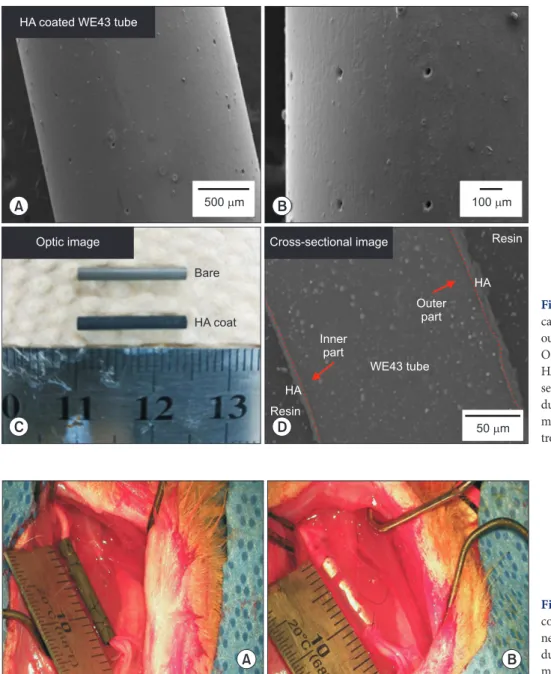
(4) Magnesium nerve conduit for peripheral nerve regeneration. HA coated WE43 tube. 500 m. A Optic image. 100 m. B. Resin. Cross-sectional image Bare. HA Outer part. HA coat Inner part. WE43 tube HA Resin. D. C. 50 m. A. B. Fig. 2. HA-WE43 nerve conduits fabri cation. (A, B) SEM image showing the outer surface with 20 µm sized pores. (C) Optic image showing bare WE43 and HA-WE nerve conduits. (D) SEM crosssectional image of HA-WE43 nerve con duit. HA-WE43, hydroxyapatite coated magnesium alloy; SEM, scanning electron microscope.. Fig. 3. Clinical photographs of nerve conduits implantation. (A) HA-WE43 nerve conduit. (B) Silicone nerve conduit. HA-WE43, hydroxyapatite coated magnesium alloy.. 9-0 Nylon (Ethicon, Livingston, UK) under a surgical. previous report [6]. Briefly, for each footprint, print length. microscope (Carl Zeiss, Oberkochen, Germany). Conduits. (PL, or the longitudinal distance between the tip of the. were flushed with heparinized saline before wound. longest toe and the heel), toe spread (TS, or the distance. closure. Muscle layer was approximated with 4/0 Vicryl. between the first and fifth toes), and the intermediate toe. (Ethicon) and skin was closed using 4/0 Dafilon (B. Braun,. spread (IT, or the distance between the second and fourth. Barcelona, Spain). Nerve regeneration was assessed over a. toes), both in the normal (N) and the experimental (E) paws. 12-week interval (Fig. 3).. were all measured. Based on these parameters, the SFI was calculated according the formula modified by Bain et al. [7].. Gait analysis with sciatic functional index (SFI) Pre- and postoperative footprints were recorded weekly until the end of the experiment interval as mentioned in a. 108. www.chosunobr.org. SFI=–38.3 (EPL–NPL/NPL)+109.5 (ETS–NTS/NTS)+13.3 (EIT–NIT/NIT)–8.8..
(5) Akram Abdo Almansoori, et al.. SFI values around –100 indicate total loss of function whereas values around 0 indicate normal function.. For simplifying axon counting, the total cross-sectional area of the nerve was measured at ×40 magnification and three sampling fields were randomly selected at ×200 magnification.. Retrograde labeling and quantification of neurons. Mean fiber density was calculated by dividing the total. After 12 weeks, 6 rats of each group were used for. number of nerve fibers within the sampling field by its area. retrograde labeling and counting of back-labeled sensory. (N/mm2). Total fibers number was estimated by multiplying. neurons as described by Geremia et al. [8] and Alrashdan. the mean fiber density by the total cross-sectional area. et al. [9]. Briefly, five sciatic nerves in each group were. of the whole nerve cross section assuming a uniform. labeled with 4% Fluorogold (FG) (Fluorochrome, LLC.,. distribution of nerve fibers across the entire section.. Denver, CO, USA), while one rat served as a negative control using DW. Sciatic nerves were sharply cut 5 mm. Statistics. distal to the distal end of the conduit and soaked in 4% FG for 20 minutes in a baseline well. The wound was closed and rats were placed back in their cages.. Data analysis was carried out using IBM SPSS statistics ver. 23 software (IBM Co., Armonk, NY, USA). All data were. 5 days later, the animals were anesthetized; transcardially. presented as mean with standard deviation of the mean.. perfused with 1% heparinized saline and fixed with 4%. For nonparametric, data comparisons were performed. paraformaldehyde solution. L4, L5, and L6 dorsal root. with One-way ANOVA on ranks (Kruskal-Wallis) test. For. ganglions (DRG) were harvested and serially frozen. parameter data, ANOVA test followed by hoc test was used. sectioned into 20-μm-thick sections using a Cryo-Cut. to compare data between different groups. p -values less. microtome (Leica CM3050S Cryostat; Leica Microsystems,. than 0.05 were considered statistically significant.. Wetzlar, Germany). A laser scanning confocal microscope (CLSM, LSM700; Carl Zeiss) was used to capture images of DRG sections. For quantification the back-labeled neurons, the largest three area sections from DRG were selected and the labeled neurons were counted, averaged and compared. RESULTS High gas formation in the preliminary pure Mg and WE43 conduits. across the groups [10]. When the preliminary implanted pure Mg and WE43 Histomorphometric analysis. conduits were re-explored 4 weeks after implantation,. After 12 weeks, 6 rats from each group were used. they were partially degraded and a large encapsulated. for histomorphometric analysis. Sciatic nerves were re-. gas was found surrounding each conduit (Fig. 1C, D).. exposed and conduits were harvested along with the inner. The histologically examined inner and surrounded tissue. regenerated nerve. The regenerated nerve segment was. revealed a fibrous tissue with invasive inflammatory cells. freed and immediately immersed into a fixation solution. and absence of any inner regenerated neural tissue (Fig. 1E, F).. containing 2.5% glutaraldehyde in PBS (pH 7.4) at 4° C for 24 hours. The regenerated nerve sample was transversely. In vitro findings. cut at the center and post-fixed with 2% osmium tetroxide. Subsequently, it was routinely processed and embedded. Cells adhesion as revealed by SEM. with Epon 812 (Nisshin EM, Tokyo, Japan). Serial sections. PC12 cells were found well adhered on HA-WE43 and. of 1 µm thickness were cut with microtome and stained. HA-Mg disks (Fig. 4A, B). Other than that, neither PC12. with 1% toluidine blue for light microscopy examination.. cells nor SCs could be visualized on the remaining disks.. Images were captured using a specialized system, SPOT RT-KE color mosaic, and digitized by SPOT software ver. 4.6 (Diagnostic Instruments, Inc., Sterling Heights, MI, USA).. 109.
(6) Magnesium nerve conduit for peripheral nerve regeneration. High proliferation of PC12 cells on HA-WE43 disks. Conduits degradation and gas formation. As assessed by EZ-CYTOX assay, modest SCs proliferation. At 12 weeks after implantation, conduits showed a. was detected as compared to the control group with no. mild degradation and became thin but maintained a well-. difference across the various disks. In contrast, PC12 cells. integrated structure. No gas formation could be observed. proliferation was higher particularly on HA-WE43 disks. in the surrounding tissue of the conduit. Grossly, silicone. where it was significantly higher than Mg and WE43 disks. conduits showed a well regenerated nerve within the. (Fig. 4C).. conduit, but a scanty neural tissue was found within the HA-WE43 conduit (Fig. 5).. In vivo evaluation of HA-WE43 nerve conduit Retrograde labeling and quantification of neurons Gait analysis with SFI. The retrograde labeled neurons were significantly lower. Preoperative SFI showed normal gait in all groups’ rats.. in HA-WE43 group in comparison with the sham and. While no change in sham group, a drop in SFI to around. silicone groups. The mean of retrograde labeled neurons. –90 to –80 in the first postoperative week was noticed. was 39.09±22.14, 33.91±17.86, and 11.80±6.59 for. and remained highly unchangeable till the 12th week. sham, silicone and HA-WE43 groups, respectively (Fig. 6).. postoperatively. No actual motor function recovery could be observed in all rats except in two rats in the silicone group. Histomorphometric analysis. where one recovered partially and the other recovered. Sham and silicone groups showed a higher axon density. almost completely. SFI mean was –78.62±21.97 for silicone. with significant difference against HA-WE43 group. Sham. group and 72.37±18.74 for HA-WE43 group (Table 1).. group revealed a higher axon density, while silicone. A. B. C 4. 3. Absorbance. *. Mg WE43 HA-Mg HA-WE43. 2. 1. 110. www.chosunobr.org. 12 PC. s SC. C. trl. 0. Fig. 4. SEM images of PC12 cells adhe sion on Mg disks. (A) HA-Mg disk. (B) HA-WE43 disk. (C) Neural cells viability and proliferation on Mg disks. Higher PC12 proliferation on HA-WE43 and HA-Mg disks. SEM, scanning electron microscope; Mg, magnesium; HA, hydroxyapatite; SCs, Schwann cells. *pvalues<0.05 vs. Mg and WE..
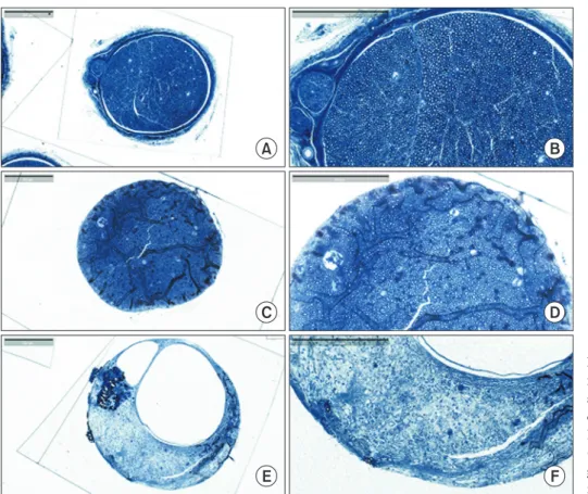
(7) Akram Abdo Almansoori, et al. Table 1. Results of SFI, axon density and retrograded labeled neurons Group Sham. SFI. Axon density (fibers/mm2). Total fibers. Retrograde labeled neurons. –6.99±2.86. 8,795.66±2,034.28. 10,485.05±7,369.65. 39.09±22.14. 13,021.32±1,718.08. 33.91±17.86. 1,198.19±1,795.06. 11.80±6.59. Silicone. –78.62±21.97. 6,975.05±347.90. HA-WE43. –72.37 ±18.74. 1,184.16±1,825.20. Values are presented as mean±standard deviation. SFI, sciatic functional index; HA-WE43, hydroxyapatite coated magnesium alloy.. A. B. C. D. E. F. Fig. 5. Clinical photos of nerve re-exploration. (A) HA-WE43 with no gas formation and mild resorption. (B) Removal of regenerated nerve within HA-WE43 conduit. (C) Regenerated nerve of 10 mm with well-formed wall but defective lumen. (D) Silicone conduit with well regenerated nerve. (F) Removal of regenerated nerve within silicone conduit. (F) Well-formed nerve of 10 mm. HA-WE43, hydroxyapatite coated magnesium alloy.. A. B. C. Fig. 6. Fluorescent photomicrographs for retrograde labeled neurons within L4-L6 dorsal root ganglions which were significantly lower in HAWE43 group in comparison with the sham and silicone groups. (A) Sham group. (B) Silicone group. (C) HA-WE43 group. HA-WE43, hydroxyapatite coated magnesium alloy. Scale bare=200 µm.. had a higher total fibers number. For HA-WE43, three. in diameter. The mean of axon density was 8,795.66±. conduits out of six showed no fibers at all, while in the. 2,034.28, 6,975.05±347.90 and 1,184.16±1,825.20 fiber/. remaining three conduits the axons were few and narrow. mm2 for sham, silicone and HA-WE43, respectively. While. 111.
(8) Magnesium nerve conduit for peripheral nerve regeneration. A. B. C. D. E. F. Fig. 7. Histological microphotographs for regenerated axons where Sham and silicone groups showed a higher axon density with significant difference against HA-WE43 group. (A, B) Sham group (C, D) Silicone group. (E, F) HA-WE43 group. HA-WE43, hydroxyapatite coated magnesium alloy. Scale bare=200 µm.. total fiber number was 10,485.05±7,369.65, 13,021.32±. a favorable growth of PC12 cells on HA-WE43 disk.. 1,718.08, and 1,198.19±1,795.06 for sham, silicone and. Unfortunately, the same high growth was not observed in. HA-WE43, respectively (Fig. 7).. case of SCs. The in vivo results revealed the formation of scanty neural tissue that made of integrated tissue wall but. DISCUSSION. of partially hollow and defected lumen. In presumption to explain this pitfall of HA-WE43 nerve conduit to regenerate. As shown in the preliminary study, bare Mg or WE43. neural tissue, is the presence of rough surface that could. were susceptible to rapid degradation and severe gas. prevent a proper adhesion of the cells, particularly SCs.. formation which countered any neural regeneration. HA-. Another possible explanation is the lack of flexibility in. WE43 was reported to be biocompatible with no toxicity. HA-WE43 nerve conduits which is unfavorable in the body. as revealed by hematological test following placement of. areas of motion as it could apply a pulling tension on both. HA-WE43 screws in rabbit’s tibia [4]. The present study’. regenerating nerve stumps. Surface modification of WE43. s use of HA-WE43 indicated its benefit in providing a. can be achieved using various materials. Recently, a micro-. well-controlled degraded conduit without any observable. textured HA and poly (l-lactic)-acid polymer composite. accumulated gas. Such findings go in accordance with Lim. coated layer on Mg implant was introduced. Such material. et al. [11] study of plates which were inserted above the. may provide the required biocompatible surface for SCs. frontal bone and proved the absence of any gas formation,. and flexibility for the conduits [13].. inflammation, infection, wound dehiscence, and/or. In conclusion, HA-WE43 nerve conduit showed a very. plate exposure over 12 weeks interval, and such period. slow controlled degradation and absence of gas formation. is the minimum required time for neural regeneration. in the surrounding tissues with scanty neural tissue. following neurotmesis [12]. The in vitro evaluation showed. regeneration.. 112. www.chosunobr.org.
(9) Akram Abdo Almansoori, et al.. ACKNOWLEDGEMENTS This research was supported by a grant from Seoul National University Dental Hospital research fund (grant number: 04-2013-0070).. CONFLICTS OF INTEREST The authors declare that they have no competing interests.. ORCID Akram Abdo Almansoori https://orcid.org/0000-0001-7134-9195 Kyung Won Ju https://orcid.org/0000-0002-2566-7405 Bongju Kim https://orcid.org/0000-0001-7309-5977 Soung Min Kim https://orcid.org/0000-0002-6916-0489 Sung-Mi Lee https://orcid.org/0000-0002-0130-1572 Jong-Ho Lee https://orcid.org/0000-0002-8843-545X. REFERENCES 1. Hoffman W. Reanimation of the paralyzed face. Oto laryngol Clin North Am 1992;25:649-667. 2. Meyer RA, Bagheri SC. Microsurgical reconstruction of the trigeminal nerve. Oral Maxillofac Surg Clin North Am 2013; 25:287-302. doi: 10.1016/j.coms.2013.01.002. 3. Deumens R, Bozkurt A, Meek MF, Marcus MA, Joosten EA, Weis J, Brook GA. Repairing injured peripheral nerves: bridging the gap. Prog Neurobiol 2010;92:245-276. doi: 10.1016/j.pneurobio.2010.10.002. 4. Lim HK, Byun SH, Lee JY, Lee JW, Kim SM, Lee SM, Kim HE, Lee JH. Radiological, histological, and hematological evaluation of hydroxyapatite-coated resorbable magnesium alloy screws placed in rabbit tibia. J Biomed Mater Res B Appl. Biomater 2017;105:1636-1644. doi: 10.1002/jbm.b.33703. 5. Yu GS, Bae YM, Choi H, Kong B, Choi IS, Choi JS. Synthesis of PAMAM dendrimer derivatives with enhanced buffering capacity and remarkable gene transfection efficiency. Bioconjug Chem 2011;22:1046-1055. doi: 10.1021/bc1004 79t. 6. Hei WH, Almansoori AA, Sung MA, Ju KW, Seo N, Lee SH, Kim BJ, Kim SM, Jahng JW, He H, Lee JH. Adenovirus vector-mediated ex vivo gene transfer of brain-derived neurotrophic factor (BDNF) tohuman umbilical cord blood-derived mesenchymal stem cells (UCB-MSCs) promotescrush-injured rat sciatic nerve regeneration. Neurosci Lett 2017;643:111-120. doi: 10.1016/j.neulet. 2017.02.030. 7. Bain JR, Mackinnon SE, Hunter DA. Functional evaluation of complete sciatic, peroneal, and posterior tibial nerve lesions in the rat. Plast Reconstr Surg 1989;83:129-138. 8. Geremia NM, Gordon T, Brushart TM, Al-Majed AA, Verge VM. Electrical stimulation promotes sensory neuron regeneration and growth-associated gene expression. Exp Neurol 2007;205:347-359. doi: 10.1016/ j.expneurol.2007.01.040. 9. Alrashdan MS, Sung MA, Kwon YK, Chung HJ, Kim SJ, Lee JH. Effects of combining electrical stimulation with BDNF gene transfer on the regeneration of crushed rat sciatic nerve. Acta Neurochir (Wien) 2011;153:2021-2029. doi: 10.1007/s00701-011-1054-x. 10. Sung MA, Jung HJ, Lee JW, Lee JY, Pang KM, Yoo SB, Alrashdan MS, Kim SM, Jahng JW, Lee JH. Human umbilical cord blood-derived mesenchymal stem cells promote regeneration of crush-injured rat sciatic nerves. Neural Regen Res 2012;7:2018-2027. doi: 10.3969/j.issn.16735374.2012.26.003. 11. Lim HK, Byun SH, Woo JM, Kim SM, Lee SM, Kim BJ, Kim HE, Lee JW, Kim SM, Lee JH. Biocompatibility and biocorrosion of hydroxyapatite-coated magnesium plate: animal experiment. Materials (Basel) 2017 Sep 30 [Epub]. https://doi.org/10.3390/ma10101149. 12. Grinsell D, Keating CP. Peripheral nerve reconstruction after injury: a review of clinical and experimental therapies. BioMed Res Int 2014 Sep 3 [Epub]. https://doi. org/10.1155/2014/698256. 13. Kim SM, Kang MH, Kim HE, Lim HK, Byun SH, Lee JH, Lee SM. Innovative micro-textured hydroxyapatite and poly (l-lactic)-acid polymer composite film as a flexible, corrosion resistant, biocompatible, and bioactive coating for Mg implants. Mater Sci Eng C Mater Biol Appl 2017;81:97-103. doi: 10.1016/j.msec.2017.07.026.. 113.
(10)
수치


+2
관련 문서
