ORIGINAL ARTICLE
대장암 환자에서 복막 전이에 대한 복수내 암태아 항원 (Carcinoembryonic Antigen)의 진단적 가치
송성은, 최바울, 김재현, 정경원, 김성은, 문원, 박무인, 박선자
고신대학교 의과대학 내과학교실
Diagnostic Value of Carcinoembryonic Antigen in Ascites for Colorectal Cancer with Peritoneal Carcinomatosis
Sung Eun Song, Paul Choi, Jae Hyun Kim, Kyoungwon Jung, Sung Eun Kim, Won Moon, Moo In Park and Seun Ja Park Department of Internal Medicine, Kosin University College of Medicine, Busan, Korea
Background/Aims: Diagnostic tests for carcinoembryonic antigen (CEA) in ascites have been performed in various malignant cases, but there is only few data on the applicability of CEA for colorectal cancer (CRC) patients with peritoneal carcinomatosis. We aimed to determine the usefulness of CEA in ascites (aCEA) as a diagnostic parameter for CRC with peritoneal carcinomatosis.
Methods: Between January 2000 and May 2013, the medical records of 259 patients who underwent paracentesis for the evaluation of ascites were retrospectively reviewed. CRC patients with ascites (n=82) and patients with non-malignant ascites (n=177) were evaluated. Patients who had other malignancies, including gastric or ovarian cancer, with ascites were excluded. The optimal diag- nostic cut-off value of aCEA for CRC with peritoneal carcinomatosis was determined using receiver operating characteristic curve analysis. The value of aCEA for predicting the occurrence of peritoneal carcinomatosis was evaluated using a logistic regression model.
Results: The optimal cut-off value of aCEA to diagnose CRC with peritoneal carcinomatosis was 3.89 ng/mL, and the area under the curve for aCEA was 0.996 (sensitivity 96.3%, specificity 100%, positive predictive value 100%, negative predictive value 98.3%).
Multivariate logistic regression analysis showed that aCEA was an independent factor predicting the occurrence of peritoneal carcinomatosis.
Conclusions: In this study, we showed that aCEA may be a useful parameter for diagnosing CRC with peritoneal carcinomatosis, and we propose an optimal aCEA cut-off value of 3.89 ng/mL. Further study that includes patients with other malignant ascites may be necessary to validate these findings. (Korean J Gastroenterol 2018;71:332-337)
Key Words: Carcinoembryonic antigen; Ascites; Peritoneal neoplasms; Colorectal neoplasms
Received December 10, 2017. Revised March 12, 2018. Accepted March 13, 2018.
CC This is an open access article distributed under the terms of the Creative Commons Attribution Non-Commercial License (http://creativecommons.org/licenses/
by-nc/4.0) which permits unrestricted non-commercial use, distribution, and reproduction in any medium, provided the original work is properly cited.
Copyright © 2018. Korean Society of Gastroenterology.
교신저자: 박선자, 49267, 부산시 서구 감천로 262, 고신대학교 의과대학 내과학교실
Correspondence to: Seun Ja Park, Department of Internal Medicine, Kosin University College of Medicine, 262 Gamcheon-ro, Seo-gu, Busan 49267, Korea. Tel:
+82-51-990-5061, Fax: +82-51-990-5055, E-mail: [email protected] Financial support: None. Conflict of interest: None.
INTRODUCTION
Liver cirrhosis (~80% of cases) is the most common cause of ascites. Ascites is the pathological accumulation of fluid in the peritoneal cavity. Malignant conditions, including peri-
toneal carcinomatosis, and benign conditions, including tu- berculous peritonitis, heart failure, pancreatic disease, and renal disease, also contribute to the development and accu- mulation of ascites. Malignant ascites account for approx- imately 10% of all cases of ascites.1 Ascites fluid analysis by
paracentesis can provide useful information for determining the cause of ascites.2,3 Several potential parameters for the diagnosis of malignant ascites have been evaluated to date, including protein levels in ascites, ascites/serum concen- tration ratio of protein (protein A/S), lactate dehydrogenase (LDH) levels in ascites, ascites/serum concentration ratio of LDH (LDH A/S),4 carbohydrate antigen 19-9 in ascites,5 se- rum-ascites albumin gradient,6 fibronectin in ascites, and cholesterol in ascites.7 However, until now, no parameter was able to completely differentiate the cause of malignant as- cites; currently, the gold standard for diagnosing malignant ascites is the presence of tumor cells in ascites.8 The specific- ity of this method is very high, but it has low sensitivity (40-60%) due to the lack of cell exfoliation, which is common to all malignancies.5 This low sensitivity sometimes leads to invasive procedures, including laparoscopy, to acquire peri- toneal tissues. The assessment of carcinoembryonic antigen in ascites (aCEA) has been suggested as an option for diag- nosing patients with ascites, and some studies have sug- gested a positive diagnostic value of aCEA.5,9,10 Such studies evaluated the diagnostic value of aCEA for various malignant cases, but there is limited data on its applicability for color- ectal cancer (CRC) patients with peritoneal carcinomatosis.
Peritoneal carcinomatosis, a complication of CRC, is the primary reason for treatment failure. When cancer patients develop ascites, a number of comorbid diseases, including liver cirrhosis, congestive heart failure, or infectious causes, should be suspected, and peritoneal carcinomatosis should be also considered. Peritoneal carcinomatosis is a common cause of death in patients treated for CRC. According to some reports, peritoneal carcinomatosis has been found in approx- imately 7% of patients during primary surgery, in approx- imately 4-19% of patients during the follow-up period after curative surgery, and in 40-80% of patients who succumb to CRC.11,12 The prognosis of CRC patients with peritoneal carci- nomatosis is poor, with reported median survival of 5.2 months.13 Recently, the overall survival rates in patients with CRC have increased as a result of improved treatment strat- egies, including target therapy. Early and precise detection of peritoneal carcinomatosis via the assessment of aCEA could also be helpful to increase long-term outcomes.
The aim of this study was to identify the clinical significance of CEA in all patients with ascites and to determine the useful- ness of aCEA as a diagnostic parameter for advanced CRC
with peritoneal carcinomatosis.
SUBJECTS AND METHODS
1. Patients
We retrospectively reviewed the medical records of 259 patients who underwent paracentesis for the evaluation of ascites at Kosin University Gospel Hospital, Busan, Korea, between January 2000 and May 2013. CRC patients with as- cites (n=82, CRC group) and those with non-malignant as- cites (n=177, benign group) were retrospectively evaluated.
Patients who had other malignancies, including gastric or ovarian cancer, with ascites were excluded. The CRC group was defined as patients with histologically proven CRC and clinically confirmed peritoneal carcinomatosis. The clinical diagnosis of peritoneal carcinomatosis was made by peri- toneal biopsy or assessment of cytology in ascites, and by ra- diological findings identified by computed tomography (CT) as follows: ascites, thickening of bowel walls, increase in the den- sity of peritoneal fat, presence of peritoneal nodules, and hydronephrosis.14 The benign group was defined as patients who had no evidence of malignancy by clinical and radio- logical findings.
2. Collection and assessment of ascites
All patients underwent paracentesis to evaluate aCEA. The collected ascites were analyzed for cytology and tumor markers. For cytologic examination, the collected peritoneal fluid was centrifuged and smeared on the slides and fixed with cytospray; Papanicolaou and Giemsa staining were performed. The levels of aCEA and serum CEA (sCEA) were measured using electrochemiluminescent immunoassay on a Cobas e-601 analyzer (Roche Diagnostics, Mannheim, Germany).
3. Statistical analyses
All statistical analyses were performed using SPSS 20.0 (IBM Co., Armonk, NY, USA). The optimal cut-off value was de- termined using receiver operating characteristic (ROC) curve analysis. Sensitivity was calculated as true positives/(true positives+false negatives), and specificity was calculated as true negatives/(true negatives+false positives). Positive pre- dictive value (PPV) and negative predictive value (NPV) deter- minations were made from the established cut-off values,
Table 1. Baseline Characteristics in Enrolled Patients Benign group
(n=177)
CRC group (n=82) Median age (range) 59 (17-87) 65 (37-47) Gender
Male 126 (71.2) 54 (65.9)
Female 51 (28.8) 28 (34.1)
Performance statusa
0 133 (75.1) 57 (69.5)
1 41 (23.2) 23 (28.0)
2 3 (1.7) 2 (2.5)
Diagnostic method of CRC
Colonoscopic biopsy 72 (87.8)
Surgery 10 (12.2)
Diagnostic method of peritoneal carcinomatosis
Cytology in ascites 9 (11.0)
CT image 73 (89.0)
Metastasis
Peritoneum only 17 (20.7)
Liver 26 (31.7)
Lung 1 (1.2)
Bone 1 (1.2)
Distant lymph node 37 (45.2)
Cytology
Positive 0 (0.0) 16 (21.1)
Negative 168 (100.0) 60 (78.9)
Values are presented as mean (range) or n (%).
CRC, colorectal cancer; CT, computed tomography.
aEvaluated by Eastern Cooperative Oncology Group (ECOG) criteria.
Fig. 1. Levels of aCEA in the benign and CRC groups. aCEA, carci- noembryonic antigen in ascites; CRC, colorectal cancer.
and were calculated as PPV=true positives/(true pos- itives+false positives) and NPV=true negatives/(true neg- atives+false negatives). A logistic regression model was used to assess the factors affecting the occurrence of peri- toneal carcinomatosis. A p value of less than 0.05 was con- sidered statistically significant.
RESULTS
1. Patient characteristics
A total of 259 patients who underwent paracentesis for cy- tologic evaluation were enrolled. The median age was 60 years (range 17-87). Of these 259 patients, 195 were male (75.3%) and 64 were female (24.7%). Patients were divided into one of two groups: the CRC group (n=82) or the benign group (n=177), based on their diagnosis. The benign group was comprised of patients with liver cirrhosis (n=155), renal disease, including chronic kidney failure and nephritic syn- drome (n=6), tuberculous peritonitis (n=6), heart failure (n=5), and pancreatitis (n=5). Among those in the CRC group,
72 patients (87.8%) were diagnosed by colonoscopic exami- nation, and 10 (12.2%) by surgical examination. Peritoneal carcinomatosis was diagnosed by cytologic evaluation in as- cites (n=9) and by computed tomography imaging (n=73).
When evaluating metastasis, 17 patients (20.7%) had meta- stasis that was limited to the peritoneum, while 65 patients (79.3%) had concurrent peritoneal and systemic organ meta- stasis, including liver, lung, bone, distant lymph node, or mul- tiple organ metastases. These results are summarized in Table 1.
2. Tumor marker assays
The median level of aCEA among all patients was 0.82 ng/mL (range 0.2-16,518 ng/mL). The median levels of aCEA in the CRC and benign groups were 778.85 ng/mL (range 0.97-16,518 ng/mL) and 0.5 ng/mL (range 0.2-3.45 ng/mL), respectively. The difference between the two groups was statistically significant (p<0.001), as shown in Fig. 1.
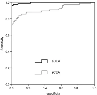
According to ROC curve analysis, the optimal cut-off value of aCEA to predict the occurrence of peritoneal carcinoma- tosis was 3.89 ng/mL. The sensitivity and specificity were 96.3% and 100%, respectively (PPV, 100%; NPV, 98.3%).
Moreover, the area under the curve for aCEA was 0.996 (p<0.001). By comparison, the optimal cut-off value for sCEA for predicting the occurrence of peritoneal carcinomatosis was 8.64 ng/mL. The sensitivity and specificity were 84.2%
and 91.3%, respectively (PPV, 81.0%; NPV, 92.9%); the AUC for sCEA was 0.914 (p<0.001). These results are summar- ized in Table 2 and Fig. 2.
Table 2. Comparison of Diagnostic Parameters
aCEA (ng/mL) sCEA (ng/mL)
Cut-off value 3.885 8.635
Sensitivity (%) 96.3 84.2
Specificity (%) 100 91.3
PPV (%) 100 81.0
NPV (%) 98.3 92.9
AUC 0.996 0.914
aCEA, carcinoembryonic antigen in ascites; sCEA, carcinoembryonic antigen in serum; PPV, positive predictive value; NPV, negative predictive value; AUC, area under the curve.
Fig. 2. ROC curve of diagnostic parameters. ROC, receiver operating characteristic; aCEA, carcinoembryonic antigen in ascites; sCEA, serum carcinoembryonic antigen.
Table 3. Factors Predicting the Occurrence of Peritoneal Carcinomatosis
Predictor Univariate analysis Multivariate analysis
OR (95% CI), p-value OR (95% CI), p-value
Age 1.049 (1.023-1.076), <0.001
Gender 0.781 (0.446-1.367), 0.386
aCEA 4.960 (1.897-12.968), 0.001 4.900 (1.878-12.783), 0.001
sCEA 1.255 (1.158-1.360), <0.001
aCA19-9 1.018 (1.011-1.025), <0.001
sCA19-9 1.003 (1.001-1.004), <0.001
SAAG 0.444 (0.304-0.649), <0.001
Protein A/S 5.367 (1.806-15.948), 0.002
LDH A/S 3.701 (2.135-6.416), <0.001
OR, odds ratio; CI, confidence interval; aCEA, carcinoembryonic antigen in ascites; sCEA, carcinoembryonic antigen in serum; aCA19-9, carbohydrate antigen 19-9 in ascites; sCA19-9, carbohydrate antigen 19-9 in serum; SAAG, serum-ascites albumin gradient; protein A/S, ascites/serum concentration ratio of protein; LDH A/S, ascites/serum concentration ratio of lactate dehydrogenase.
We evaluated the linear correlation between aCEA and sCEA, and found that they were not correlated (correlation co- efficient of -0.017, p=0.884). According to the cytology tests performed in the CRC group (76 patients) showed that 16 pa- tients (21.1%) were positive and 60 (78.9%) were negative.
The mean level of aCEA in the CRC group with negative cytol- ogy was higher than that in the CRC group with positive cytol- ogy; but this difference was not statistically significant (2075.5 ng/mL vs. 1093.6 ng/mL, p=0.06).
Univariate and multivariate analyses using logistic re- gression were performed to evaluate the factors predicting the occurrence of peritoneal carcinomatosis. In univariate analysis, age, aCEA, sCEA, carbohydrate antigen 19-9 in as- cites, carbohydrate antigen 19-9 in serum, serum-ascites al- bumin gradient, protein A/S, and LDH A/S were significant predictors for the occurrence of peritoneal carcinomatosis.
However, in the multivariate analysis, aCEA was the only sig- nificant predictor for the occurrence of peritoneal carcinoma- tosis (odds ratio 4.900, 95% confidence interval 1.878-12.783, p=0.001) (Table 3).
DISCUSSION
This study showed that sensitivity, specificity, PPV, and NPV of aCEA were high, and aCEA was a significant factor for predicting the occurrence of peritoneal carcinomatosis. Our results suggest that aCEA may be a useful parameter for the diagnosis of CRC with peritoneal carcinomatosis, and that aCEA of 3.89 ng/mL could be considered as a cut-off value.
CRC is the third most common cancer in Korea. Peritoneal
carcinomatosis is the second most frequent metastatic pat- tern in advanced CRC.15 Peritoneal fluid cytology is the gold standard to confirm peritoneal carcinomatosis due to its high specificity. However, the low positive detection rate is its limi- tation in clinical practice. In advanced ovarian cancer with peritoneal dissemination, the detection rate of malignant cells in ascites has been reported to be as high as 89%,16 and in advanced gastric cancer with peritoneal dissemination, the detection rate has ranged from 42.3-59%.17,18 However, in advanced CRC with peritoneal dissemination, the de- tection rate of malignant cells in ascites, using peritoneal cy- tology assays, has been as low as 5.8-35.5%.18-21 Radiological findings, including ascites, thickening of bowel walls, in- creased density of peritoneal fat, presence of peritoneal nod- ules, and hydronephrosis, are also used to make clinical diag- nosis of peritoneal carcinomatosis.14,22 According to Gerbes et al.23 aCEA has been proposed as a helpful marker for de- tecting malignant ascites.There have been a few studies evaluating the diagnostic value and cut-off level of CEA in pa- tients with ascites. Nystrom et al. 24 used an empiric cut-off value, and Loewenstein and Zamcheck9 reported a cut-off value for aCEA in malignant ascites of 10 ng/mL (the highest aCEA level of the benign group). Recently, Kaleta et al.5 re- ported that the optimal cut-off value of aCEA to differentiate the causes was >3.5 ng/mL when the cut-off value was se- lected to achieve a specificity of 95.2% by ROC curve analysis.
In previous studies, the sensitivity and specificity of aCEA in patients with advanced CRC ranged from 31.5-48.3% and from 95.2-100%, respectively.5,9 In our study, we assessed aCEA in patients with advanced CRC and determined an opti- mal cut-off value using a ROC curve. We identified an optimal cut-off value of 3.89 ng/mL for aCEA, and the sensitivity and specificity were 96.3% and 100%, respectively.
Our study showed that only 21.1% of patients had positive cytology, even though all patients had clinical peritoneal carcinomatosis. The mean aCEA value of the CRC group was higher in patients with negative cytology than in those with positive cytology.25,26 Hence, these results show a low level of positive cytology in patients with aCEA. There are several possible mechanisms for this. First, this might be due to the low peritoneal metastatic potential of CRC cells.27 Second, delayed examination could yield false-negative results due to lysis of tumor cells.28 Third, peritoneal inflammation could make the difference between malignant cells and atypical or
reactive mesothelial cells that are ambiguous in body fluids.21
We hypothesized that the level of aCEA would positively correlate with the level of sCEA and investigated the relation- ship between them. However, our results did not show any correlation between aCEA and sCEA. There are several limi- tations in this study. First, our study did not include patients who had other malignant ascites, such as gastric or ovarian cancer with ascites. Therefore, our study does not provide re- alistic and clinically available data. Second, there was no CRC patient who had ascites but did not had peritoneal carcino- matosis in this study. Therefore, whether there was an ele- vation of aCEA in CRC patients without peritoneal car- inomatosis remains unclear.
In conclusion, our study demonstrated that aCEA may have predictive value for the occurrence of peritoneal carcinoma- tosis, and this finding suggests that aCEA may be helpful in the initial diagnosis of peritoneal carcinomatosis. According to the results of this study, aCEA may be a useful parameter for diagnosing peritoneal carcinomatosis in advanced CRC patients, with a suggested cutoff value of 3.89 ng/mL.
Further study that includes patients with other malignant as- cites may be necessary to validate these findings.
REFERENCES
1. Runyon BA. Care of patients with ascites. N Engl J Med 1994;
330:337-342.
2. Runyon BA, Hoefs JC, Morgan TR. Ascitic fluid analysis in malig- nancy-related ascites. Hepatology 1988;8:1104-1109.
3. Salerno F, Restelli B, Incerti P, et al. Utility of ascitic fluid analysis in patients with malignancy-related ascites. Scand J Gastroenterol 1990;25:251-256.
4. Boyer TD, Kahn AM, Reynolds TB. Diagnostic value of ascitic fluid lactic dehydrogenase, protein, and WBC levels. Arch Intern Med 1978;138:1103-1105.
5. Kaleta EJ, Tolan NV, Ness KA, O'Kane D, Algeciras-Schimnich A.
CEA, AFP and CA 19-9 analysis in peritoneal fluid to differentiate causes of ascites formation. Clin Biochem 2013;46:814-818.
6. Paré P, Talbot J, Hoefs JC. Serum-ascites albumin concentration gradient: a physiologic approach to the differential diagnosis of ascites. Gastroenterology 1983;85:240-244.
7. Archimandritis A, Kapsalas D, Douvara M, Tjivras M, Tsirantonaki M, Fertakis A. Value of ascitic fibronectin and cholesterol concen- tration in the differentiation between malignancy-related and non-malignant ascites. Ann Med Interne (Paris) 1996;147:145- 150.
8. Saif MW, Siddiqui IA, Sohail MA. Management of ascites due to gastrointestinal malignancy. Ann Saudi Med 2009;29:369-377.
9. Loewenstein MS, Zamcheck N. Carcinoembryonic antigen (CEA) levels in benign gastrointestinal disease states. Cancer 1978;
42(3 Suppl):1412-1418.
10. Tuzun Y, Yilmaz S, Dursun M, et al. How to increase the diagnostic value of malignancy-related ascites: discriminative ability of the ascitic tumour markers. J Int Med Res 2009;37:87-95.
11. Jayne DG, Fook S, Loi C, Seow-Choen F. Peritoneal carcinoma- tosis from colorectal cancer. Br J Surg 2002;89:1545-1550.
12. Koppe MJ, Boerman OC, Oyen WJ, Bleichrodt RP. Peritoneal carci- nomatosis of colorectal origin: incidence and current treatment strategies. Ann Surg 2006;243:212-222.
13. Sadeghi B, Arvieux C, Glehen O, et al. Peritoneal carcinomatosis from non-gynecologic malignancies: results of the EVOCAPE 1 multicentric prospective study. Cancer 2000;88:358-363.
14. Yajima K, Kanda T, Ohashi M, et al. Clinical and diagnostic sig- nificance of preoperative computed tomography findings of as- cites in patients with advanced gastric cancer. Am J Surg 2006;
192:185-190.
15. Hess KR, Varadhachary GR, Taylor SH, et al. Metastatic patterns in adenocarcinoma. Cancer 2006;106:1624-1633.
16. Shen-Gunther J, Mannel RS. Ascites as a predictor of ovarian malignancy. Gynecol Oncol 2002;87:77-83.
17. Burke EC, Karpeh MS Jr, Conlon KC, Brennan MF. Peritoneal lav- age cytology in gastric cancer: an independent predictor of outcome. Ann Surg Oncol 1998;5:411-415.
18. Vogel P, Rüschoff J, Kümmel S, et al. Prognostic value of micro- scopic peritoneal dissemination: comparison between colon and gastric cancer. Dis Colon Rectum 2000;43:92-100.
19. Gozalan U, Yasti AC, Yuksek YN, Reis E, Kama NA. Peritoneal cytol- ogy in colorectal cancer: incidence and prognostic value. Am J
Surg 2007;193:672-675.
20. Hase K, Ueno H, Kuranaga N, Utsunomiya K, Kanabe S, Mochizuki H. Intraperitoneal exfoliated cancer cells in patients with color- ectal cancer. Dis Colon Rectum 1998;41:1134-1140.
21. Yamamoto S, Akasu T, Fujita S, Moriya Y. Long-term prognostic value of conventional peritoneal cytology after curative resection for colorectal carcinoma. Jpn J Clin Oncol 2003;33:33-37.
22. González-Moreno S, González-Bayón L, Ortega-Pérez G, González- Hernando C. Imaging of peritoneal carcinomatosis. Cancer J 2009;15:184-189.
23. Gerbes AL, Jüngst D, Xie YN, Permanetter W, Paumgartner G.
Ascitic fluid analysis for the differentiation of malignancy-related and nonmalignant ascites. Proposal of a diagnostic sequence.
Cancer 1991;68:1808-1814.
24. Nystrom JS, Dyce B, Wada J, Bateman JR, Haverback B.
Carcinoembryonic antigen titers on effusion fluid. A diagnostic tool? Arch Intern Med 1977;137:875-879.
25. Jung M, Jeung HC, Lee SS, et al. The clinical significance of ascitic fluid CEA in advanced gastric cancer with ascites. J Cancer Res Clin Oncol 2010;136:517-526.
26. Lee IK, Kim DH, Gorden DL, et al. Prognostic value of CEA and CA 19-9 tumor markers combined with cytology from peritoneal flu- id in colorectal cancer. Ann Surg Oncol 2009;16:861-870.
27. Hara M, Nakanishi H, Jun Q, et al. Comparative analysis of intra- peritoneal minimal free cancer cells between colorectal and gas- tric cancer patients using quantitative RT-PCR: possible reason for rare peritoneal recurrence in colorectal cancer. Clin Exp Metastasis 2007;24:179-189.
28. Runyon BA. Malignancy-related ascites and ascitic fluid "humoral tests of malignancy". J Clin Gastroenterol 1994;18:94-98.