I. 서론
세균성 치태가 치아 우식증, 치은염 및 치주염을 유발시킨다고 밝혀짐에 따라치태 형성을 억제하거 나 효과적으로 제거하는 치태관리에 대한 연구가 계 속되어 왔다. 치태를 예방하고 치주 건강을 관리하 기 위해서는 적절한 치태조절이 필요하며, 일반적으 로 기계적 방법과 화학적 방법으로 나뉜다1,2). 기계 적 방법로 칫솔질이 중요시되는데 잘 훈련된 사람이 칫솔질에 의하여 치은연상 치태를 완전히 제거할 때 한 번의 칫솔질만으로도 24-48시간 동안 치은 건강 이 유지되지만 대부분의 경우 치태 제거도가 낮아 건강 유지 기간이 훨씬 짧다3). 성인에서 평균적인 칫 솔질 시간은 약 2분이며 이때 약 40%의 치태가 제거 되고, 1일 2회 이상으로 칫솔질 횟수를 늘리면 치주 건강이 더욱 개선된다고 알려져 있다4). 그러나 최근 조사에서 미국인의 57% 정도가 1일 2회 칫솔질을 하 지만 1회만 하는 인구도 30% 정도에 이른다고 보고 되었다5). 이는 치태 관리가 제대로 이루어지지 않는 부분이 상당히 존재할 수 있음을 의미한다. 이와 같 이 칫솔이나 보조적인 도구를 이용한 기계적인 치태 관리는 개인 차이가 심하고 치태 제거가 불완전하기 때문에 이러한 기계적 치태제거 방법의 한계를 극복 하고 보다 효과를 증대시키기 위해 화학적 치태 관
리법이 제안되었다6,7). 또한 칫솔질의 물리적 효과와 더불어 항균 및 항염효과를 얻기 위해 여러 가지 물 질들이 치약에 첨가되어 왔는데, 이러한 물질 중에는 chlorhexidine gluconate, phenolic compounds, plant extracts, essential oils, fluoride 등이 포함되었 으며,항균제재가 첨가된 구강세정제도 항치태 작용 을 통하여 치주 질환을 방지하고 치주 치료에 효과 적이라고 보고되었다8-13).
이 중 임상에서 가장 일반적으로 사용되는 구강세 정제 성분으로서 chlorhexidine gluconate는 1970년 Löe와 Schiott의 보고14)이래 가장 강력하고 안정된 치태억제 및 항균효과를 나타내는 화학 요법제로 인 정받고 있으나, 장기간 사용하였을 때 치아 및 혀의 변색, 미각이상,사용자 중10% 이하에서 점막자극 과 민증 유발 등을 야기하므로15)이를 보완해 줄 수 있 는 다른 세정제의 개발이 요구된다.
한편, 치태내 세균중 Streptococcus mutans등의 그람 양성균은 세포외 다당류를 생성하는데 주로 수 용성α(1-6) linked glucan인 dextran과 불용성 α(1- 3) linked glucan인 mutan으로 구성되어 치태세균의 부착과 증식에 기여한다. 이들 치태간 기질의 분해 에 기여하는 효소로서 dextranase는 불용성 glucan 인 mutan의 합성을 억제하며16), 치면에 연쇄상 구균 의 부착을 억제하고16,17) 불용성 glucan인 mutan을
Dextranase 함유 구강 세정액의 치태 억제 및 치은염 예방 효과에 관한 임상적 연구
송우성·손은주·김도만a·정현주
전남대학교 치과대학 치주과학교실 및 치의학연구소, a공과대학 생물화공과
대한치주과학회지 : Vol. 31, No. 2, 2001
교신저자: 정현주, 광주광역시 동구 학1동 5번지 전남대학교 치과대학 치주과학교실, 우편번호: 501-757
부분적으로 분해하여18)치태 형성과 우식 발생을 저 하시킨다고 알려져 있다19,20). Fitzgerald 등21)은 동물 실험을 통하여 Penicillium funiculosum에서 분리된 dextranase로 치태 형성이 억제되었음을 보고하였 다. 다양한 Streptococcus에 의해 형성된 dextran은 주어진 dextranase에 대해 서로 다른 반응을 보이는 데, dextranase는Actinomyces viscosus에 의해 형성 된 nondextranous plaque의 분산에는 효과적이지 않으며, 치태를 효과적으로 제거하기 위해서는 한가 지 이상의 효소가 필요하다고 보고되어있다22). 반면 김 등23)과 류 등24)은Lipomyces starkeyi KSM 22에서 분리된 dextranase가 mutanolytic, dextranolytic, amylolytic 복합 활성을 지니며Penicillium funiculo- sumdextranase에 비해 matan 분해능이우수하고 치 질을 구성하는 수산화인회석과의 결합력이 높아 구 강내에서도 높은 항치태 작용을 나타낼 수 있는 근 거를 제시하였다.
이에 본 연구에서는 이 Lipomyces starkeyiKSM 22 dextranase를 함유한 구강세정제가 건강인에서 칫솔질과 병용시 chlorhexidine gluconate을 함유한 세정제와 비교하여 어느 정도 치태 형성을 억제하고 치은염을 예방하는지 평가하고자 하였다.
II. 실험 대상 및 방법
1. 실험 대상 및 재료연구 종료시까지 참여하기로 동의서 서명한 자 중 에서 검사할 수 있는 치아가 25개 이상인 18-25세의 전신적으로 건강한 39명을 대상으로 연구를 시행하 였다. 이때 피검자는 연구 기시전에 Turesky- Quigley-Hein plaque index가 1.5이상이고 Löe -Sil- ness gingival index가 1.0 이상이며, 교정장치나 가 철성 국소의치, 구강내 병소, 심한 치주질환이 없고 연구 시점으로부터 2주전에 항생제를 투여한 적이 없으며 약제에 대한 과민 반응이나 알러지 병력이 없는 자 중 선정하였다.
실험재료로서 dextranase 효소함유액(Lipomyces starkeyi KSM 22 dextranase 1 IU/ml of 0.02M Sodi-
um phosphate buffer, pH 7.0), 0.12% chlorhexidine gluconate 용액(Hexamedine , 부광약품), 효소제제 제조시 사용한 완충액(0.02M Sodium phosphate buffer, pH 7.0)을 placebo로 사용하였다. 피검자들 은 번호표를 주어 실험참여 순서에 따라 사용할 구 강 세정액을 할당하여 3개군으로 구분하여 효소 함 유 세정제를 사용한 13인을 실험군,0.12% chlorhexi- dine을 사용한 13인을 양성대조군, placebo액을 세 정제로 사용한 13인을 음성대조군으로배정하였다.
2. 실험과정
실험전의 원래 상태를 검사, 기록한 후 피검자들에 게 치은 연상치태와 치석을 완전히 제거하고 치면세 마를 시행하였다. 철저한 기계적 치태관리가 이루어 지도록 교육한 2주 후착색제를 이용하여 치태가 효 과적으로 제거되고 염증없는 건강한 치은을 확인하 였다. 실험과정 중 각 피검자들에게 미리 coding된 구강 세정액을 검사자가 아닌 제삼자가 공급함으로 써 검사자는 이들이 어느 군에 속하는지 모르게 하 였다. 칫솔질은 원래 사용법에 따라 1일 2회 시행하 고 칫솔질 후 세정액 20cc를 30초간 함수하게 하였 다. 그 외의 치간 청결용 기구는실험 기간동안 사용 을 금지하였다.피검자들에게는 일정한 간격으로 세 정제와 구강 청정용품(세정액 1000cc/개월, 치약 250gm/2개월, 칫솔(중등모 강모의 성인용 칫솔: But- ler 409/3개월)을 공급하였으며. 교환시 이전 용품 의 사용 상태를 확인하여 참가자의 순응도를 점검하 였다.
피검자들을 1개월, 3개월, 6개월 후 임상검사를 위 하여 재소환하여 실험 시작 전에 평가한 동일한 검 사자에 의해 치태 및 치은 염증 지수를 평가하였다.
이때 모든 참가자는 구강내 연조직 및 경조직 상태 를 점검받았다.
3. 평가
1) 치태 축적도 평가
처음 내원시, 실험 기시전, 실험 1, 3, 6개월 후 제3
대구치나 치경부 수복물이 있는 경우를 제외한 모든 치아에서 Turesky-Quigley-Hein plaque index를 사 용하여 치은연상 치태 축적상태를 관찰하였다. 각 치아 순설면에 15초간 적색 착색제(Erythrosin , Sul- tan,USA)로 착색후 치은연상 치태축적 상태를 치아 당 6부위(순면, 원심순면, 근심순면, 설면, 원심설면, 근심설면)에서 점수화하였으며, 각 피검자자당 전체 구강점수는 각 치면 점수의 평균치로 구하였다.
추가적으로 구강내 Turesky- Quigley-Hein Plaque index가 3∼5인 치면의 비율을 점수화하여 치태 심 도 지수(Plaque severity index)26)로 산정하였다.
2) 치은 염증도 평가
처음 내원시, 실험 기시전, 실험 1, 3, 6개월 후 제3 대구치나 치경부 수복물이 있는 경우를 제외한 모든 치아에서 Löe-Silness gingival index로서 치은염증 상태를 치아당 6부위(순면, 원심순면, 근심순면, 설 면, 원심설면, 근심설면)에서 점수화하였다. 각 피검 자당 전체 구강점수는 각 치면 점수의 평균치로 구 하였다.
추가적으로구강내 Löe-Silness Gingival index가 2
∼3인 치면의 비율을 점수화하여 치은염 심도 지수 (Gingivitis severity index)26)로 산정하였다.
3) 치면 착색도 평가
Raber의 Discoloration index system28)과Surface coverage percentage(%)를 결합한 Lang 등의 Area and severity index(AVI) system29)을 이용하여 각치 면의 착색도를 산정하였으며, 전처치 전 협설면으로 나누어 측정하여 정상적인 외인성 착색정도를 기록 하고 실험 기시전, 실험 1, 3, 6개월 후에 검사하였으 며, 구강별 평균 착색도는 평가된 치면 점수의 평균 치로 산출하였다.
4) 연조직 부작용에 대한 평가
모든 참가자는 구강내 연조직 및 경조직 상태를 점 검받고 궤양, 혀의 침착물, 상피탈락, 혀나 점막의 작 열감, 미각이상과 같은 세정제의 부작용이나 이상 작 용이 있었는지에 대한 문항에 답하게 하였다.
5. 통계처리
각군의 실험 전 및 실험 후 측정치의 시기별 비교 와 군간의 비교에는 ANOVA를 이용하였다. 실험 결 과 분석을 위하여 SPSS ver 7.5(SPSS Inc., Chicago, USA)를 사용하였고 5% 유의수준에서 각 시기별 군 간의 차이 여부와 각군의 시기별 차이를 Duncan grouping을 이용하여 검정하였다.
III. 결과
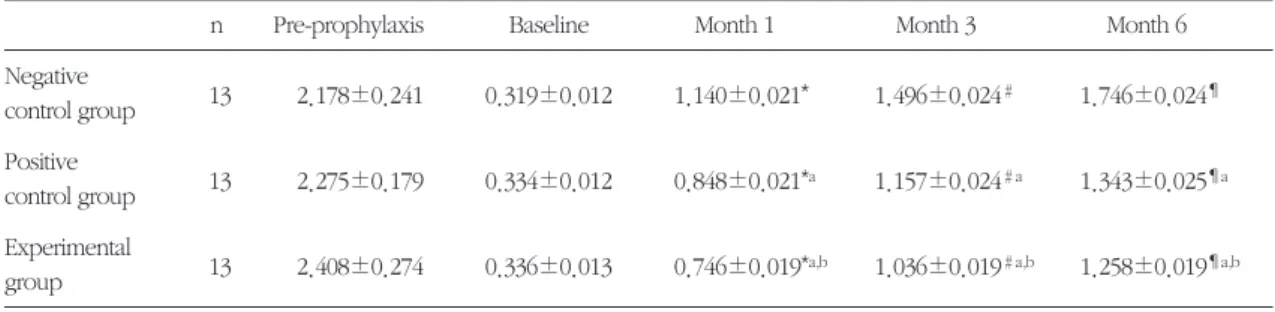
1. 치태 축적도의 변화1) 치태 지수
치태 지수는 처치전과 실험전(baseline)에는 세 군 이 모두 유사하였으나, 실험 후에는 음성대조군에서 수치 증가가 급속하였고 양성대조군 및 실험군은 음 성대조군에 비해 유의하게 낮았다(p<0.05). 또한 실 험기간 내내 실험군은 양성대조군에 비하여 유의하 게 낮았다(p<0.05). 6개월의 실험기간 중 세 군이 모 두 실험기간에 따라 baseline 등, 이전의 검사 시기에 비하여 계속 유의하게 증가하였지만 6개월 이후의 치태 축적도는 처음 내원시에 비해 여전히 낮게 나 타났다(Table 1, 2, Figure 1).
모든 치아 부위에서 각 기간별로 실험 1개월∼6개 월에 걸쳐 세 군 모두 치태 지수가 유의하게 증가하 였으며(p<0.05), 양성대조군과 실험군 모두 음성대 조군에 비해 유의하게 낮았다. 한편, 실험군은 하악 전치부에서는 실험 1개월 후와 3개월 후에, 하악 구 치부에서는 실험 1개월 후와 6개월 후에 양성대조군 에 비해 유의하게 낮았다(p<0.05)(Table 3).
모든 치면에서 세 군 모두 실험 1개월∼6개월에 걸쳐 유의하게 증가하였으며(p<0.05), 양성대조군은 실험 3개월 후부터 음성대조군에 비해 유의하게 낮 았다(p<0.05). 한편, 실험군은 협측 인접면에서는 실 험 3개월 후에, 설측 인접면과 설면에서는 실험 3개 월 후부터 양성대조군에 비하여 유의하게 낮았다 (p<0.05)(Table 4).
Table 2. Increase in Plaque index score during the experiment period
Baseline~Month 1 Month 1~Month 3 Month 3~Month 6
Negative
control group 0.821±0.021 0.356±0.021 0.249±0.012
Positive
control group 0.514±0.020a 0.309±0.021 0.186±0.012a
Experimental
group 0.410±0.017a,b 0.291±0.022a 0.222±0.015
Values are expressed in Mean±SE.
aSignificantly different from Negative control group at p<0.05 by Duncan grouping
bSignificantly different from Positive control group at p<0.05 by Duncan grouping
Figure 1. Change in Plaque index score during the experiment period.
2.5
2
1.5
1
0.5
0
score
Pre-prophylaxis Baseline Month 1 Month 6
Experiment period
Negative control group Positive control group Experimental group
Month 3 Table 1. Plaque index score during the experiment period
n Pre-prophylaxis Baseline Month 1 Month 3 Month 6
Negative
control group 13 2.178±0.241 0.319±0.012 1.140±0.021* 1.496±0.024# 1.746±0.024¶ Positive
control group 13 2.275±0.179 0.334±0.012 0.848±0.021*a 1.157±0.024#a 1.343±0.025¶a Experimental
group 13 2.408±0.274 0.336±0.013 0.746±0.019*a,b 1.036±0.019#a,b 1.258±0.019¶a,b n; No of subjects in each group. No. of subjects for Pre-prophylaxis score is 9.
Values are expressed in Mean±SE.
*Significantly different from Baseline at p<0.05 by Duncan grouping
#Significantly different from Month 1 at p<0.05 by Duncan grouping
¶Significantly different from Month 3 at p<0.05 by Duncan grouping
aSignificantly different from Negative control group at p<0.05 by Duncan grouping
bSignificantly different from Positive control group at p<0.05 by Duncan grouping
2) 치태심도 지수(Plaque severity index)
구강내 Turesky-Quigley-Hein Plaque index가 3∼
5인 치면의 비율인 치태심도 지수는 baseline시에 세 군이 유사하였으나, 음성대조군과 실험군에서 1개월 과 3개월 후 실험 전에 비해 유의하게 증가하였으며 6개월 후에는 3개월 이전에 비하여 비교적 완만하게 증가하였다. 양성대조군에서는 6개월에 걸쳐 거의 같은 비율로 증가하였다. 한편, 음성대조군에 비해
양성대조군은 1개월 후부터, 실험군은 3개월 이후부 터 유의하게 낮았다(p<0.05)(Table 5, Figure 2).
2. 치은 염증도의 변화
1) 치은염 지수
치은염 지수는 baseline에서는 세 군이 유사하였으 나, 실험 1개월 후에 음성대조군과 양성대조군이 실
Table 3. Six-month study of mouthrinses as adjunct to toothbrushing: Plaque index score according to the tooth position
PI Negative control group Positive control group Experimental group
n Mean±SE n Mean±SE n Mean±SE
A
Allll tteeeetthh 362 356 363
at baseline 0.319±0.012 0.334±0.012 0.336±0.013
after 1 Month 1.140±0.021* 0.848±0.021*a 0.746±0.019*a,b
after 3 Month 1.496±0.024# 1.157±0.024#a 1.036±0.019#a,b
after 6 Month 1.746±0.024¶ 1.343±0.025¶a 1.258±0.019¶a,b
U
Uppppeerr aanntteerriioorr tteeeetthh 78 76 78
at baseline 0.301±0.023 0.283±0.024 0.295±0.024
after 1 Month 1.086±0.044* 0.645±0.039*a 0.557±0.032*a
after 3 Month 1.318±0.050# 0.844±0.045#a 0.795±0.039#a
after 6 Month 1.589±0.049¶ 1.081±0.048¶a 1.154±0.041¶a
U
Uppppeerr ppoosstteerriioorr tteeeetthh 104 101 104
at baseline 0.405±0.028 0.414±0.029 0.449±0.031
after 1 Month 1.135±0.040* 0.903±0.044*a 0.881±0.041*a
after 3 Month 1.512±0.046# 1.259±0.051#a 1.141±0.037#a
after 6 Month 1.759±0.045¶ 1.390±0.051¶a 1.274±0.035¶a
LLoowweerr aanntteerriioorr tteeeetthh 76 77 77
at baseline 0.241±0.022 0.266±0.022 0.226±0.021
after 1 Month 1.053±0.046* 0.777±0.047*a 0.651±0.037*a,b
after 3 Month 1.448±0.059# 1.138±0.049#a 0.918±0.038#a,b
after 6 Month 1.648±0.058¶ 1.282±0.051a 1.147±0.044¶a
LLoowweerr ppoosstteerriioorr tteeeetthh 104 102 104
t baseline 0.301±0.021 0.343±0.021 0.337±0.021
after 1 Month 1.250±0.038* 0.998±0.036*a 0.822±0.034*a,b
after 3 Month 1.649±0.039# 1.302±0.043#a 1.201±0.034#a
after 6 Month 1.919±0.039¶ 1.614±0.046¶a 1.403±0.036¶a,b
n; No. of teeth included in each group.
*Significantly different from Baseline at p<0.05 by Duncan grouping
#Significantly different from Month 1 at p<0.05 by Duncan grouping
¶Significantly different from Month 3 at p<0.05 by Duncan grouping
aSignificantly different from Negative control group at p<0.05 by Duncan grouping
bSignificantly different from Positive control group at p<0.05 by Duncan grouping
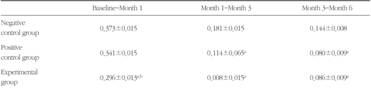
험군에 비해 유의하게 크게 증가하였고, 실험 3, 6개 월 후에는 세 군간에모두차이를 보여, 음성대조군에 비해 양성대조군이, 양성대조군에 비해 실험군이 유 의하게 낮았다(P<0.05). 세 군이 모두 실험 기간동안 내내 계속증가하였지만 6개월 이후에도 최초 내원시 치은염증 상태보다는1/2 정도 낮은 치은 염 지수를 보였다(Table 6,7, Figure 3).
각 실험 기간별로 치은염증도는 baseline∼1개월 간에는 세 군 모두 다른 기간에 비해 급격한 수치의 증가를 보였으며, 실험군이 음성대조군과 양성대조 군에 비해 유의하게 낮은 증가율을 나타내었다. 또 한 1개월∼3개월과 3개월∼6개월에는 음성대조군이 가장 많은 증가를 보였고, 양성대조군과 실험군간에 유의한 차이가 없이 유사한 증가율을 보였다 Table 4. Six-month study of mouthrinses as adjunct to toothbrushing: Plaque index score according to the
tooth surfaces
PI Negative control group Positive control group Experimental group
n Mean±SE n Mean±SE n Mean±SE
A
Allll tteeeetthh 2172 2136 2177
at baseline 0.319±0.012 0.334±0.012 0.336±0.013
after 1 Month 1.140±0.021* 0.848±0.021*a 0.746±0.019*a,b
after 3 Month 1.496±0.024# 1.157±0.024#a 1.036±0.019#a,b
after 6 Month 1.746±0.024¶ 1.343±0.025¶a 1.258±0.019¶a,b
B
Buuccccoo--pprrooxxiimmaall 724 712 726
at baseline 0.361±0.023 0.344±0.023 0.415±0.025
after 1 Month 1.191±0.038* 0.798±0.035* 0.861±0.036*a
after 3 Month 1.500±0.043# 1.059±0.037#a 1.206±0.036#a,b
after 6 Month 1.739±0.042¶ 1.258±0.039¶a 1.402±0.033¶a
B
Buuccccaall 362 356 363
at baseline 0.320±0.033 0.287±0.032 0.394±0.038
after 1 Month 1.014±0.056* 0.537±0.048* 0.700±0.050*a
after 3 Month 1.271±0.062# 0.787±0.058#a 0.871±0.048#a
after 6 Month 1.526±0.063¶ 1.006±0.064¶a 1.077±0.049¶a
LLiinngguuoo--pprrooxxiimmaall 724 712 725
at baseline 0.301±0.019 0.362±0.021 0.295±0.017
after 1 Month 1.214±0.033* 1.069±0.038* 0.748±0.028*
after 3 Month 1.655±0.038# 1.412±0.042#a 1.072±.0.028#a,b
after 6 Month 1.885±0.038¶ 1.559±0.043¶a 1.313±0.031¶a,b
LLiinngguuaall 362 356 363
at baseline 0.268±0.025 0.303±0.026 0.204±0.023
after 1 Month 1.017±0.049* 0.819±0.05* 0.559±0.040*
after 3 Month 1.396±0.058# 1.214±0.064# 0.790±0.044#a,b
after 6 Month 1.698±0.057¶ 1.419±0.066¶a 1.041±0.049¶a,b
n; No. of teeth included in each group.
*Significantly different from Baseline at p<0.05 by Duncan grouping
#Significantly different from Month 1 at p<0.05 by Duncan grouping
¶Significantly different from Month 3 at p<0.05 by Duncan grouping
aSignificantly different from Negative control group at p<0.05 by Duncan grouping
bSignificantly different from Positive control group at p<0.05 by Duncan grouping
Table 5. Plaque severity index score during the experiment period
n Pre-prophylaxis Baseline Month 1 Month 3 Month 6
Negative
control group 13 0.412±0.057 0.012±0.006 0.099±0.011* 0.168±0.024# 0.182±0.022 Positive
control group 13 0.437±0.057 0.011±0.006 0.063±0.015a 0.104±0.029#a 0.125±0.026a Experimental
group 13 0.484±0.078 0.013±0.003 0.068±0.004* 0.095±0.006#a 0.112±0.008a
n; No of subjects in each group. Values are expressed in Mean±SE.
*Significantly different from Baseline at p<0.05 by Duncan grouping
#Significantly different from Month 1 at p<0.05 by Duncan grouping
aSignificantly different from Negative control group at p<0.05 by Duncan grouping
score
Pre-prophylaxis Baseline Month 1 Month 6
Experiment period
Negative control group Positive control group Experimental group
Month 3 0.5
0.4
0.3
0.2
0.1
0
Figure 2. Change in Plaque severity index score during the experiment period
Table 6. Gingival index score during the experiment period
n Pre-prophylaxis Baseline Month 1 Month 3 Month 6
Negative
control group 13 1.265±0.069 0.159±0.008 0.532±0.014* 0.712±0.015# 0.856±0.015¶ Positive
control group 13 1.344±0.097 0.159±0.008 0.501±0.014* 0.616±0.014#a 0.696±0.013¶a Experimental
group 13 1.178±0.091 0.158±0.008 0.454±0.012*a,b 0.530±0.012#a,b 0.619±0.013¶a,b n; No of subjects in each group. No. of subjects in Pre-prophylaxis score is 9.
Values are expressed in Mean±SE.
*Significantly different from Baseline at p<0.05 by Duncan grouping
#Significantly different from Month 1 at p<0.05 by Duncan grouping
¶Significantly different from Month 3 at p<0.05 by Duncan grouping
aSignificantly different from Negative control group at p<0.05 by Duncan grouping
bSignificantly different from Positive control group at p<0.05 by Duncan grouping
(P>0.05)(Table 7).
각 치아 부위별로 세 군 모두 실험 1개월∼6개월 에 걸쳐 유의하게 증가하였으며, 양성대조군은 실험 3개월 후부터 음성대조군에 비해 유의하게 낮았다 (p<0.05). 한편, 실험군은 상악 구치부에서는 실험 3 개월 후부터 하악 구치부에서는 실험 1개월 후부터 양성대조군에 비해 유의하게 낮았다(p<0.05)(Table 8).
각 치면별로 협측 인접면과 설측 인접면에서 양성 대조군과 실험군 모두 실험 1개월∼6개월에 걸쳐 유 의하게 증가하였으며, 협면과 설면에서는 1개월 후 와 6개월 후에 유의하게 증가하였다(p<0.05). 한편,
실험군은 실험 3개월 후부터 협면을 제외한 다른 치 면 에 서 양 성 대 조 군 보 다 유 의 하 게 낮 았 다 (p<0.05)(Table 9).
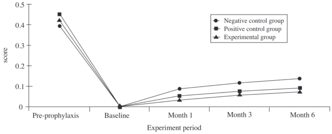
2) 치은염 심도지수(Gingivitis severity index) 구강내 Löe - Silness Gingival index가 2∼3인 치면 의 비율로 산정되는 심도지수는 세 군 모두 1개월 후 baseline에 비해, 6개월 후에는 1개월 후에 비해 유의 하게 증가하였으며(P<0.05), 실험군과 양성대조군은 염증 심도지수가 동일 기간중 유사하게 증가하였으 나, 음성대조군에 비해서는 두 군 모두 유의하게 낮 았다(P<0.05)(Table 10, Figure 4).
score
Pre-prophylaxis Baseline Month 1 Month 6
Experiment period
Month 3 Negative control group Positive control group Experimental group 1.5
1
0.5
0
Figure 3. Change in Gingival index score during the experiment period.
Table 7. Increase in Gingival index score during the experiment period
Baseline~Month 1 Month 1~Month 3 Month 3~Month 6
Negative
control group 0.373±0.015 0.181±0.015 0.144±0.008
Positive
control group 0.341±0.015 0.114±0.065a 0.080±0.009a
Experimental
group 0.296±0.013a,b 0.008±0.015a 0.086±0.009a
Values are expressed in Mean±SE.
aSignificantly different from Negative control group at p<0.05 by Duncan grouping
bSignificantly different from Positive control group at p<0.05 by Duncan grouping
3. 치면 착색도
착색도는 baseline시에 비해 음성대조군은 실험기 간 내내 거의 증가하지 않고 그대로 유지되었으나 양성대조군은 실험 3개월 및 6개월 후에는 baseline 에 비해, 그리고 동일 기간 중 음성대조군에 비해 유 의하게 증가하였다. 반면, 실험군은 3개월 후에는 baseline에 비하여 증가하였고, 음성대조군에 비해서
는 6개월 후 유의하게 증가하였다. 따라서 6개월 후 착색도는 양성대조군, 실험군, 음성대조군 순으로 낮 아져 각 군간에 유의한 차이를 나타내었다(P<0.05) (Table 11, Figure 5).
4. 구강내 부작용
구강내 부작용에 대한 평가시 음성대조군에서는 Table 8. Six-month study of mouthrinses as adjunct to toothbrushing: Gingival index score according to the
tooth position
GI Negative control group Positive control group Experimental group
n Mean±SE n Mean±SE n Mean±SE
A
Allll tteeeetthh 362 356 363
at baseline 0.159±0.008 0.159±0.008 0.158±0.008
after 1 Month 0.532±0.014* 0.501±0.014* 0.454±0.012*a,b
after 3 Month 0.712±0.015# 0.616±0.014#a 0.530±0.012#a,b
after 6 Month 0.856±0.015¶ 0.696±0.013¶a 0.619±0.013¶a,b
U
Uppppeerr aanntteerriioorr tteeeetthh 78 76 78
at baseline 0.139±0.346 0.134±0.341 0.135±0.348
after 1 Month 0.429±0.624* 0.373±0.564* 0.345±0.506*a
after 3 Month 0.607±0.647# 0.485±0.625#a 0.449±0.506#a
after 6 Month 0.756±0.658¶ 0.583±0.591¶a 0.517±0.553¶a
U
Uppppeerr ppoosstteerriioorr tteeeetthh 104 101 104
at baseline 0.181±0.386 0.190±0.393 0.193±0.403
after 1 Month 0.644±0.708* 0.618±0.706* 0.569±0.615*
after 3 Month 0.838±0.711# 0.741±0.664#a 0.630±0.540#ab
after 6 Month 0.960±0.699¶ 0.817±0.737a 0.718±0.600¶ab
LLoowweerr aanntteerriioorr tteeeetthh 76 77 77
at baseline 0.112±0.316 0.108±0.318 0.117±0.322
after 1 Month 0.436±0.619* 0.418±0.791* 0.375±0.519*
after 3 Month 0.594±0.639# 0.478±0.602#a 0.418±0.552a
after 6 Month 0.726±0.644¶ 0.600±0.595¶a 0.511±0.607¶a,b
LLoowweerrppoosstteerriioorr tteeeetthh 104 102 104
t baseline 0.188±0.391 0.185±0.389 0.170±0.376
after 1 Month 0.566±0.677* 0.562±0.663* 0.480±0.564*a,b
after 3 Month 0.752±0.681# 0.693±0.635# 0.575±0.611#a,b
after 6 Month 0.922±0.695¶ 0.747±0.597a 0.675±0.655¶a,b
n; No. of teeth included in each group.
*Significantly different from Baseline at p<0.05 by Duncan grouping
#Significantly different from Month 1 at p<0.05 by Duncan grouping
¶Significantly different from Month 3 at p<0.05 by Duncan grouping
aSignificantly different from Negative control group at p<0.05 by Duncan grouping
bSignificantly different from Positive control group at p<0.05 by Duncan grouping
2.7%, 양성대조군에서는 19.4%, 그리고 실험군에서 는 8.3%에서 혀의 침착 및 변색을 보였다. 한편 양성 대조군에서는 8.3%에서, 실험군에서는 2.7%에서 미 각 이상을, 양성대조군의 5.5%에서는 상피 탈락을 보였다(Table 12, Figure 6).
IV. 고찰
치주질환의 원인으로서국소 요인 중 치태내 세균
이 병인으로서 주된 역할을 하므로 치주질환의 예방 과 치료를 위하여 적절한 치태조절이 필수적이다.
치태조절법은 기계적 방법과 화학적 방법으로 나뉘 는데 기계적 방법으로서 중요한 칫솔질은 일반적으 로 1일 2회, 3분씩 시행하는 것이 권장되고 있으나 대다수의 경우 이를 제대로 지키지 못하는 것이 현 실이다. 이와 같이 일반적으로 이용하는 기계적인 치태 관리법만으로는 치태의 완전한 제거가 어려우 므로 보다 안전하고 효과적인 항치태 기능을 가진 Table 9. Six-month study of mouthrinses as adjunct to toothbrushing: Gingival index score according to the
tooth surfaces
GI Negative control group Positive control group Experimental group
n Mean±SE n Mean±SE n Mean±SE
A
Allll tteeeetthh 2172 2136 2177
at baseline 0.159±0.008 0.159±0.008 0.158±0.008
after 1 Month 0.532±0.014* 0.501±0.041* 0.454±0.012*a,b
after 3 Month 0.712±0.015# 0.616±0.014#a 0.530±0.012#a,b
after 6 Month 0.856±0.015¶ 0.696±0.013¶a 0.619±0.013¶a,b
B
Buuccccoo--pprrooxxiimmaall 724 712 726
at baseline 0.144±0.013 0.149±0.014 0.163±0.014
after 1 Month 0.586±0.025* 0.528±0.024* 0.497±0.021*a
after 3 Month 0.822±0.025# 0.674±0.023#a 0.596±0.021#ab
after 6 Month 0.938±0.025¶ 0.735±0.022¶a 0.716±0.023¶a
B
Buuccccaall 361 356 363
at baseline 0.139±0.018 0.144±0.019 0.141±0.019
after 1 Month 0.368±0.033* 0.312±0.024* 0.303±0.028*
after 3 Month 0.450±0.034 0.332±0.029a 0.322±0.027a
after 6 Month 0.622±0.037¶ 0.447±0.031¶a 0.408±0.030¶a
LLiinngguuoo--pprrooxxiimmaall 724 712 725
at baseline 0.164±0.014 0.194±0.015 0.183±0.014
after 1 Month 0.619±0.025* 0.602±0.025* 0.551±0.022*
after 3 Month 0.858±0.024# 0.784±0.025#a 0.656±0.021#a,b
after 6 Month 0.989±0.024¶ 0.847±0.026¶a 0.733±0.022¶a,b
LLiinngguuaall 362 356 363
t baseline 0.202±0.021 0.126±0.018 0.113±0.017
after 1 Month 0.412±0.034* 0.389±0.033* 0.324±0.027*
after 3 Month 0.464±0.035 0.447±0.033 0.355±0.027#a,b
after 6 Month 0.660±0.036¶ 0.565±0.032¶a 0.405±0.029¶a,b
n; No. of teeth included in each group.
*Significantly different from Baseline at p<0.05 by Duncan grouping
#Significantly different from Month 1 at p<0.05 by Duncan grouping
¶Significantly different from Month 3 at p<0.05 by Duncan grouping
aSignificantly different from Negative control group at p<0.05 by Duncan grouping
bSignificantly different from Positive control group at p<0.05 by Duncan grouping
Table 10. Gingival severity index score during the experiment period
n Pre-prophylaxis Baseline Month 1 Month 3 Month 6
Negative
control group 13 0.404±0.029 0.001±0.001 0.086±0.015* 0.125±0.019 0.138±0.015# Positive
control group 13 0.453±0.058 0.001±0.001 0.052±0.010*a 0.075±0.014a 0.090±0.013#a Experimental
group 13 0.425±0.061 0.004±0.002 0.034±0.005*a 0.059±0.009a 0.072±0.009#a
n; No of teeth included ineach group. Values are ecpressed in Mean±SE
*Significantly different from Baseline at p<0.05 by Duncan grouping
#Significantly different from Month 1 at p<0.05 by Duncan grouping
aSignificantly different from Negative control group at p<0.05 by Duncan grouping
score
Pre-prophylaxis Baseline Month 1 Month 6
Experiment period
Month 3
Negative control group Positive control group Experimental group 0.5
0.4
0.3
0.2
0.1
0
Figure 4. Change in Gingivitis severity index score during the experiment period.
Table 11. Discoloration index score during the experiment period
n Baseline Month 1 Month 3 Month 6
Negative
control group 13 8.4±0.7 8.8±0.7 9.2±0.7 9.8±0.7
Positive
control group 13 8.2±0.7 9.7±0.8 11.6±0.6*a 14.1±0.5¶a
Experimental
group 13 8.4±0.7 9.3±0.6 10.1±0.6* 11.9±0.7¶ab
n; No of teeth included in each group. Values are expressed in Mean±SE.
*Significantly different from Baseline at p<0.05 by Duncan grouping
¶Significantly different from Month 3 at p<0.05 by Duncan grouping
aSignificantly different from Negative control group at p<0.05 by Duncan grouping
bSignificantly different from Positive control group at p<0.05 by Duncan grouping
세정제 개발에 대한 필요성이 대두되었으며, 이를 위 한 방법으로서 항균 또는 항염 기능을 가진 약물을 치약이나 세정제에 넣어 사용하고 있다30-32).
Chlorhexidine gluconate는 강력하고 안정된 치태 억제 및 항균효과를 나타내는 화학 요법제로 임상에 서 많이 이용되고 있다.Segreto 등33)과 Grossman 등 Table 12. Prevalence(%) of side effect in oral cavity during the experiment period
Ulcer Tongue accumulation Burning sense Bad taste Desquamation Total Negative
control group 0 2.7 0 0 0 2.7
Positive
control group 0 19.4 0 8.3 5.5 33.3
Experimental
group 0 8.3 0 2.7 0 11.1
score
Baseline Month 1 Month 6
Experiment period Month 3 Negative control group Positive control group Experimental group 16
12
8
Figure 5. Change in Discloration index score during the experiment period.
40
30
20
10
0 (%)
Nagative control group Positive control group Experimental group Group
Tongue deposition Bad taste Desquamation
Figure 6. Side effects in oral cavity during the experiment period.
34)은 6개월에서 2년까지의 장기간 임상 실험을 통해 0.12%의 chlorhexidine을 하루에 2회 함수하게 함으 로써 치태 및 치은염증이 유의하게 감소함을 보여 주었다. 이러한 약물의 치태 억제 효과는 농도보다 는 함수하는 양이 더 중요하지만 국소적 부작용은 농도에 좌우되기 때문에 장기간 임상 실험에서는 적 은 농도를 사용하게 되어35,36)이 연구에서는 0.12%
의 세정제 20cc 함수가 권장되고 있다. 하지만 장기 간 사용하였을 때 치아 및 혀의 변색, 미각이상, 10%
이하의 사용자에서 점막자극 과민증 유발 등을 야기 하므로 이를 보완해 줄 수 있는 다른 세정제의 개발 을 위한 노력이 이루어지고 있다32,37,38).
치태내 세균은 세포외 다당류를 생성하는데 주로 수용성 α(1-6) linked glucan인 dextran과 불용성 α(1-3) linked glucan인 mutan으로 구성되며, dex- tranase 효소를 이용하여 이들을 분해하기 위한 시도 가 이루어진 바 있다16-23). Dextranase 세정액의 임상 적 사용시 치태제거 및 치은염 예방 효과에 대해 이 견이 있는데, Fitzgerald 등21)과 Block 등39)은in vitro 실험과 동물 실험을 통하여Penicillium funiculosum 에서 분리된 dextranase에 의하여 치태형성이 억제 되었다고 보고한 반면, Caldwell 등40)은 임상적 연구 를 통하여 이러한 치태 감소를 증명하지 못하였다.
Keyes 등22)은 충분한 농도의 dextran 분해효소를 충 분한 기간 충분한 횟수로 사용하는 경우 치면에 dex- tran성 세균축적물의 분산을 촉진하며, dextranase의 빈번한 사용시 치면에 dextran성 치태 형성이 지연 된다고 하였다. 한편 다양한 Streptococcus에 의해 만들어진 dextran은 주어진 dextranase에 대해 서로 다른 반응을 보이며, dextranase는Actinomyces vis- cosus에 의해 형성된 nondextranous plaque의 분산 에는 효과적이지 않으므로, 치태를 효과적으로 제거 하기 위해서는 한가지 이상의 효소가 필요하다고 제 안되었다41). Dextranase는 수용성 glucan인 dextran 은 쉽게 분해하지만 불용성인 mutan을 분해하는데 는 한계가 있기 때문에 이를 해결하기 위한in vitro, 동물 및 임상 실험 연구가 이루어졌다. 최근 김 등23) 과 류 등24)은 Lipomyces starkeyi KSM 22에서 dex- tranase와 amylase의 복합 활성도를 보이는 효소
(DXAMase)를 정제하여, 독성 및 알러지 반응이 없 으며 항치태효과를 가질 수 있는 특성을 가진다고 보고하였다.
본 연구는 dextranase 효소함유 구강 세정액을 칫 솔질에 보조적으로 병용시 어느 정도 치태 및 치은 염 억제 효과를 가지는지 평가하기 위하여 시행하였 다. 전신적으로 건강한 대학생 39인을 치석제거 및 치면세마하여 건강한 치은조직을 갖게 하고 1일 2회 정상적인 칫솔질을 하게 하였다. 2주 후 칫솔질과 함 께 세정액(dextranase 함유 세정제, 0.12% Chlorhex- idine액, Placebo액) 20cc를 30초간 1일 2회 함수하게 하면서 1, 3, 6개월 후 치태 축적도, 치은 염증도, 치 면 착색도의 변화, 그리고 구강내 부작용의 발생 빈 도를 평가하고 서로 비교하였다. 6개월의 실험기간 중 세 군이 모두 실험기간중 치태 축적도와 치은염 증도가 계속 유의하게 증가하였지만 6개월 이후의 상태는 처치 및 실험전 처음 내원시에 비해 여전히 양호하게 나타났다. Brecx 등42)은 위와 같은 연구결 과에 대해 치태의 전문적인 제거 외에 연구에 참여 한 사람들이 연구에 참여한다는 사실만으로 그들의 구강위생습관의 개선에 대한 동기부여와 연구원들 이 낮은 점수를 기대할 것이라 생각하여 철저한 구 강위생을 시행하기 때문에 가능하다고 하였다.
치태축적도는 실험 기간 내내 양성대조군이 음성 대조군에 비해 유의하게 낮았으며, 실험군인 dex- tranase 세정군에서 다른 두 군에 비해 유의하게 낮 았다(P<0.05). 이는 dextranase 양치시 치태의 건조 된 무게는 감소시키나 치태의 면적을 유의하게 변화 시키지는 못했다고 보고한Lobene 등41)의 임상 실험 과 는 약 간 다 른 결 과 를 보 인 다 . 그 이 유 는 Lipomyces starkeyi KSM 22 dextranase가Penicilli- um funiculosum dextranase와는 달리 칫솔질을 병 행하여 성숙치태가 제거된 상태에서 신생 치태의 형 성을 억제하기 때문이라 여겨진다. 기간별 증가율을 보면 실험군이 baseline∼1개월에 양성대조군에 비 해 치태 축적도 및 치은 염증도가 유의하게 적어 dextranase 양치에 의한 효과가 치태형성 및 성숙과 정의 초기에 가능하다는 증거를 제공하였다. 치태 심도 지수, 치은염 지수 및 치은염 심도 지수의 경우
에도 실험군이 양성대조군과 유사하게 낮아 dex- tranase 양치에 의한 치태 축적 및 치은염증 억제 효 과가 가장 큼을 보여주었다.
치면착색도는 baseline시에 비해 음성대조군은 실 험기간 내내 거의 증가하지 않고 유지되었으나 양성 대조군은 실험 3개월 및 6개월 후에는 baseline에 비 해, 각 기간별로 음성대조군에 비해 유의하게 증가하 였다. 반면 실험군은 3개월 후에는 baseline에 비하 여 증가하였고, 음성대조군에 비해서는 6개월 이후 유의하게 증가하였다. 따라서 6개월 후 착색도는 양 성대조군, 실험군, 음성대조군 순으로 서로 유의하게 차이를 보였다(P<0.05). Brecx 등42)은 chlorhexidine 사용시 착색도 지수는 처음 1개월까지 급증하여 3개 월까지도 계속 증가한다고 하였지만 이 실험에서는3 개월 이후 baseline이나 음성대조군에 비해 유의하 게 증가하여 피검자들의 구간관리에 대한 관심도 및 동기 증가가 영향을 주었을 것으로 추정된다.Dex- tranase 함유 껌의 사용시, 치태 축적 및 치은염 발생 의 감소는 현저했으나 혀의 작열감 및 미각 이상과 같은 약간의 부작용이 보고되어있다43). 그러나 이 연 구에서는 구강내 부작용도 dextranase 세정제 시용 시 혀의 침착, 미각 이상 등이 있었으나 그외에도 상 피 탈락등이 빈번한 chlorhexidine 세정제에 비해 낮 게 나타났다.
이상의 결과로 볼 때 칫솔질과 함께 장기간 함수시 dextranase 함유 세정제는 0.12% chlorhexidine 세정 제보다 나은 치태 축적 및 치은 염증 억제 효과를 가 지고 있었으며, 치면 착색 및 구강 점막에 대한 부작 용이 적어 치주 질환의 예방 및 치료에 이 효소함유 세정제가 매우 효과적임을 시사하므로 이에 따른 상 품화가 추진되어야하겠다.
V. 결론
본 연구는 dextranase 함유 구강 세정액을 칫솔질 에 보조적으로 병용시 치태 및 치은염 억제 효과를 어느 정도 가지는지 평가하기 위하여 시행되었다.
전신적으로 건강한 성인 39인을 선택하여 치석제거 및 치면세마를 시행하여1일 2회 정상적인 칫솔질을
하게 하였다. 2주 후 칫솔질과 함께 세정액[Dex- tranase 함유 세정제 : 실험군, 0.12% Chlorhexidine 액 : 양성대조군, 실험군과 동일한 완충액(0.02M Sodium phosphate buffer) : 음성대조군] 20cc를 30 초간 1일 2회 함수하게 하면서 1, 3, 6개월 후 치태 축 적도(Turesky-Quigley-Hein plaque index) 및 치은 염증도(Löe-Silness gingival index), 치면 착색도 (Area and severity index system by Lang et al)의 변 화, 그리고 구강내 부작용의 발생 빈도를 비교 평가 하였다.
치태 축적도에서는 치태 지수 및 치태심도 지수 모 두 실험군이 양성대조군 및 음성대조군에 비해 유의 하게 낮아 dextranase 양치에 의한 치태 축적 억제 효과가 가장 컸다. 치은염 지수 및 치은염 심도 지수 도 실험군이 양성대조군 및 음성대조군에 비해 유의 하게 낮아 dextranase 양치에 의한 치은 염증 억제 효과가 가장 컸다. 치면 착색도는 실험 3개월과 6개 월 후에 양성대조군이 음성대조군에 비해 유의하게 증가하였으나, 실험군은 3개월까지는 음성대조군과 유사하였고 6개월 이후에도 양성대조군에 비해 유의 하게 낮았다. 구강내 부작용도 실험군은 혀의 침착, 미각 이상 및 상피 탈락 등이양성대조군에 비해 낮 게 나타났다.
이상의 결과로 볼 때 칫솔질과 함께 장기간 임상적 적용시 dextranase 함유 세정제는 0.12% chlorhexi- dine 액보다 나은 치태 축적 및 치은 염증 억제 효과 를 가지고 있었으며,치면 착색 및 구강 점막에 대한 부작용이 적어, 이 효소 함유 세정제가 치주 질환의 예방 및 치료에매우 효과적임을 시사하였다.
VI. 참고문헌
1. Slots J : Subgingival microflora and periodontal disease. J Clin Periodontol, 6 : 351-382, 1979.
2. Axelsson P, Lindhe J : The effect of a preventive programme on dental plaque, gingivitis and caries in school children. Results after one and two years. J Clin Periodontol, 1 : 126-138, 1974.
3. Lang NP, Cumming BR, Löe H : Tooth brushing
frequency as it related to plaque development and gingival health. J Periodontol,44 : 396-405, 1973
4. De La Rosa MR, Guerra JZ, Johnston DA, Radike Aw : Plaque growth and removal with daily toothbrushing. J Periodontol,50 : 661-664, 1979.
5. Stoltenberg JL, Osborn JB, Philstrom BL : Preva- lence of periodontal disease in a health mainte- nance organization and comparisons to national survey of oral health. J Periodontol, 64 : 853-858, 1993.
6. Mckendrick AJW, Barbenel LMH, Mchugh WD : The influence of time of examination, eating, smoking and frequency of brushing on oral debris index. J Periodont Res,5 : 205-209, 1970.
7. Binney A, Addy M, Newcombe RG : The effect of a number of commercial mouthrinses com- pared with toothpaste on plaque regrowth. J Periodontol, 63 : 839-842, 1992.
8. Johnes AA, Kornman KS, Newbold OA, Maxwell MA : Clinical and microbiological of controlled- release locally delivered minocycline in peritoni- tis. J Peridontol65 : 1058-1066, 1944.
9. Mandel ID : Chemotherapeutic agents for con- trolling plaque and gingivitis. J Clin Periodontol 15 : 488-498, 1988.
10. 배규현, 설양근, 류인철, 한수부, 최상묵, 정종평 : 불화나트륨을 함유한 저작성 정제의 치태 제거 및 치은염 완화 효과에 대한 임상시험. 대한치주 과학회지 제 29권 : 433-444, 1999.
11. 정종평, 구영, 배기한 : 호박 및 은행잎 추출물질 의 항균, 항염 및 세포활성도의 미치는 영향. 대 한치주과학회지 제 25권 : 478-485, 1995.
12. 류인철 : 수종의 상용 세치제들의 항균 및 항염 효과 비교 연구. 대한치주과학회지, 제26권 : 557-566, 1996.
13. 유승한, 홍성우, 김탁, 박영채, 김흥식, 유용욱, 유 형근, 신형식 : 수종의 생약재제가 함유된 치약이 치주 질환에 미치는 영향. 대한치주과학회지 제
29권 : 737-748, 1999.
14. Löe H, Schiott CR : The effect of mouthrinses and topical application of chlorhexidine on the development of dental plaque and gingivitis in man. J Periodont Res5 : 79-83, 1970.
15. Gjermo P : Chlorohexidine and related sub- stances.J Dent Res, 68 : 1602-1608, 1989.
16. Hamada S, Mizuno J, Murayama Y, Ooshima T, Masuda M, Sobue S : Effect of dextranase on the extracellular polysaccharide synthesis of Strepto- coccus mutans: chemical and scaning electron microscopy. Infect Immun, 12 : 1415-1425, 1975.
17. Wenham DG, Davis RM, Cole JA : Insoluble glu- can synthesis by mutansucrase as a determinat of the cariogenecity of Streptococcus mutans . J Gen Microbiol, 127 : 407-415, 1981.
18. Walker GJ : Metabolism of the polysaccharides of human dental plaque : release of dextranase in batch cultures of Streptococcus mutans. J Gen Microbiol, 127 : 201-208, 1981.
19. Goldstein-Lifschitz B, Bauer B : Comparison of dextranase of their possible use in eliminating dental plaque.J Dent Res, 55 : 886-892, 1976.
20. Schachtele CF, Staat RH, harlande SK : Dex- tranase from oral bacteria : Inhibition of water- insoluble glucan production and adherence to smooth surfaces by Streptococcus mutans. Infect Immun,12 : 309-317, 1975.
21. Fitzgerald RJ, Keyes PH, Stoudt TH, Spinell DM : The effects of a dextranase preparations on plaque and caries in hamsters. A preliminary report. J Am Dent Assoc, 76 : 301-304, 1968.
22. Keyes PH, Hicks MA, Goldman BM, McCabe RM, Fitzgerald RJ : Dispersion of dextranous bac- terial plaques on human teeth with dextranase. J Am Dent Assoc, 82 : 136-141, 1971.
23. Kim D, Ryu SJ, Hoe SJ,Kim DW,Kim HS :Charac- terization of a novel carbohydrase from Lipomyces starkeyiKSM 22 for dental applica-
tion. J Microbiol Biotechnol,9 : 260-264, 1999.
24. Ryu SJ, Kim D, Ryu HJ : Purification and partial characterization of a novel glucanhydrolase from Lipomyces starkeyi KSM 22 and its use for inhi- bition of insoluble glucan formation. Biosci.
Biotechnol. Biochem, 64 : 223-228, 2000.
25. Turesky S, Gilmore ND, Glickman I : Reduced plaque formation by the chloromethyl analogue of vitamin C. J Periodontol 41:41-43, 1970.
26. Allen DR, Davies R, Ellwood R : Efficacy of a mouthrinse containing 0.05% cetylpyridinium chloride for the control of plaque and gingivitis : A 6-month clinical study in adults.
Compendium, 19 : 20-26, 1998.
27. Löe H, Silness J : Periodontal disease in pregnan- cy. Acta Odontologica Scandinavia, 21 : 533-551, 1963.
28. Lang NP, Hotz P, Graf H, Geering AH, Saxer UP : Effects of supervised chlorhexidine mouthrins- es in children. J Periodont Res, 17 :101-111, 1982.
29. Lang NP, Raber K : Use of oral irrigators as vehi- cle for the application of antimicrobial agents in chemical plaque control. J Clin Peridont, 8 : 177- 188, 1981.
30. Claydon NCA, Addy M : A 24-h regrowth study to evaluate the plaqueinhibitory properties of a proprietary liquid dentifrice. J Clin Peridont, 26:286-288, 1999.
31. Deasy MJ : Chemotherapy. Dental Clinics of North America, 34 : 1-9, 1990.
32. Gjermo P : Chlorhexidine and Related Com- pounds. J Dent Res, 68 : 1602 -1608, 1989.
33. Segreto VA, Collins EM, Beiswanger BB, De La Rosa MR, Isaacs RL, Lang NP, Mallatt ME, Meckel AH : A comparison of mouthrinses containing two concentrations of Chlorhexidine. J Peri- odont Res, 21 : 23-32. 1986.
34. Grossman E, Reiter G, Sturzenberger OP, De La
Rosa MR, Dickenson TD, Ferretti GA, Ludlam GE, Meckel AH : Six-month study of the effects of a Chlorhexidine mouthrinse on gingivitis in adults. J Periodont Res, 21 : 33-43, 1986.
35. Briner W, Buckner R, Rebitski G : Effect of two years' use of 0.12% chlorhexidine on plaque bacteria. J Dent Res, 68 : 1719-1721, 1989.
36. Banting D, Bosma M, Bollmer B : Clinical effec- tiveness of a 0.12% chlorhexidine mouthrinse over two years. J Dent Res, 68 : 1716-1718, 1989.
37. Haskel E, Esquenasi J, Yussim L : Effects of sub- gingival chlorhexidine irrigation in chronic mod- erate periodontitis. J Periodontol, 57 : 305-310, 1986.
38. Scheie A : Modes of action of currently known chemical antiplaque agents other than chlorhexi- dine. J Dent Res, 68 : 1609-1617, 1989.
39. Block PL, Dooley CL, Howe EE : The retardation of spontaneous periodontal disease and the pre- vention of caries in hamsters with dextranase. J Peridontol, 40 : 105-109, 1969.
40. Caldwell RC, Sandham HJ, Mann Jr WV, Finn SB, Formucola AJ : The effect of a dextranase mouthwash on dental plaque in young adults and childrens. J Am Dent Assoc, 82 : 124-131, 1971.
41. Lobene RR : A clinical study of the effect of dex- tranase on human dental plaque. J Am Dent Assoc, 82 : 132-135, 1971.
42. Brecx M, MacDonald LL, Legary K, Cheang M, Forgay MGE : Long-termeffects of Meridal and chlorhexidine mouthrinses on plaque, gingivitis, staining and bacterial vitality. J Dent Res, 72 : 1194-1197, 1993.
43. Keltrup J, Holm-Pedersen P, Poulsen S : Reduc- tion of the formation of dental plaque and gin- givitis in humans by crude mutanase. Scandina- vian journal of Dental Research, 86 : 93-102, 1978.
-Abstract-
A Clinical Trial of Dextranase-Containing Mouthwash on the Inhibition of Plaque Formation and Gingivitis
Woo-Sung Song, Eun-Ju Son, Do-Man Kima, Hyun-Ju Chung
Department of Periodontology, College of Dentistry and Dental Science Research Institute,
aDepartment of Biochemical Engineering, College of Engineering, Chonnam National University
A novel glucanhydrolase(DXAMase) from a mutant of Lipomyces starkeyi(KSM 22) has been shown effective in hydrolysis of mutan, reduction of mutan formation by Streptococcus mutansand removal pre-formed sucrose-dependentadherent microbial film and DXAMase has been strongly bound to hydroxyapatitie. These in vitroproperties of Lipomyces starkeyi DXAMase are desirable for its application as a dental plaque control agent.
This study was performed to determine the adjunctive oral hygiene benefits and safety of dextranase(Lipomyces starkeyi KSM 22 DXAMase)-containing mouthwash when used alongside normal tooth- brushing. This 6-month clinical trial was placebo-controlled double-blind design evaluating 1U/ml dextranase mouthwash and 0.12% chlorhexidine mouthwash. A total 39 systemically healthy subjects, who had moderate levels of plaque and gingivitis were included. At baseline, 1, 3 and 6 months, subjects were scored for plaque accumulation(Turesky modification of Quingley-Hein's plaque index), gingivitis status(Löe and Silness gingival index), and tooth stain(Area and severity index system by Lang et al). Additionally, oral mucosal examinations were performed and subjects questioned for adverse symptoms. Two weeks after pre-experiment examina- tions and a professional prophylaxis, the subjects provided with allocated mousewash and instructed to use 20- ml volumes for 30s twice daily after toothbrushing.
All the groups showed significant increase in plaque accumulation since 1 month of experiment. During 6 months' period, the Dextranase mouthwash group showed the least increase in plaque accumulation, com- pared to the Chlorhexidine mouthwash and placebo groups. As for gingival inflammation, all the groups showed significant increase during 6 months of experiment. The Experimental group(Dextranase mouthwash) also showed the least increase in gingival index score, compared to the Positive control(Chlorhexidine mouth- wash) as well as the Negative control(placebo) groups. Whereas the tooth stain was increased significantly in the Positive control group, compared to the baseline score and the Negative controlgroup since 3 months of mouthrinsing. It was significantly increased after 6 months in the Experimental group, still less severe than the Positive control group. As for the oral side effect, the Experimental group showed less tongue accumulation, bad taste, compared to the Positive control group .
From these results, mouthrinsing with Lipomyces starkeyi KSM 22 dextranase provided adjunctive benefits to toothbrushing, comparable to 0.12% chlorhexidine mouthwash in inhibition of plaque accumulation and gingi-
val inflammation and local side effects were if anything less frequent and less intense than chlorhexidine, with long-term use of the mouthwash.
All data had provided positive evidence for Lipomyces starkeyi KSM 22 dextranase as an antiplaque agent and suggested that further development of dextranase formulations for plaque control are warranted.