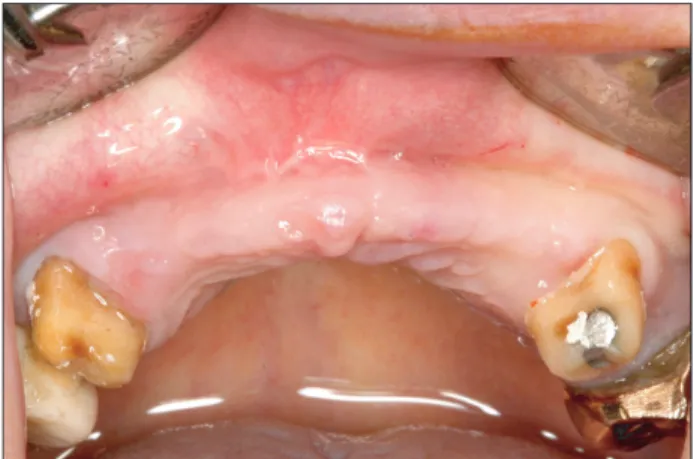
Familial tooth bone graft for ridge and sinus augmentation: a report of two cases
전체 글
수치


관련 문서
Experimental alveolar ridge augmentation by distraction osteogenesis using a simple device that permits secondary implant placement. Anteroinferior distraction of
Clinical and histomorph ometric evaluation of extraction sockets treated with an autologous bone marro w graft... Barone A, Aldini NN, Fini M, Giardino R, Calvo Guirado
The OSFE (osteotome sinus floor elevation) technique has been used for maxillary sinus augmentation.. The implants were clinically and radiographically followed
success rates of dental implants placed at the time of or after alveolar ridge augmentation with an autogenous mandibular bone graft and titanium mesh: a 3-to
LIPUS therapy is a recently developed method for application of mechanical stress, and used clinically to promote bone healing. Author evaluated the effect
In 4-week group, the group filled with bone graft with decortication revealed larger new bone formation area than shown in the group that had a defect area
The rationale for augmentation of the residual alveolar socket at the time of tooth removal such as socket reservation, socket augmentation and ridge
The result of using PLGA nanofiber membrane with bone graft material showed that the PLGA nanofiber membrane in the experimental group of 2 weeks were ten times more new