Paper
Role of Electron Acceptor-donor on Elemental Mercury Removal Using Nano-silver-plated Activated Carbons Complexes
Hyo In Lee *, Yoon-Ji Yim*, Kyong-Min Bae*, Soo-Jin Park* †
ABSTRACT: In this study, the elemental mercury removal behaviors of silver-plated porous carbons materials were investigated. The pore structures and total pore volumes of the hybrid materials were analyzed by N
2adsorption/
desorption analysis at 77 K. The pore structures and surface morphologies of the hybrid materials were characterized by XRD and SEM, respectively. The elemental mercury adsorption capacities of all silver-plated porous carbons hybrid materials were higher than those of the as-received samples, despite the fact that the specific surface areas and total pore volumes decreased with increasing metal loading time. It was found that silver nanoparticles showed excellent elemental mercury removal behaviors in carbonaceous hybrid materials.
Key Words: Elemental mercury removal, Silver-plated carbonaceous materials, Porous carbons, Textural properties
1. INTRODUCTION
Mercury is a major toxic metal and an important source of air pollution—addressed in the 1990 Clean Air Act Amend- ments (CAAA)—due to its volatility, persistence, bioaccumu- lation in the environment, and impact on neurological health.
Although mercury is one of the least abundant trace metals in coal, the volume of coal consumed globally is now over 5 bil- lion ton per year and increasing (China □ 2.7 billion ton, United States □ 1.7 billion ton, and India □ 0.5 billion ton).
Coal-fired utility boilers are recognized as the dominant anthropogenic source of mercury. As a result, attempts to establish a fundamental understanding of the behavior of mer- cury in coal-fired power plants and to explore cost-effective methods for removing mercury from flue gases have received increasing attention over the past decade [1-4]. Mercury is car- cinogenic, mutagenic, and teratogenic, and promotes tyrosin- emia. A high concentration of mercury causes impairment of pulmonary and kidney function, chest pain, and dyspnea. Sev- eral important ecological accidents caused by mercury—nota- bly between 1953 and 1956 in Minamata’s bay in Japan—are known.
The adsorption process of mercury in coal-fired flue gas is
complicated. A number of studies have been carried out to understand the mercury adsorbing mechanism of activated carbons (ACs) [5-7]. For example, sulfur trioxide was recently found to have a strong inhibitory effect on Hg
0adsorption [5].
In the presence of NO
2, Hg
0was catalytically oxidized on the surface to generate nonvolatile nitrate Hg(NO
3)
2, which was attached to basic sites on the carbon. Adsorption of Hg
0by ACs is determined by many factors such as ACs surface char- acteristics, temperature, and composition of the flow gas.
Numerous physical and chemical separation processes such as solvent extraction, ion-exchange, precipitation, membrane separation, reverse osmosis, coagulation, and photoreduction have been applied for the effective reduction of mercury con- centrations from aqueous solutions. Normally, activity depends on the nature of the support, which may modify the properties of the active phase. Both physical and chemical properties of the support—its acidity, reducibility, and the extent of inter- action with the active metal—play important roles in the com- plex chemistry of supported metal catalysts [8-12].
The objective of this study is to prepare silver-plated porous carbons hybrid materials by a known plating technique and to evaluate the efficiency of elemental mercury removal as a function of the plated silver contents. Silver is known to have a
Received 22 February 2018, received in revised form 26 April 2018, accepted 29 April 2018
* *
†Department of Chemistry, Inha University
Department of Chemistry, Inha University, Corresponding author (E-mail: [email protected])
negative charge in aqueous solutions opposite to that of ele- mental mercury, and this property improve the ability to remove elemental mercury by electrical adsorption [22]. The mercury removal ability in enhanced by the combination of physical adsorption of activated carbons utilizing the large specific surface area and electrical adsorption of silver.
2. EXPERIMENTAL
2.1 Sample preparation
Porous carbons (PCs) were supplied by Hanil Green Tech of Korea. The silver-plated PCs procedure used here is identical to that in a previous study [13,14]. Before deposition, PCs were immersed in 10 wt% nitric acid for 30 min at room tem- perature (to remove impurities) and rinsed with distilled water. The purified PCs were then sequentially activated in tin chloride (SnCl
2) and a palladium chloride (PdCl
2) solution.
Sn
2+ions were initially deposited on the surfaces of the PCs and increases the reactivity of the next step. Pd
2+ions were reduced to Pd
0by Sn
2+to form catalytic centers that silver can react and be reduced. Silver-plated PCs were obtained by immersing the palladium-loaded PCs in a silver plating bath for 5, 10, and 25 min. The silver-plated PCs are named Ag- PCs-t and t is plating time (5, 10, and 25 min). Conditions of the silver plating bath was shown in Table 1. The pH of the sil- ver plating solution was adjusted to about 5.0 and the tem- perature was 90
oC and a schematic explanation of the preparation process is shown in Fig. 1.
2.2 Surface characterization
The silver content was characterized by atomic absorption spectrophotometry (AAS). To study the surface structures of the Ag-PCs, X-ray diffraction (XRD) patterns were charac- terized with a Rigaku model MAX 2200v diffraction meter
(Cu-Kα).
XRD patterns were obtained in the 2θ range between 20°
and 80° at a scanning rate of 0.2° s
-1. Scanning electron micros- copy (SEM, S-4200, Hitachi, Japan) was used to observe the surface morphologies of the Ag-PCs. An energy-dispersive X- ray spectroscope (EDS, AN-10000/85S, LINK system, Japan) was used to evaluate the silver contents in the Ag-PCs.
2.3 Textural properties
N
2adsorption isotherms were obtained using a Belsorp Max (BEL, Japan) at 77 K. The specimens were degassed at 573 K for 10 h to a residual pressure of lower than 10
-6Torr. The amount of N
2adsorbed on a specimen was used to calculate the specific surface area through the Brunauer-Emmett-Teller (BET) equation [15]. The micropore volume was calculated using the Dubinin-Radushkevitch (D-R) equation [16]:
log(W) = log(W
0) + M(log
2(P
0/P)) (1) where W is the amount of adsorbate adsorbed at equilibrium, W
0the micropore volume calculated from the intercept of the log(W) − log
2(P
0/P) plot, and M is the slope of the best-fit line.
2.4 Elemental mercury removal efficiency Textural prop- erties
Experiments were performed using a lab-scale apparatus.
Using this apparatus, a gas stream with the required tem- perature and elemental mercury concentration was produced, and the capture of elemental mercury on a fixed bed of adsor- bent material was performed. The 12.7 mm diameter quartz reactor was placed inside a temperature-controllable tubular furnace. For each experiment, 1.0 g of sample was loaded and packed inside a quartz tube. A carrier gas was fed into the adsorption apparatus at a flow rate of 100 mL min
-1. Elemental mercury gas was generated using elemental mercury perme- ation tubes (Dynacalibrator
®Model 150, VICI Metronics Inc., USA) and the concentration of elemental mercury gas was maintained at 800 μg m
-3during the experimental process.
The inlet and outlet concentrations of elemental mercury were measured using a mercury analyzer (VM-3000, Mercury Instruments, Germany). Sulfur was used to capture elemental mercury from the effluent gas.
3. RESULTS AND DISCUSSION
3.1 Surface morphology analysis
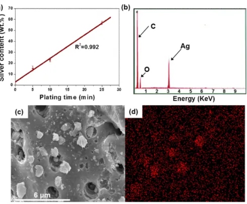
Fig. 2(a) shows the silver content of the Ag-PCs studied as a function of the treatment time. The correlation coefficient for metal contents with deposition time was 0.992 (R
2), indicating that the silver contents of the Ag-PCs was proportional to the plating time. Fig. 2(b) shows the EDS result of the Ag-PCs-10 sample. Carbon, oxygen, and silver were detected in the Ag- PCs-10 sample. Fig. 2(c) and (d) shows SEM images and image mapping results of the Ag-PCs-10 sample to examine Fig. 1. Schematic illustration of Ag-PCs preparation process by
electroless plating method and removal process of ele- mental mercury by silver-plated PCs
Table 1. Composition of the silver plating bath Compositions
AgCN 5 g/L
Na
2CO
33 g/L
NaCN 12 g/L
the surface morphologies of the Ag-PCs before and after silver plating. The silver particles completely covered the surfaces of Ag-PCs and the silver particles were dispersed well, indicating that micro- and mesopores of the PCs can be blocked due to the metal loading.
An understanding of the surface structure can be achieved by wide-angle XRD. Fig. 3 shows wide-angle XRD diffraction results of the Ag-PCs; the peaks around 2θ = 38°, 44°, 64°, and 77° correspond to the (111), (200), (220), and (311) planes of silver, respectively [17,18]. Intensities of the silver peaks and
crystallinities of the Ag-PCs strengthened with increasing plat- ing time.
The N
2adsorption isotherms of the Ag-PCs are shown in Fig. 4. All of the samples exhibited Type-I isotherms according to the IUPAC classification, having well-developed micropores [19]. Most of the pore volume of the samples was filled below a relative pressure of ~0.1, indicating high microporosity. After the adsorbed volume sharply increased to 0.1 relative pressure, the isotherms showed very small increases of the pore volume Fig. 2. (a) Silver contents of Ag-PCs as a function of the plating time (R
2is the correlation coefficient between metal content and plating
time). (b) EDS image of the Ag-PCs-10 sample. (c) SEM image of the Ag-PCs-10 sample. (d) Image mapping result of the Ag-PCs- 10 sample
Fig. 3. XRD patterns of Ag-PCs as a function of the plating time
Fig. 4. Adsorption isotherms of N
2/77 K on Ag-PCs as a function
of the plating time
to ~1.0 relative pressure with no further adsorption. This result indicates that silver particles had some influence on the blocking or filling of pores on the PC surfaces.
Table 2 displays the textural properties of the samples as cal- culated from the N
2/77 K adsorption isotherms. The PCs had specific surface area of 1158 m
2g
-1, total pore volume of 0.553 cm
2g
-1, and micropore volume of 0.476 cm
2g
-1. Ag-PCs exhibited decreasing specific surface area and decreasing total and micropore volumes as the plating time increased. This result indicates that the adsorption capacities of the PCs can be diminished by the silver nanoparticles produced, due to the blocking or filling of the micropores in the PCs.
Fig. 5 displays the BJH and HK pore size distributions of
Ag-PCs. Firstly, mesopore peaks of Ag-PCs were observed at around 2.5, 28, and 66 nm. Secondly, the pore size distribution of Ag- PCs decreased relative to the PCs. Thirdly, the pore vol- umes of Ag-PCs at □ 76 nm decreased dramatically. As a result, it was found that the micro- and mesopores of PCs can be blocked by plated silver metal.
3.2 Removal efficiency
Fig. 6 shows the removal of elemental mercury by PCs and Ag-PCs as a function of the plating time. All tests were con- ducted at 423 K for 80 min in the elemental mercury adsorp- tion apparatus (the internal temperature of the apparatus was maintained in the range 418-428 K). As mentioned in the experimental section, the initial concentration of elemental was approximately 800 μg m
-3(balanced by N
2gas). Normally, exhaust mercury concentration is strictly regulated to lower than 100 μg m
-3. Therefore, in the present study, outlet con- centration >90% (less 10% removal by the filter media) is spec- ified as the breakthrough of the Ag-PCs. The outlet elemental mercury concentration is reported in dimensionless form (Hg
out/Hg
in) as the ratio between the outlet and inlet elemental mercury concentrations (breakthrough curves).
The PCs showed breakthrough after just 7 min of reaction time, indicating that the PCs could not remove elemental mer- cury. In contrast, all Ag-PCs showed better removal efficien- cies than those of the PCs, with Ag-PCs-10 showing the maximum. The Ag-PCs-10 sample showed breakthrough at 62 min. The removal capacity listed in Table 3 was calculated using the breakthrough time and the equation is represented below:
(2)
where E
removalis the elemental mercury removal efficiency of prepared samples, C
Hgis the concentration of elemental mer- cury gas, Q is the volumetric flow rate, t
totalis the total flow
E
removalC
HgQt
total--- w Hg
outHg
in---
⋅
= Table 2. Textural properties of the Ag-PCs as a function of the
plating time Samples
a
S
BET(m
2/g)
b
V
Total(cm
3/g)
c
V
Micro(cm
3/g)
d
D
p(nm)
PCs 1158 0.553 0.476 1.91
Ag-PCs-5 864 0.389 0.343 1.85
Ag-PCs-10 823 0.379 0.337 1.84
Ag-PCs-25 810 0.371 0.333 1.83
a
Specific surface area
b
Total pore volume
c
Micropore volume
d