Copyright © 2016. Anatomy & Cell Biology
Infants and small children are more exposed to acrylamide toxicity. The toxicity is due to low body weight and high consumption of snacks (their acrylamide intake is estimated to be 2- to 3-fold higher than adults) [5]. Dietary acrylamide intake caused reproductive toxicity [6], genotoxicity, and neurotoxicity [7]. It may increase the risks of kidney and breast cancer [8]. Neurotoxic effects of acrylamide have been established in humans and animals [9]. Many researchers proved the effect of high-dose acrylamide exposure on the development of the nervous system; however, few study proved the effect of it on the development of the spinal cord [10]. There are several reports that antioxidant agents could rescue neurotoxicity induced by acrylamide via increasing antioxidant activity [11]. Rosemary (Rosmarinus officinalis) is a herb, composed of dried leaves and flowers, commonly used as a spice and flavouring agents in food processing for its desirable flavor [12]. It has anti-inflammatory [13] and antispasmodic [14] effects. In addition, it has antimicrobial [12] and antitumor [15] activities. Carnosic acid, a rosemary
Introduction
From 2002 onward, Food and Drug Administration (FDA) warned crunchy, fried strips of potato consumers from excess use as it contained carcinogen material [1]. Frying, roasting, or baking of foods at temperatures above 120oC resulted in a formation of a high amount of acrylamide’s toxic compound [2]. Acrylamide has multiple chemicals and industrial applications [3]. These included gel electrophoresis, papermaking, and the manufacture of permanent press fabrics, manufacture of dyes and other monomers [4].
Corresponding author:
Hanaa Zakaria Nooh
Department of Anatomy and Embryology, Faculty of Medicine, Menoufia University, Gamal Abd El Naser St., Shebin El Kom, Egypt Tel: +20-1096288260, Fax: +20-48-2317502-2317508,
E-mail: drhanaanooh@gmail.com
Protective effect of rosemary on acrylamide motor neurotoxicity in spinal cord of rat
offspring: postnatal follow-up study
Marwa A. Al-Gholam, Hanaa Zakaria Nooh, Abeer E. El-Mehi, Abd El-Moneum El-Barbary, Ahmed Zo El Fokar
Department of Anatomy and Embryology, Faculty of Medicine, Menoufia University, Shebeen El-Kom, Egypt
Abstract: The direct interactive effects of rosemary and acrylamide on the development of motor neurons in the spinal cord remains unknown. Our goal is to confirm the protective effects of rosemary against motor neuronal degeneration induced by acrylamide in the developing postnatal rat spinal cord using a postnatal rat model. We assigned the offspring of treated female rats into control, rosemary; acrylamide group; and recovery groups. This work depended on clinical, histopathological, morphometrically, immunohistochemical and genetic methods. In the acrylamide group, we observed oxidation, motor neuron degeneration, apoptosis, myelin degeneration, neurofilament reduction, reactive gliosis. Whoever, concomitant rosemary intake and withdrawal of acrylamide modulate these effects. These findings proof that dietary rosemary can directly protect motor neuron against acrylamide toxicity in the mammalian developing spinal cord.
Key words: Acrylamide, Motor neuron, Rosemary, Development, Spinal cord
Received January 21, 2016; 1st Revised February 20, 2016; 2nd Revised February 26, 2016; Accepted February 29, 2016
phenolic component has been protecting cortical neurons from glutamate and the brain from middle cerebral artery occlusion/reperfusion injury [16]. Rosemary extracts exhi- bited very high antioxidant activity, almost equal to that of synthetic antioxidants [17]. The main objective of this work was to address the oxidative stress of acrylamide on the development of the spinal cord motor neurons in new-born rats and protective role rosemary as antioxidant.
Materials and Methods
ChemicalsAcrylamide was obtained from a product of leucon Sto SPP (Sigma-Aldrich, St. Louis, MO, USA). It was available in the form of white powder (99% purity) and dissolved in 1 ml distilled water [18]. It was given to rat as 10 mg/kg/day (i.e., 2 mg; 0.2 ml/rat) by oral gavage [19].
Rosemary was obtained from a local market as green leaves. The air dried leaves were powdered. Ten grams of dried plants was dissolved in 100 ml of distilled water after boiling for 5 minutes. After cooling and passing through filter paper, a clear solution was obtained [20]. During 24 hours of preparation, the extract was given to rats as (220 mg/kg, i.e., 44 mg; 0.44 ml/rat) by oral gavage twice weekly [21].
Animals
Twenty-five sexually mature female albino rats and five male albino rats (for mating) of Sprague-Dawley strain, weighing between 200–250 g (8–9 months of age) were obtained from El Helw animal house, Tanta, Egypt. The rats will be kept in cages with particular care and hygiene, artificial light/dark cycle 12 hours, at room temperature (25±2oC) and standard laboratory chow and water ad labium.
The procedure approved by the ethics committee on the animal experiment of the Faculty of Medicine, Menoufia University according to the international regulations on care and use of laboratory animals. Each five of them were housed overnight with a sexually mature male albino rat for mating, and every morning vaginal smears were taken and microscopically examined for the presence of sperms. The first day of gestation was corresponding to the detection of sperms in the smears.
144 new born babies were labelled into four groups as follows.
Group I (control group): pregnant rats were given saline (normal group). The new-born were sacrificed at the age of
1-, 7-, 14-, 21-, and 42-day postnatal (6 rats each) and this corresponding to subgroups Ia, Ib, Ic, Id, and Ie, respectively.
Group II (rosemary group): pregnant rats were adminis- tered a rosemary from day 7 of gestation until day 28 after birth. The new-born were sacrificed at the age of 1-, 7-, 14-, 21-day postnatal (6 rats each) and this corresponding to subgroups IIa, IIb, IIc, and IId, respectively.
Group III (acrylamide group): pregnant rats were adminis- tered acrylamide from day 7 of gestation until day 28 after birth. The new-born were sacrificed at the age of 1-, 7-, 14-, and 21-day postnatal (6 rats each) and this corresponding to subgroups IIIa, IIIb, IIIc, and IIId, respectively.
Group IV (protected group): pregnant rats were adminis- tered acrylamide and rosemary from day 7 of gestation until day 21 after birth. The newborn were sacrificed at the age of 1-, 7-, 14-, and 21-day postnatal (6 rats each) and this corresponding to subgroups IVa, IVb, IVc, and IVd, respectively.
Group V (recovery group): pregnant rats were adminis- tered acrylamide and rosemary from day 7 of gestation until day 21 after birth. After weaning 6 rats new-borns were separated from their mothers and allowed for free water untill the age of 42 days.
Postnatal investigations
The newborns were investigated by the experimenter, and the following notes were recorded in each group (6 new- borns).
Daily:
- The appearing time of fur.
- The opening time of ear.
- The opening time of eye.
Weekly:
- Weights in gram.
- Head length, head-rump length and tail length in centi- metre [22].
- Gait score examination: rats were placed in a clear glass box and observed for 3 minutes. Following observation, a gait score was assigned from 1 to 4 (Table 1) [23].
Light microscopic study
At the end of each detected period. The rats were anesthe- tized by diethyl ether inhalation. Lumbar segment of the spinal cord was fixed in 10% buffered formalin (pH 7.4) for 48 hours.
The tissue was dehydrated in ascending ethyl alcohol followed by two changes of xylene. The tissue was impregnated in
paraffin wax and then embedded in paraffin wax. Sections (5 µm) were cut, dewaxed, hydrated and stained with hematoxylin and eosin, silver and toluidine blue stain [24].
Immunohistochemical study
For the immunohistochemical study, the spinal cord para ffin sections were deparaffinized and rehydrated in descending grades of alcohol. Following blocking of endo- genous peroxidase activity with 3% H2O2 in methanol and nonspecific binding sites with a protein blocker, the primary antibody (1:500, neurofilament [NF]; 1:300, myelin basic protein; Abcam, Cambridge, MA, USA) added with overnight incubation in a cold room. On day 2, the biotinylated secon- dary antibody (Vector, Peterborough, UK) was added at a concentration of 2% for 30 minutes (37oC) followed by addition of the avidin-biotin-peroxidase complex (Vector).
All steps were performed at room temperature in a humidity chamber [24].
Biochemical assay
For determination of antioxidant enzymes, segments from lumbar part of the spinal cord were removed and homo- genized in potassium phosphate buffer solutions (50 mM, pH 7.5) using a Potter-Elvehiem homogenizer to give a 10%
homogenate. Homogenates were centrifuged at 1,500 ×g for 10 minutes at 4oC; the supernatant was recovered, placed on ice, and immediately used for the determination of peroxidase and superoxide dismutase (SOD). The activity of SOD was determined calorimetrically according to the method of Marklund and Marklund [25]. While peroxidase activity was determined according to the method of Kar and Mishra [26].
Molecular study: Comet assay for motor neuron cell [27]
Preparation of motor neuron cell suspensions:
Spinal cords were isolated from rats that were anesthetized deeply with a mixture of enflurane:oxygen:nitrous oxide (1:33:
66) and then decapitated. After removal of the pia matter, lumbar/cervical enlargements were dissected segmentally under a surgical microscope and then the segments were microdissected into gray matter columns of the ventral horn without appreciable contamination of dorsal horn and surrounding white matter funiculi. Gray matter tissue columns from spinal cord were collected and rinsed in a cell culture dish on ice containing dissection medium (1Ca2+ and Mg2+-free Hanks balanced salt solution [Gibco BRL, Grand Island, NY, USA] supplemented with glucose and sucrose).
These tissues were used to prepare motor neuron cell suspen- sions.
Encapsulation:
A volume of 50 μl cell suspension (containing ~4.4×10⁴ motor neurons) was added to 200 µl 0.7% low melting point agarose at 4oC, and then layered onto a pre-coated micro- scopic slide with 100 µl of low-melting-point agarose and covered with a coverslip. The agarose was gelled at 4oC, and then the coverslip was removed. The slides were immersed in lysing solution (2.5 M NaCl, 100 mM EDTA, 10 mM Tris- HCl buffer, pH 10, 1% sodium sarcosinate with 1% Triton X-100 and 10% DMSO; Sigma-Aldrich) for ~1 hour.
Electrophoresis:
The slides were washed with distilled water to remove all salts and then placed in a horizontal gel electrophoresis unit (CBS Scientific, San Diego, CA, USA) filled with fresh electro- phoretic buffer (1 mM disodium EDTA and 200 mM NaOH, pH 13).
Electrophoresis was conducted for 20 minutes at 25 V and 300 mA. Slides were then stained with ethidium bromide (Sigma-Aldrich). Slides were examined with a Carl Zeiss fluorescent micro scope (Jena, Germany) equipped with a 510 nm excita tion filter and a barrier filter of 590 nm. In damaged cells, breaks appear as fluorescent tails extending from the core towards the anode. The tail length reflects the amount of DNA breakage in the cell [28].
Quantitative study
By using Image analyzer software (Image J 1.47v, National Institute of Health, Bethesda, MD, USA) (Department of Anatomy and Embryology, Faculty of Medicine, Menoufia University) the following parameters were calculated.
(1) Number of motor neurons and the number of neuroglia cells (the neurons were differentiated from glial cells by glial
Table 1. Criteria for gait scoring
Score Behavioral index
1 A normal, unaffected gait
2 A slightly affected gait (foot splay, slight hind limb weakness, and spread)
3 A moderately affected gait (foot splay, moderate hind limb weakness, and moderate limb spread during ambulation)
4 A severely affected gait (footsplay, severe hind limb weakness, dragging hind limbs, and inability to rear)
nuclei diameter <5 µm).
(2) Color intensity of Nissl granules and the surface area of the brown color of NF and myelin basic protein (MBP) immunohistochemistry.
(3) The migrated nuclear DNA was considered as a dam- aged DNA spot. The migration was evaluated by measuring the basal nuclear DNA and migrating DNA. In 50 randomly selected cells per sample, the used comet parameters for the evaluation are tail length, tail DNA% and tail moment.
The tail length was measured from the center of the nucleus towards the end of the tail, the percentage of DNA in the tail DNA% and tail moment (Tail moment=Tail length×Tail DNA%) [29].
Statistical analysis
SPSS version 20 (IBM Co., Armonk, NY, USA) was used for the statistical analyzes. The data were analyzed using Mann-Whitney test and Kruskal-Wallis test followed by Post hoc test to compare various groups with each other. Results were expressed as mean±SD. The level of significance was expressed as P>0.05 for insignificantly [30].
Results
General developmental observations
Signs of acrylamide toxicity were observed postnatally in the treated mothers. It represented by ataxia, splayed hind limbs, weakness of the hind limb muscles, and paralysis. The limb weakness decreased both food and water consumption.
There were no significant changes in all tested parameters of the offspring of control/rosemary groups. Neither congenital anomalies nor deaths were reported in-between offspring.
Body weight:
There was a significant increase in body weight with a progression of age (P<0.05). In an acrylamide-treated group, the increase in body weight was significantly lower than a control group (P<0.05). While supplementation of aqueous rosemary extract with acrylamide led to an extremely signifi- cant increase in body weight with age (P<0.001) when compared with acrylamide-treated rats. Also, acrylamide withdrawal resulted in significant increase in body weight (P<0.05). However, it was still significantly lower than the control group (Fig. 1).
Developmental landmarks:
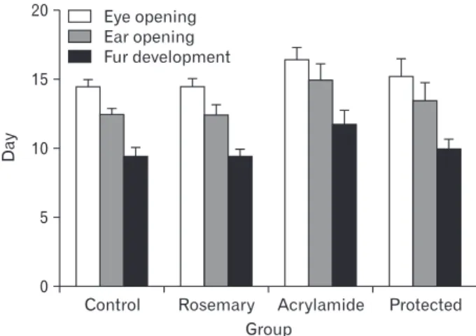
At birth, the newborns of all groups were hairless. The time of fur appearing and both ear and eye opening were signi ficance retarded in the acrylamide-treated group (P<0.05) when compared with that of the control group. In the protected group, the developmental parameters showed signi- ficantly earlier development (P<0.05) when compared with an acrylamide-treated group (Fig. 2).
Gait scores:
At birth, all rats were unable to walk except at the age of the 12–13 day postnatal. Rats of the acrylamide-treated group showed a significant increase in gait score from day 14 to day 21 (P<0.05) when compared with control group. However, this change significantly decreased (P<0.05) when rats treated by rosemary with acrylamide, it was significantly low at day 14. Also, withdrawal of acrylamide for three weeks led to a decline in gait score but still significantly higher when com- pared with control group (Fig. 3).
7 28 42
100
80
60
40
20
0 1
Weight(g)
Days
14 21 35
Control Rosemary Acrylamide Protected Recovery
Fig. 1. Assessment of weight gain with age in different groups.
Rosemary 20
15
10
5
0
Control
Day
Group
Acrylamide Protected Eye opening
Ear opening Fur development
Fig. 2. Developmental landmarks in different groups.
Skeletal landmarks:
Both control and rosemary groups showed a steady increase in head length, cervical rump length and tail length with age. In acrylamide group, the head length of acrylamide rats significantly decrease (P<0.05) than the control group at the age of 14 days. The cervical rump was significantly decrease (P<0.05) from control group from day one to day 14. It became pronounced at day 21 (P<0.001) and tail length significantly decrease (P<0.05) at day 21. Rosemary supplementation was significantly increase (P<0.05) in the tail length cervical rump in acrylamide rats at day 1 and that increase (P<0.001) from day 7 inward. Also, withdrawal of acrylamide for three weeks led to increasing in three parameters but still significantly lower when compared with control group (Table 2).
Tissue biochemical results
Both control and rosemary treated animals showed no sig- nificant differences (P>0.05). With the advancement of age, both groups showed a significant decrease in the spinal levels
of an antioxidant marker (SOD) and increased in the content of peroxidase as compared with controls (P<0.05).
With the advancement of age and compared with controls group, the acrylamide-treated rats showed a significant de- crease in the spinal levels of an antioxidant marker (SOD) and an important increase in the content of peroxidase (P<
0.001). Rosemary administration to acrylamide-treated (pro- tected group) rats resulted in a significant rise in SOD and a
7 28 42
4
3
2
1
0 1
Gaitscore
Days
14 21 35
Control Rosemary Acrylamide Protected Recovery
Fig. 3. Assessment of gait score changes with age in different groups.
Table 2. Mean skeletal landmarks in newborn rats in the different studied groups Group Subgroup Head length
(cm)
Cervical rump length (cm)
Tail length (cm) Control Ia 1.74±0.114 2.48±0.0837 1.56±0.0894
Ib 2.38±0.084 3.56±0.1817 3.14±0.3507a) Ic 2.58±0.083 4.84±0.2408a) 5.24±0.4450a) Id 2.74±0.055 5.56±0.1517a) 6.42±0.2588a) Ie (day 28) 2.81±0.062 6.84±0.1346a) 9.14±0.3250a) Ie (day 35) 2.85±0.131 7.83±0.2132a) 10.32±0.2360a) Ie (day 42) 2.90±0.152 8.68±0.1517a) 12.42±0.2588a) Rosemary IIa 1.68±0.109 2.42±0.0837 1.57±0.1517
IIb 2.42±0.130 3.52±0.2280 3.15±0.5119 IIc 2.60±0.071 4.90±0.2345 5.23±0.3937 IId 2.70±0.071 5.34±0.1817 6.40±0.3082 Acrylamide IIIa 1.54±0.114 2.28±0.0837b) 1.34±0.2302 IIIb 2.28±0.084 2.66±0.1673a),b) 2.90±0.3391a) IIIc 2.46±0.055 3.20±0.1871a),b) 4.68±0.2775a) IIId 2.50±0.071a),b) 4.10±0.1304a),d) 5.72±0.3493a),b) Protected IVa 1.70±0.123 2.46±0.1517c) 1.46±0.0548
IVb 2.340±0.055 3.58±0.2280a),e) 2.99±0.3421a) IVc 2.50±0.114 4.26±0.1517a),e) 5.08±0.3647a) IVd 2.67±0.084c) 5.18±0.1643a),e) 6.01±0.2881a),c) Recovery V (day 28) 2.59±0.058f) 5.10±0.1245f) 6.52±0.3410f)
V (day 35) 2.62±0.076b) 6.01±0.1421b) 7.910±0.4653a),b) V (day 42) 2.67±0.084b) 6.80±0.3912b) 9.31±0.6419a),b) Values are presented as mean±SD. a)A significant difference between successive days (P<0.05). b)A significant difference from control (P<0.05). c)A significant difference from acrylamide (P<0.05). d)A significant difference from control (P<0.001). e)A significant difference from acrylamide (P<0.001). f)A significant difference from control (P<0.001).
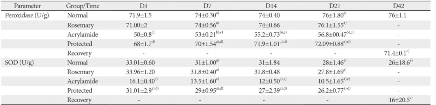
Table 3. Mean peroxidase, SOD in spinal cord tissue of rats’ newborn in the different studied groups
Parameter Group/Time D1 D7 D14 D21 D42
Peroxidase (U/g) Normal 71.9±1.5 74±0.30a) 74±0.40 76±1.80a) 76±1.1
Rosemary 71.00±2 74±0.56a) 74±0.66 76.1±1.55a) -
Acrylamide 50±0.8c) 53±0.21b),c) 55.2±0.73b),c) 56.8±00.47b),c) -
Protected 68±1.7d) 70±1.54a),d) 71.9±1.01a),d) 72.09±0.88a),d) -
Recovery - - - - 71.4±0.1c)
SOD (U/g) Normal 33.01±0.60 31±1.00a) 31±1.84 28±1.46a) 26±18.6a)
Rosemary 33.96±1.20 31.8±0.40a) 31.8±0.48 27.8±1.69a) -
Acrylamide 16.1±0.40c) 13.5±1.60c) 12±0.50a),c) 10.5±1.65a),c) -
Protected 31.01±2.9a),d) 29±0.95a),d) 27±2.39a),d) 26.2±0.77a),d) -
Recovery - - - - 16±20.5c)
Values are presented as mean±SD. SOD, superoxide dismutase. a)A significant difference between successive days. b)A significant difference between successive days (P<0.001). c)Significant difference from control. d)Significant difference from acrylamide (P<0.001).
significant decrease in the peroxidase level when compared with acrylamide-treated rats (P<0.001). Withdrawal of acry- lamide (recovery group) ameliorated both parameters but still a significant difference from control (P<0.001) (Table 3).
Histopathological results
Control and rosemary treated groups:
At birth, histological sections of the control, as well as the rosemary groups, revealed that anterior horn cell of the spinal cord showed numerous well-differentiated motor neurons in-between scattered small capillaries and different forms of neuroglia. With age advanced, the neurons became more basophilic. It showed a significantly increase in number and size (P<0.05) while neuroglia cells showed significantly decrease in number (P<0.05) (Table 4, Fig. 4A–E).
The motor neurons of anterior horn cells had a central vesicular nucleus with an eccentric nucleolus, partially myeli- nated axons with no-dendrites. The oligodendroglia, astro- cyte, and microglia represented the presented neuroglia.
With advanced age, the neurons acquired small dense gra- nules, long branched dendrites, and long myelinated and nodded axons. Many microglia were reported in early age
while oligodendroglia became more numerous with the advancement of age (Fig. 5A–E). With advanced age, the neurons acquired small dense granules, long branched dendrites and long myelinated and nodded axons. Many microglia were reported in early age while oligodendroglia became more numerous with the advancement of age (Fig.
5A–E).
Acrylamide group:
In comparing with control, the anterior horn cell sections at day 1 revealed non-significant decrease (P>0.05) in differentiated motor neurons number and size and significant increase (P<0.05) in neuroglia cells. The neuropil showed small vacuolation, hemorrhage, dilated congested capillaries.
With the increase in age, the neurons showed a steady decrease in number and size marked at day 21 (P<0.001 and P<0.05, respectively). The neuropil showed a significant increase in neuroglia that became marked (neurogliosis) at day 21 (P<0.001). A concomitant increase in vacuolation, congestion and neuronophagia were noticed (Table 4, Fig.
4F–I).
Acrylamide rats motor neurons at day 1 showed either neurofibrillary tangle or early central chromatolysis (eccentric nucleus and homogenous cytoplasm) and segmental demyeli- nated, slight swollen axons. With an increase in age, the motor neuron showed extensive pathological future as: end stage chromatolysis, degenerated pyknotic neurons, attenuated den drites, and giant swollen destructed axons with an irre- gular myelin pattern. A remarkable number of astrocytes, many microglia cells also noticed (Fig. 5F–I).
Protected group:
In compare with acrylamide group, rats of protected group showed, with age increase, significant increase and a decrease (P<0.05) in the number of motor neurons and neuroglia respectively starting from day 1. From day 14, the neurons size began to show significant increase (P>0.05). The archi- tecture of ventral horn showed noticed improvement with few areas of vacuolation, vascular congestion, and few dege- nerated cells (Table 4, Fig. 4J–M). Some neurons showed with short dendrites, and slight swollen destructed and demyeli- nated axons (Fig. 5J–M).
Recovery group:
In compare with acrylamide group, the recovery rats showed significantly increased number and size of motor
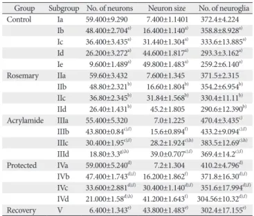
Table 4. Mean neuron and neuroglia parameters in the different studied groups Group Subgroup No. of neurons Neuron size No. of neuroglia Control Ia 59.400±9.290 7.400±1.1401 372.4±4.224
Ib 48.400±2.704a) 16.400±1.140a) 358.8±8.928a) Ic 36.400±3.435a) 31.440±1.304a) 333.6±13.885a) Id 26.200±3.272a) 44.600±1.817a) 293.3±3.162a) Ie 9.600±1.489a) 49.800±1.483a) 259.2±6.140a) Rosemary IIa 59.60±3.432 7.600±1.345 371.5±2.315
IIb 48.80±2.321b) 16.60±1.804b) 354.2±6.954b) IIc 36.80±2.345b) 31.84±1.568b) 330.4±11.11b) IId 26.40±1.431b) 45.2±1.805 290.6±12.390b) Acrylamide IIIa 55.400±5.320 7.0±1.225 470.4±3.435c)
IIIb 43.800±0.84c),f) 15.6±0.894f) 433.2±9.094c),f) IIIc 30.400±1.95c),f) 28.2±1.924c),h) 383.5±12.69c),h) IIId 18.80±3.3g),h) 39.0±0.707c),f) 369.4±14.2c),f) Protected IVa 59.000±5.240d) 7.2±1.304 410.2±4.796d) IVb 47.400±1.743d),f) 16.200±1.862f) 371.8±16.30d),f) IVc 33.600±2.881d),f) 30.400±1.140d),f) 351.6±17.994d),f) IVd 21.000±1.58d),h) 41.200±1.643f) 304.56±10.32d),f) Recovery V 6.400±1.343e) 43.800±1.483e) 302.4±17.155e)
a)A significant difference between control groups. b)A significant difference between different ages rosemary group. c)A significant difference of acrylamide from control group (P<0.05). d)A significant difference of protected from acrylamide group (P<0.05). e)A significant difference of recovery from control group (P<0.05). f)A significant difference between two successive ages. g)A significant difference of acrylamide from control group (P<0.001). h)A significant difference between two successive ages (P<0.001).
neurons (P<0.05) and significant decrease in neuro glia number (P<0.05). Although the general architecture showed improve ment great areas of vacuolation, vascular congestion, and few degenerated cells were still observed (Table 4, Fig.
4N) and neurons with short dendrites and visible swollen destructed and demyelinated axons (Fig. 5N).
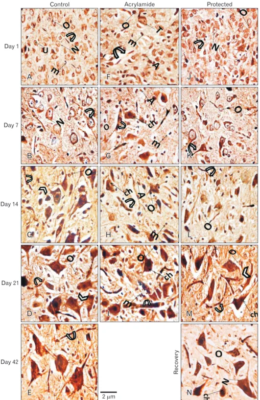
Histochemical and immunohistochemical results Toluidine blue staining:
At birth, anterior horn control section stained with tolui- dine blue showed motor neurons with round body, defined
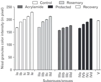
nucleus and clear cytoplasm containing Nissl granule. The latter appears as fine scanty faint basophilic granules inside the cytoplasm and proximal part of dendrites. Both control and rosemary rats showed no significant difference (P>0.05) in Nissl granules content and intensity which increased sig- nificantly (P<0.05) with age advancement. Compared with the control group at the same age, chronic acrylamide admin- istration showed significantly reduction of Nissl granules content and intensity at the age of 21 days (P<0.001). On day 7 and age forward, protected group showed steady significant increase in the Nissl granule content and intensity (P<0.05).
Withdrawal of acrylamide for 21 days after acrylamide stop-
Control Acrylamide Protected
Day 1
Day 7
Day 14
Day 21
Day 42
Recovery
A
B
C
D
E
F
G
H
I
J
K
L
M
20 m N
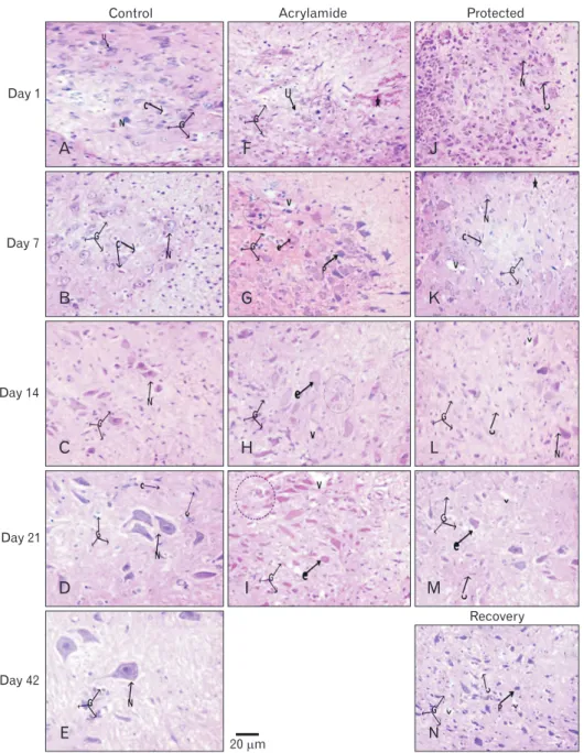
Fig. 4. Hematoxylin and eosin-stained (×400) lumbar anterior horn transverse sections in rats with advancement of age. (A–E) Control rat shows apparent increase in number and size of basophilic dif ferentiated motor neurons (N) and decrease in undifferentiated one. In addition to, decrease in different form of neuroglia (G) and scattered small capillaries (C). (F–I) Acrylamide rat shows decrease in differentiated and increase in degenerated neurons (esi- nophilic [e], pyknotic [p]). In ad dition to, neuropil hemorrhage (asterisk) vacuolation neuropil (v), dilated con- gested capillaries, and neurogliosis (G) and neuronophagia (dashed circle).
Protected (J–M) and recovery (N) rats, with age advancement, shows show similar picture to that of the control with few vacuolation, degenerated neu- rons and dilated congested capillaries.
page showed an increase in Nissl granules content and inten- sity but still significantly lower than the control group (P<0.05) (Figs. 6, 7).
NF immunostaining:
At birth, the pattern of NF expression immunohistochemi- stry stain expression (specific for axonal neurofilament), in the anterior horn control sections, appeared as fine scanty parallel lines. Both control and rosemary rats showed no
Control Acrylamide Protected
Day 1
Day 7
Day 14
Day 21
Day 42
Recovery
2 m
A A
B B
C C
D D
E E
F F
G G
H H
I I
J J
K K
L L
M M
N N
Fig. 5. Sliver staine d (×1,000) lumbar anterior horn transverse sections in in rats with advancement of age. (A–E) Control rat shows the motor neu rons with central vesicular nucleus, small eccentric nucleoli ac- quires small dense granules (N), long branched dendrites (arrowheads) and the myelinated nodded axons (cur- ved arrow). Oligodendroglia (O), astrocyte (A), and microglia (m) are also increased. Immature neuron with indistinct nucleus (U). (F–
I) Acry lamide rat shows increase in neuron cen tral chromatolysis (ch), swollen demyelination axons axon, and short dendrites. The degenerated oligodendroglia (o), astrocytes (A), and microglia (m) are also increased.
Protected (I–M) and recovery (N) rats shows similar picture to that of the control group with few reported central chromatolysis (ch) es pecially in recovery rat.
significant difference (P>0.05) in NF protein expression (area
%) which increased significantly (P<0.05) age advancement.
Compared with control group, chronic acrylamide adminis- tration showed steady significant decreased in NF expression with age and became marked (P<0.001) at the age of 21 days.
Compared with acrylamide group, at the same age, concomi- tant administration of rosemary with acrylamide significantly increase (P<0.001) in NF protein at the age of 7 days. While the withdrawal of acrylamide increased NF protein but still significantly lower (P<0.05) than a control group (Figs. 8, 9).
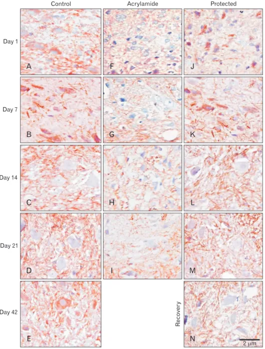
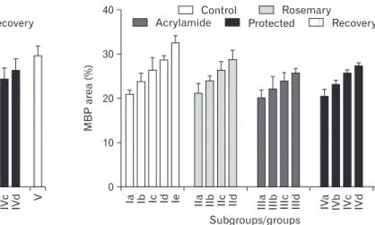
MBP immunostaining:
At birth, the pattern of MBP immunohistochemistry stain expression (specific for myelin sheath warping axon and oligodendroglia) in anterior horn control section appeared scanty small fine parallel brown lines or rings. Both control and rosemary rats showed no significant difference (P>0.05) in MBP content (area %) which increased significantly (P<0.05) with age advancement. Compared with the control group at the same age, chronic acrylamide administration showed significant reduction (P<0.001) of MBP content (area
%) at the age of 14 days.
Compared with acrylamide group, at the same age, conco-
Control Acrylamide Protected
Day 1
Day 7
Day 14
Day 21
Day 42
Recovery
2 m
A
B
C
D
E
F
G
H
I
J
K
L
M
N
Fig. 6. Toluidine blue (×1,000) stained lumbar anterior horn transverse sec- tions in in rats with advancement of age. (A–E) Control rat shows dra- matically increase in Nissl granules. (F–
I) Acrylamide rat shows reduction in Nissl granules. Protected ( J–M) and recovery (N) rats show upregulation in Nissl granules.
mitant administration of rosemary with acrylamide signifi- cantly increase MBP content in the ventral horn (P<0.05).
While acrylamide withdrawal showed an increase in MBP content but still significantly lower than the control group (P<0.05) (Figs. 10, 11).
Single comet assays result
At birth, the pattern single motor neuron comet assay was detected morphologically by a faint small tail, morpho- metrically by few % DNA in the tail and mathe matically by small tail moment value. Both control and rosemary rats showed no significant difference (P>0.05) in previous men- tioned three parameters which increased significantly (P<
0.05) with age advancement. Compared with control groups, acrylamide administration showed a steady, significant increase in DNA damage (P<0.001) with age. The significant in crease in tail length, % DNA in tail and tail moment re- presented DNA damage. Compared with acrylamide group cumulative concomitant administration of rosemary signi- ficantly improved in DNA by decreasing in tail length, % DNA in tail and tail moment. This improvement was more pronounced in younger ages (P<0.001) than in older ages (P<0.05). Acrylamide withdrawal led to a reduction of DNA damage but the tail moment still significantly lower than control of the same age (P<0.05) (Table 5, Fig. 12).
Discussion
Great numbers of the population are exposed to acryla- mide toxicity as it formed during baking, grilling, or frying of
starchy foods [8]. Although the oxidative stress of acrylamide in the central nervous systems has been reported [31], its effect on the neurons developments takes little attention in spite of its crossing the placental barrier [32, 33] and ex- pressed in milk during lactation [34]. As a powerful phenolic natural agent, rosemary possessing a protective activity on nervous system [16]. In this study, we investigated the effect of acrylamide on postnatal development of spinal cord motor neurons in new born rats and the possible protective effect of rosemary.
In this study, the maternal acrylamide exposure during the gestation and lactation periods proved its toxic developmental effects, as it delayed fur appearing and ear and eye opening [35]. In addition to its potential teratogenic effects, as it and decreased head, crown-rump, and tail lengths [36].
As reported in previous studies [37, 38], our study re- ported important clinical signs in acrylamide intoxicated rat.
This was represented by a decrease in body weight and gait disorder. The decrease in the body weight might be due to losing of appetite resulted from leptin transport disorders, accompanied acrylamide toxicity [39] as well as reduction of prolactin of affected mother [40]. While gait disorder might be due to the development of neuropathic syndrome (ataxia and dragging of hind limb) that recorded to acrylamide poison. In our work, this could be supported through (1) histopathological changes, neural axon and myelin sheath degenerations and accumulation of the neurofilament, (2) biochemical increase in oxidation enzyme [41].
The obvious toxic effect of acrylamide on motor neuron proved in our study (undifferentiation, degeneration, and chromatolysis) might also explain the increased gait score and the retardation of sensorimotor reflexes as observed by others [42, 43]. As motor activity regulated by the functional integration of neuronal activity in various regions of the brain and the spinal cord [44].
In acrylamide group, the chronic destructive effect on new born neuronal axons that accompanied with decreased myelin and NF protein were in agreeing with Fleck [45]. Also, it disagrees with Takahashi et al. [46] who didn’t observe acrylamide developmental neural toxicity in sciatic nerve of a new born of a perinatal exposed mother. They assumed this to the high plasticity of the nervous tissue and decrease toxic through decrease milk of the mother. This discrepancy might be due to the different animal species and different plasticity of central from the peripheral nervous system.
In this study, we also approved the apoptotic effect of
250
200
150
100
50
0
Subgroups/groups
Nisslgranulescolorintensity(inpixel) Ia Ib Ic Id Ie IIa IIb IIc IId IIIa IIIb IIIc IIId IVa IVb IVc IVd V
Control Rosemary
Acrylamide Protected Recovery
Fig. 7. Mean area (%) of toluidine blue in different groups.
acrylamide on developing motor neurons in the form of cen- tral chromatolysis and significant DNA destruction by comet assay. This might be due to the high-affinity acrylamide metabolic glycinamide derivative to form DNA adducts [47].
The destructive effect acrylamide on RNA and DNA added a more explanation to increased score gait by a decrease in new-born's activity through impairing neural protein pro- duction as mentioned by others [40, 48]. This manifested in our work through a significant decrease in intracellular Nissl granules.
In acrylamide group, a concomitant significant decline of
antioxidant enzymes with the developing spinal cord tissue damage proved oxidative stress of acrylamide substance. This recorded by others [43] who attributed its oxidation effect to change in the neuron cytoskeleton or its necrotic membrane effect and mitochondrial dysfunction. This developmental acrylamide motor neuron toxicity could be cleared by known its mode of action as previously mentioned [49]. They said:
as a soft electrophile acrylamide acts by forming Michael adducts with soft nucleophilic sulfhydryl thiolate sites on proteins. In our work, this could be obvious in nerve terminal protein targets in this work: NF and myelin protein. The
Control Acrylamide Protected
Day 1
Day 7
Day 14
Day 21
Day 42
Recovery
A
B
C
D
E
F
G
H
I
J
K
L
M
N 2 m
Fig. 8. Neurofilament (NF) neuro- filament (×1,000) immune-stained lumbar anterior horn transverse sections in rats with advancement of age. (A–
E) Control rat shows dramatically increase in NF neurofilament. (F–I) Acrylamide rat shows reduction in NF neurofilament. Protected ( J–M) and recovery (N) rats show upregulation in NF neurofilament.
40
30
20
10
0
Subgroups/groups
NFarea(%) Ia Ib Ic Id Ie IIa IIb IIc IId IIIa IIIb IIIc IIId IVa IVb IVc IVd V
Control Rosemary Acrylamide Protected Recovery
Fig. 9. Mean neurofilament content area (%) in different groups.
Control Acrylamide Protected
Day 1
Day 7
Day 14
Day 21
Day 42
Recovery
A
B
C
D
E
F
G
H
I
J
K
L
M
N 2 m
Fig. 10. Myelin basic protein (MBP) neurofilament (×1,000) immune-stained lumbar anterior horn trans verse sec- tions in in rats with ad vance ment of age. (A–E) Control rat shows dra ma- tically increase in MBP neuro filament.
(F–I) Acrylamide rat shows reduction in MBP neurofilament. Protected (J–
M) and recovery (N) rats show upregu- lation in MBP neurofilament.
40
30
20
10
0
Subgroups/groups
MBParea(%) Ia Ib Ic Id Ie IIa IIb IIc IId IIIa IIIb IIIc IIId IVa IVb IVc IVd V
Control Rosemary
Acrylamide Protected Recovery
Fig. 11. Mean myelin basic protein content area (%) in different groups.
change in the latter might be relevant to the mechanisms of the neuro-filamentous axonopathies induced by acrylamide as proved by others [50]. Wei et al. [51] added the changes of calpain activity as a cause of axonopathy produced by acry- lamide. Collectively, rapidly absorption and distribution of acrylamide through the tissue [52] might explain its multi- toxicity observed in this study.
Our result revealed that rosemary extract was able to act as a neuroprotective agent against acrylamide-induced motor neuron toxicity not only on the through biochemical antioxidant effect as well as histomorphological protection
effect and anti-apoptotic effect. A significant enhancement of performance rosemary on central nervous system has been proved by others [53]. Rosemary exerted protective effects against acrylamide-induced oxidative damage via its detected antioxidant properties and decreased lipid peroxidation and hence tissue damage. Rosemary might exert its antioxidant through the ability to protect cell membranes against attack by reactive species through its high concentration of carnosic and rosmarinic acid recognized as natural antioxidants.
Carnosic acid as phytopolyphenol can trap oxygen/nitrogen- based free radicals. However, as a polyphenol, carnosic acid
Control Acrylamide Protected
Day 1
Day 7
Day 14
Day 21
Day 42
Recovery
A
B
C
D
E
F
G
H
I
J
K
L
M
N 100 m
D
Fig. 12. Ethidium bromide stained comet assay (×40) in motor neuron in lumbar anterior horn in rats with advancement of age. (A–E) Control rat with normal neurons. (F–I) Acrylamide rat in increase of damaged neurons.
Protected ( J–M) and recovery (N) rats with downregulation of damaged neurons.
has several enol sites which can ionize to a nucleophilic enolate that can scavenge electrophiles such as acrylamide and the unsaturated aldehydes (e.g., acrolein, 4-hydroxy-2- nonenal) that mediate oxidative stress [54].
A significant proliferation of neurons detected in the protected group might occur due to rosemary neural prolife- rative effect [55]. The regeneration of axon accompanied with an increase in NF and myelin protein might be due to the enhancement effect of the rosemary extract constitution, carnosic acid and carnosol, on nerve growth [15]. As rose- mary, phenolic diterpene produces enhancement of reper- fusion circulation injury [16] and prevents acetylcholine breakdown that enhances transmission [56]. The recorded improvement in the recovery group comes in line with others [57] who observed human neural recovery from acrylamide after several months to a year of cessation of exposure. The absence of postnatal maternal acrylamide exposure might play a role. As well as, the catalyzed of its glutathione conju- gation (N-acetyl-S-cysteine) in the liver, brain, and skin both enzymatically and non-enzymatically and its short half-life span, as it eliminated in rats urine after about two hours in rats [58]. Smaller amounts eliminated via faeces and exhaled CO2 [59].
From the results of the present work, we can infer that acrylamide has deleterious effects on the postnatal develop-
ment of spinal cord motor neurons as clarified by biochemical, histopathological, immunohistochemical, and molecular changes. Also, rosemary was found to have a protective effect against acrylamide toxicity. Therefore, we highly recom- mended rosemary as a promising neuroprotective natural agent especially when added to starch frying food.
References
1. Godoy M. Remember 'French fries cause cancer'? Here's the acrylamide update. Eating and health. The salt what is in your plate [Internet]. NPR; 2013 [cited 2016 Jan 1]. Available from:
http://www.npr.org/blogs/thesalt/2013/11/19/246188051/
remember-death-by-french-fries-here-s-the-story.
2. Carere A. Genotoxicity and carcinogenicity of acrylamide: a critical review. Ann Ist Super Sanita 2006;42:144-55.
3. Giese JH. Acrylamide in foods. Food Technol 2002;56:71-5.
4. Chen W, Feng L, Shen Y, Su H, Li Y, Zhuang J, Zhang L, Zheng X.
Myricitrin inhibits acrylamide-mediated cytotoxicity in human Caco-2 cells by preventing oxidative stress. Biomed Res Int 2013;
2013:724183.
5. WHO/IPCS. 2006. Summary and conclusions of the sixty- fourth meeting of the Joint FAO/WHO Expert Committee on Food Additives (JECFA) Rome, 8-17 February 2006 [Internet].
Geneva: World Health Organization; 2006 [cited 2016 Jan 1].
Available from: http://www.who.int/ipcs/food/jecfa/summaries/
en/i.
Table 5. Mean DNA damage of the anterior horn cell in the different studied groups
Group Subgroup Tail length (µm)
x+SD
% DNA in tail x+SD
Tail moment x+SD
Control Ia 1.20±0.447 3.1±2.005 0.04±0.010
Ib 1.30±0.542 3.14±0.181 0.0558±0.024884
Ic 1.86±0.702 3.72±0.661 0.0692±0.034079
Id 2.40±0.224 4.02±0.661 0.0965±0.033398
Ie 3.86±0.829 4.74±0.932 0.1829±0.082522
Rosemary IIa 1.10±0.28 1.20±0.384 0.0134±0.013
IIb 1.20±0.135 1.31±0.175 0.016±0.017
IIc 1.75±0.31 3.620±0.108 0.0534±0.022
IId 2.36±0.452 3.98±0.238 0.080±0.025
Acrylamide IIIa 8.148±1.173882e) 10.8±1.30384e) 0.9062±0.137085a)
IIIb 15.28±4.211532d),e) 14.4±1.140175d),e) 2.2224±0.727214d),e)
IIIc 24.43±2.729102d),e) 19.1±1.35708d),e) 4.3642±0.732322d),e)
IIId 32.26±1.762952d),e) 22.2±1.923538d),e) 5.932131±0.950927d),e)
Protected IVa 3.4±0.474342f) 5.74 ±0.944987b) 0.19424±0.03526b)
IVb 7.9±1.951922f) 6.08±1.648332f) 0.78528±0.19378f)
IVc 13.78±4.134247b),d) 9.5±2.236068d),f) 1.7009±0.513141b)
IVd 16.66±3.004663b),d) 13.48±1.482228b),d) 2.82472 ±0.357898b),d)
Recovery V 22.32±3.506708g) 16.04±1.270039g) 3.66126±0.820564c)
a)A significant difference of acrylamide from control (P<0.05). b)A significant difference of protected from acrylamide (P<0.05). c)A significant difference of recovery from control (P<0.05). d)A significant difference between two successive ages (P<0.001). e)A significant difference of acrylamide from control (P<0.001). f)A significant difference of protected from acrylamide (P<0.001). g)A significant difference of recovery from control (P<0.001).
6. Watson C, Kayalioglu G. The organization of the spinal cord. In:
Watson C, Paxinos G, Kayalioglu G, editors. The Spinal Cord:
A Christopher and Dana Reeve Foundation Text and Atlas. San Diego, CA: Academic Press; 2010. p.1-7.
7. Mojska H, Gielecinska I, Szponar L, Oltarzewski M. Estimation of the dietary acrylamide exposure of the Polish population.
Food Chem Toxicol 2010;48:2090-6.
8. Hogervorst JG, Baars BJ, Schouten LJ, Konings EJ, Goldbohm RA, van den Brandt PA. The carcinogenicity of dietary acryla- mide intake: a comparative discussion of epidemiological and experimental animal research. Crit Rev Toxicol 2010;40:485-512.
9. Motamedshariaty VS, Amel Farzad S, Nassiri-Asl M, Hossein- zadeh H. Effects of rutin on acrylamide-induced neurotoxicity.
Daru 2014;22:27.
10. El-Bakry AM, Abdul-Hamid M, Allam A. Prenatal and perinatal exposure of acrylamide disrupts the development of spinal cord in rats. World J Neurosci 2013;3:17-31.
11. Hosseinzadeh H, Tabeshpur J, Mehri S. Effect of Saffron extract on acrylamide-induced toxicity: in vitro and in vivo assessment.
Chin J Integr Med (in press).
12. Issabeagloo E, Kermanizadeh P, Taghizadieh M, Forughi R.
Antimicrobial effects of rosemary (Rosmarinus officinalis L.) essential oils against Staphylococcus spp. Afr J Microbiol Res 2012;6:5039-42.
13. Inoue K, Takano H, Shiga A, Fujita Y, Makino H, Yanagisawa R, Kato Y, Yoshikawa T. Effects of volatile constituents of rosemary extract on lung inflammation induced by diesel exhaust particles.
Basic Clin Pharmacol Toxicol 2006;99:52-7.
14. Peter KV. Handbook of herbs and spices. Cambridge: Wood head Publishing; 2004. p.250.
15. Kosaka K, Yokoi T. Carnosic acid, a component of rosemary (Rosmarinus officinalis L.), promotes synthesis of nerve growth factor in T98G human glioblastoma cells. Biol Pharm Bull 2003;
26:1620-2.
16. Satoh T, Kosaka K, Itoh K, Kobayashi A, Yamamoto M, Shimojo Y, Kitajima C, Cui J, Kamins J, Okamoto S, Izumi M, Shirasawa T, Lipton SA. Carnosic acid, a catechol-type electrophilic com- pound, protects neurons both in vitro and in vivo through acti- vation of the Keap1/Nrf2 pathway via S-alkylation of targeted cysteines on Keap1. J Neurochem 2008;104:1116-31.
17. Chen X, Zhang Y, Zu Y, Yang L, Lu Q, Wang W. Antioxidant effects of rosemary extracts on sunflower oil compared with synthetic antioxidants. Int J Food Sci Technol 2014;49:385-91.
18. El-Bakary NA, Mousa AM. Light and electron microscopic study on the effect of acrylamide on the cerebellar cortex of adult albino rat and the possible protective role of vitamin B6. Egypt J Histol 2006;29:179-92.
19. Tyl RW, Marr MC, Myers CB, Ross WP, Friedman MA. Relation- ship between acrylamide reproductive and neurotoxicity in male rats. Reprod Toxicol 2000;14:147-57.
20. Zohrabi M, Ashtiyani SC, Hajihashemi S, Hassanpoor A, Hosseini N. The study of 24 h post treatment effects of the aqueous extract of Rosmarinus officinalis after renal ischemia/
reperfusion in rat. J Physiol Pathophysiol 2012;3:12-9.
21. Sakr SA, Lamfon HA. Protective effect of rosemary (Rosmarinus officinalis) leaves extract on carbon tetrachloride-induced nephrotoxicity in albino rats. Life Sci J 2012;9:779-85.
22. Dhungel S, Mukerjee B. Longitudinal study on the effect of chronic stresses on postnatal growth of the body and its cons- tituent parts in male albino rat. J Anat Soc India 2007;56:18-24.
23. LoPachin RM, Ross JF, Reid ML, Das S, Mansukhani S, Lehning EJ. Neurological evaluation of toxic axonopathies in rats:
acrylamide and 2,5-hexanedione. Neurotoxicology 2002;23:95- 110.
24. Suvarna KS, Layton C, Bancroft JD. Bancroft’s theory and practice of histological techniques. 7th ed. Philadelphia, PA:
Churchill Livingstone/Elsevier; 2013. p.172-214, 318-434.
25. Marklund S, Marklund G. Involvement of the superoxide anion radical in the autoxidation of pyrogallol and a convenient assay for superoxide dismutase. Eur J Biochem 1974;47:469-74.
26. Kar M, Mishra D. Catalase, peroxidase, and polyphenoloxidase activities during rice leaf senescence. Plant Physiol 1976;57: 315- 9.
27. Liu Z, Martin LJ. Isolation of mature spinal motor neurons and single-cell analysis using the comet assay of early low-level DNA damage induced in vitro and in vivo. J Histochem Cytochem 2001;49:957-72.
28. Singh NP, McCoy MT, Tice RR, Schneider EL. A simple technique for quantitation of low levels of DNA damage in indi- vidual cells. Exp Cell Res 1988;175:184-91.
29. Wojewodzka M, Buraczewska I, Kruszewski M. A modified neutral comet assay: elimination of lysis at high temperature and validation of the assay with anti-single-stranded DNA antibody.
Mutat Res 2002;518:9-20.
30. De Muth JE. Basic statistics and pharmaceutical statistical applications. 3rd ed. Boca Raton, FL: CRC Press; 2002. p.61-95.
31. El-Beltagi HS, Badawi MH. Comparison of antioxidant and anti- microbial properties for Ginkgo biloba and rosemary (Rosmarinus officinalis L.) from Egypt. Not Bot Horti Agrobot Cluj Napoca 2013;41:126-35.
32. Annola K, Karttunen V, Keski-Rahkonen P, Myllynen P, Segerbäck D, Heinonen S, Vähäkangas K. Transplacental trans- fer of acrylamide and glycidamide are comparable to that of antipyrine in perfused human placenta. Toxicol Lett 2008;182:
50-6.
33. von Stedingk H, Rydberg P, Törnqvist M. A new modified Edman procedure for analysis of N-terminal valine adducts in hemoglobin by LC-MS/MS. J Chromatogr B Analyt Technol Biomed Life Sci 2010;878:2483-90.
34. Sorgel F, Weissenbacher R, Kinzig-Schippers M, Hofmann A, Illauer M, Skott A, Landersdorfer C. Acrylamide: increased con- centrations in homemade food and first evidence of its variable absorption from food, variable metabolism and placental and breast milk transfer in humans. Chemotherapy 2002;48:267-74.
35. Allam A, El-Gareeb A, Ajarem J, Abdul-Hamid M, El-Bakry A.
Effect of acrylamide on the development of medulla oblongata in albino rat: biochemical and morphological studies. Afr J Pharm Pharmacol 2013;7:1320-31.