De Vega Annuloplasty for Functional Tricupsid Regurgitation:
Concept of Tricuspid Valve Orifice Index to Optimize Tricuspid Valve Annular Reduction
We evaluated long-term results of De Vega annuloplasty measured by cylindrical sizers for functional tricuspid regurgitation (FTR) and analyzed the impact of measured annular size on the late recurrence of tricuspid valve regurgitation. Between 2001 and 2011, 177 patients (57.9 ± 10.5 yr) underwent De Vega annuloplasty for FTR. Three cylindrical sizers (actual diameters of 29.5, 31.5, and 33.5 mm) were used to reproducibly reduce the tricuspid annulus. Long-term outcomes were evaluated and risk factor analyses for the recurrence of FTR ≥ 3+ were performed. Measured annular diameter indexed by patient’s body surface area was included in the analyses as a possible risk factor. Operative mortality occurred in 8 patients (4.5%). Ten-year overall and cardiac death-free survivals were 80.5% and 90.8%, respectively. Five and 10-yr freedom rates from recurrent FTR were 96.5% and 93.1%, respectively. Cox proportional hazard model revealed that higher indexed annular size was the only risk factor for the recurrence of FTR (P = 0.006). A minimal P value approach demonstrated that indexed annular diameter of 22.5 mm/m2 was a cut-off value predicting the recurrence of FTR. De Vega annuloplasty for FTR results in low rates of recurrent FTR in the long-term. Tricuspid annulus should be reduced appropriately considering patients’ body size to prevent recurrent FTR.
Key Words: Tricuspid Valve insufficiency; Cardiac Valve Annuloplasty Ho Young Hwang,1 Hyoung Woo Chang,1
Dong Seop Jeong,2 and Hyuk Ahn1
1Department of Thoracic and Cardiovascular Surgery, Seoul National University Hospital, Seoul;
2Department of Thoracic and Cardiovascular Surgery, Samsung Medical Center, Seoul, Korea Received: 29 June 2013
Accepted: 22 October 2013 Address for Correspondence:
Hyuk Ahn, MD
Department of Thoracic and Cardiovascular Surgery, Seoul National University Hospital, 101 Daehak-ro, Jongno-gu, Seoul 110-744, Korea
Tel: +82.2-2072-3349, Fax: +82.2-762-3566 E-mail: ahnhyuk@snu.ac.kr
http://dx.doi.org/10.3346/jkms.2013.28.12.1756 • J Korean Med Sci 2013; 28: 1756-1761 Cardiovascular Disorders
INTRODUCTION
Previous studies demonstrated that tricuspid annuloplasty us- ing annular sutures or insertion of a prosthetic ring resulted in favorable results for patients with functional tricuspid regurgi- tation (TR) (1-6). Among those, De Vega procedure is a well- known technique of suture annuloplasty correcting TR (1). How- ever, there has been a concern that long-term results of De Vega annuloplasty are less satisfactory than those of annuloplasty with prosthetic rings (7, 8). In our institution, cylindrical sizers have been used during the De Vega annuloplasty to reproduc- ibly measure the reduced tricuspid annular diameter, as sug- gested by another author (9). The aims of this study were 1) to evaluate the long-term results of measured De Vega annulo- plasty and 2) to analyze the impact of the measured diameter of the tricuspid annulus on the late recurrence of TR.
MATERIALS AND METHODS Patient characteristics
From January 2001 to December 2011, 177 patients (male:female
= 63:114) who underwent De Vega procedure with or without concomitant cardiac surgery by a single surgeon were enrolled
in the present study. Patients who had a history of previous tri- cuspid valvuloplasty and those with organic tricuspid valve pa- thology were excluded. Mean age at the operation was 57.9 ± 10.5 yr. Hypertension was the most common co-morbidity (Ta- ble 1).
Surgical procedures
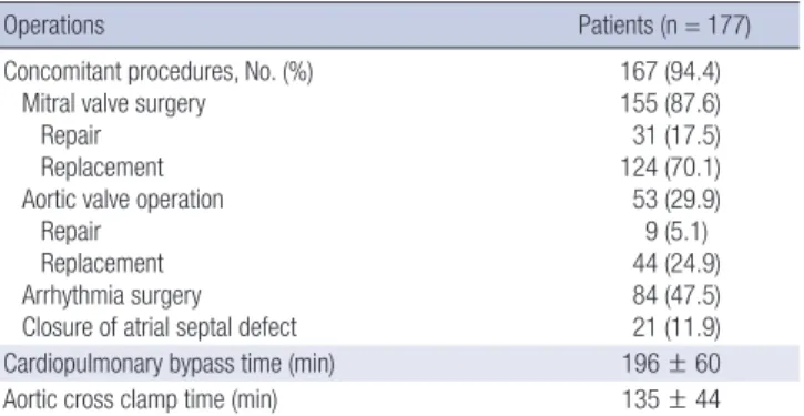
All operations were performed under a routine aorto-bicaval cannulation, moderate hypothermia and cold blood cardiople- gic arrest via a median sternotomy. Mean cardiopulmonary by- pass and aortic cross clamp times were 196 ± 60 and 135 ± 44 min, respectively. One hundred and sixty seven patients (94.4%) underwent concomitant cardiac procedures including mitral valve operation (n = 155), arrhythmia surgery (n = 84), and aor- tic valve procedure (n = 53) (Table 2).
De Vega annuloplasty was performed with a 3-0 extended polytetrafluoroethylene (e-PTFE) suture. Reduced annular size was measured with commercially available cylindrical valve sizers. Three sizers with actual diameters of 29.5, 31.5, and 33.5 mm (labeled sizes of 27, 29, and 31 mm) were used. Annular re- duction was performed by tying down the plication suture while the cylindrical sizer was inserted in the tricuspid valve orifice.
In early study period, a sizer with an actual diameter of 33.5 mm
was used due to a concern of too tight reduction (n = 37). How- ever, since 2003, a sizer with diameter of 31.5 mm was used in the majority of the patients (n = 129) by the operating surgeon’s choice. During the study period, De Vega technique was exclu- sively used for all patients of the operating surgeon who needed tricuspid annuloplasty.
Evaluation of early and long-term clinical outcomes Patients underwent regular postoperative follow-up through the outpatient clinic at 3 or 4 month intervals, and were con- tacted by telephone for confirmation of their condition if the last clinic visit was not conducted at the scheduled time. Clini- cal follow-up was closed on December 31, 2012. Follow-up was complete in 96.6% (171 of 177) of patients, with a follow-up du- ration of 72 ± 36 months. Operative mortality was defined as death within 30 days or during the same hospitalization after the surgery. Cardiac death was defined as any death related to cardiac events, including sudden death during the follow-up.
Valve-related complications were recorded according to the previous guidelines (10). Tricuspid valve-related events (TVRE) included the following: 1) Cardiac death, 2) recurrent TR ≥ 3+, 3) tricuspid valve endocarditis, 4) tricuspid valve reoperation, and 5) congestive heart failure needed readmission.
Echocardiographic evaluation
An initial postoperative echocardiographic evaluation (7 [1-46]
days after surgery) was performed before discharge in all but 3 patients who died early after the surgery. Follow-up echocar- diograms were performed at the discretion of the operating sur- geon or referring physicians during the follow-up. At least 1 fol- low-up echocardiogram after the early postoperative period was performed in 94.1% (159 of 169) of the survivors. The last
follow-up echocardiogram was performed 57 ± 34 months af- ter the surgery. Tricuspid regurgitation was graded from 0 to 4+;
0 = no, 1+ = mild, 2+ = moderate, 3+ = moderately severe, and 4+ = severe. Recurrent TR was defined as TR of equal or more than grade 3 after the surgery.
Statistical analysis
Statistical analysis was performed using the SAS statistical soft- ware (SAS system for Windows, version 9.2; SAS institute, Cary, NC, USA) and the SPSS software package (Version 12.0, SPSS Inc, Chicago, IL, USA). Data were expressed as mean ± standard deviation, median with ranges, or proportions. Comparisons of categorical and continuous variables were performed with the chi-squared test or Fisher exact test and Student t test, respec- tively. Multivariate analysis to identify risk factors for operative mortality was performed with logistic regression analysis. Sur- vival rates were estimated using the Kaplan-Meier method. The Cox proportional hazard model was adopted for analysis of risk factors for recurrent TR. The proportional hazard property was tested using the restricted cubic spline (11, 12). All independent variables in the Cox regressions were met the proportional haz- ards assumption. When the separation occurred in univariate analysis, hazard ratio (HR) was estimated using penalized max- imum likelihood test. It was demonstrated with 95% confidence interval (CI). Multicollinearity was controlled using backward stepwise regression. Variables with a P value of less than 0.2 were entered into multivariate analyses. To analyze the impact of selection of the diameter of the cylindrical sizers, the diame- ter of the sizers (= reduced annular diameter) was included in the analysis. In addition, tricuspid valve orifice index (TVOI) was defined as a reduced annular diameter divided by the pa- tient’s body surface area and included in the analysis. The mini- mal P value approach was used to estimate an optimal cut-off value of a continuous variable predicting a time-related event (13). A P value of less than 0.05 was considered statistically sig- nificant.
Ethics statement
The study protocol was approved by the institutional review Table 1. Preoperative characteristics and risk factors of the study patients
Characteristics Patients (n = 177)
Age (yr) 57.9 ± 10.5
Male:Female 63:114
Body surface area (m2) 1.58 ± 0.18
Risk factors, No. (%) Smoking Hypertension Diabetes mellitus
Overweight (BMI ≥ 25 kg/m2) History of stroke
Coronary artery disease Chronic renal failure NYHA class ≥ 3 Atrial fibrillation History of cardiac surgery
16 (9.0) 36 (20.3) 16 (9.0) 25 (14.1) 23 (13.0) 6 (3.4) 4 (2.3) 116 (65.5) 155 (87.6) 50 (28.2) Echocardiographic data
TR ≥ moderate, No. (%) LVEF (%)
Estimated sPAP (mmHg)
120 (67.8) 56.1 ± 9.1 50.3 ± 15.0
BMI, body mass index; NYHA, New York Heart Association; TR, tricuspid regurgitation;
LVEF, left ventricular ejection fraction; sPAP, systolic pulmonary artery pressure.
Table 2. Operative data of the study patients
Operations Patients (n = 177)
Concomitant procedures, No. (%) Mitral valve surgery
Repair Replacement Aortic valve operation
Repair Replacement Arrhythmia surgery Closure of atrial septal defect
167 (94.4) 155 (87.6) 31 (17.5) 124 (70.1) 53 (29.9) 9 (5.1) 44 (24.9) 84 (47.5) 21 (11.9)
Cardiopulmonary bypass time (min) 196 ± 60
Aortic cross clamp time (min) 135 ± 44
board (IRB) of our hospital (IRB No. H-1204-022-403). Informed consent was waived by the IRB.
RESULTS Early results
The operative mortality rate was 4.5% (8 of 177). Postoperative morbidities included low cardiac output syndrome (n = 24, 13.6%), postoperative bleeding requiring reoperation (n = 11, 6.2%), stroke (n = 9, 5.1%), and acute renal failure (n = 5, 2.8%) (Table 3). Multivariate analysis revealed that combined coro- nary artery disease was the only significant risk factor for early mortality (Table 4). Early postoperative echocardiography re- vealed TR grades of 2+ and 3+ in 5 and 2 patients, respectively (Fig. 1).
Long-term survival
Among 169 survivors, late death occurred in 19 (11.2%) patients including 8 cardiac deaths. Overall survival rates at 5 and 10 yr were 85.5% and 80.5%, respectively. Age (HR [95% CI] = 1.062 [1.023-1.103], P = 0.001), smoking (HR [95% CI] = 3.835 [1.522- 9.664], P = 0.004) and coronary artery disease (HR [95% CI] = 5.232 [1.136-24.087], P = 0.034) were associated with overall survival.
Freedom rates from cardiac death at 5 and 10 yr were 94.3%
and 90.8%, respectively. Chronic renal failure (HR [95% CI] = 13.255 [2.204-79.740], P = 0.005), coronary artery disease (HR [95% CI] = 14.951 [2.265-98.689], P = 0.005) and smoking (HR [95% CI] = 6.189 [1.499-25.555], P = 0.012) were risk factors for long-term cardiac death.
Tricuspid valve-related events
Tricuspid valve regurgitation was found in 7 patients during the follow-up period. Freedom rates from TR ≥ 3+ at 5 and 10 yr were 96.5% and 93.1%, respectively (Fig. 2). None of the study patients had significant tricuspid valve stenosis during the fol- low-up. Mean transvalvular pressure gradient of repaired tri- cuspid valve was 1.06 ± 0.45 mmHg.
Tricuspid valve reoperation was performed in 2 patients. One patient underwent mitral and tricuspid valve replacement at 1 month after the initial mitral repair and tricuspid annuloplasty due to persistent severe mitral regurgitation and moderately severe TR. The other patient underwent tricuspid valve replace- ment 29 months after the surgery due to severe TR. Reoperative findings in both patients revealed intact initial De Vega sutures.
The patients’ TVOIs were 22.64 and 23.16 mm/m2, respectively.
Readmission owing to congestive heart failure was needed in 9 Table 3. Early mortality and postoperative complications of the study patients
Mortality and complications Total (n = 177)
Mortality, No. (%) 8 (4.5)
Morbidities, No. (%)
Low cardiac output syndrome Bleeding reoperation Respiratory complication Stroke
Acute renal failure Mediastinitis
24 (13.6) 11 (6.2) 10 (5.7) 9 (5.1) 5 (2.8) 1 (0.6) Table 4. Risk factor analysis for early mortality
Variables
Univariate
analysis Multivariate analysis P value Odds ratio [95% CI] P value
Age 0.049 - NS
Coronary artery disease 0.025 10.13 [1.35-76.24] 0.050
Hypertension 0.055 - NS
Chronic renal failure 0.170 - NS
Cardiopulmonary bypass time 0.021 1.011 [0.999-1.023] 0.066 CI, confidence interval; NS, not significant.
Fig. 1. Early postoperative changes in grades of tricuspid regurgitation.
Number of patients
Preoperative grade Early postoperative grade 140
120 100 80 60 40 20
0 0
57 86
10 24
118
49
5 2 0 Grade 0 Grade 1 Grade 2 Grade 3 Grade 4
Fig. 2. A Kaplan-Meier curve for freedom from tricuspid regurgitation (TR) ≥ 3+.
Freedom from TR (%)
Duration (months)
0 24 48 72 96 120 100
80
60
40
20
0
5-yr freedom from TR: 96.5%
10-yr freedom from TR: 93.1%
Patients at risk
174 137 119 104 82 70 52 36 25 9 3
Table 5. Changes in echocardiographic data at last follow-up
Echocardiography Preoperative data Last follow-up P value
LVESD (mm) 37.1 ± 8.3 34.4 ± 7.9 < 0.001
LVEDD (mm) 55.2 ± 10.1 51.9 ± 7.5 < 0.001
Left ventricular ejection fraction (%) 56.1 ± 9.1 55.3 ± 9.7 0.412 Estimated sPAP (mmHg) 50.3 ± 15.0 37.0 ± 10.2 < 0.001 LVESD, left ventricular end-systolic diameter; LVEDD, left ventricular end-diastolic di- ameter; sPAP, systolic pulmonary artery pressure.
Table 6. Results of Cox proportional hazard analysis to identify risk factors for recur- rent tricuspid regurgitation (≥ 3+)
Variables
Univariate analysis
Multivariate analysis P value Hazard ratio
[95% CI] P value
Age 0.178 - NS
Female sex 0.174 - NS
Body surface area 0.010
Body mass index 0.053 - NS
Chronic renal failure 0.032 - NS
Diameter of a cylindrical sizer 0.489
TVOI 0.006 1.80 [1.18-2.75] 0.006
Preoperative TR grade 0.023 - NS
Left ventricular ejection fraction 0.516 Recurrence of left-sided valve disease 0.302 Atrial fibrillation at follow-up 0.398
CI, confidence interval; TVOI, tricuspid valve orifice index; TR, tricuspid regurgitation.
Fig. 3. A Kaplan-Meier curve for freedom from tricuspid regurgitation (TR) ≥3+ ac- cording to the cut-off value of tricuspid valve orifice index (TVOI).
Freedom from TR (%)
Duration (months)
0 24 48 72 96 120 100
80
60
40
20
0
Patients with TVOI < 22.5
Patients with TVOI ≥ 22.5
Patients at risk
TVOI<22.5 139 112 96 83 63 54 42 31 22 8 3 TVOI ≥22.5 35 25 23 21 19 16 10 5 3 1
patients. Although 3 patients experienced infective endocardi- tis, those were not related with the tricuspid valve. No patient suffered from thromboembolism from the right heart chamber.
Five- and 10-yr TVRE-free survivals were 91.3% and 81.9%, re- spectively. Age (HR [95% CI] = 1.090 [1.037-1.146], P = 0.001) and coronary artery disease (HR [95% C I] = 9.977 [1.781-55.870], P = 0.009) but not TVOI were associated with TVRE-free survival.
Long-term results other than tricuspid valve
During the follow-up, left-sided valve-related events occurred in 10 patients including severe regurgitation of repaired mitral valve (n = 1), structural valve deterioration of a bioprosthetic mitral valve (n = 1), paravalvular leak of prosthetic mitral valves (n = 4), and infective endocarditis (n = 4). Among those, 7 pa- tients underwent mitral and aortic valve reoperations. At last follow-up, atrial fibrillation remained in 74 patients including 22 of 84 patients who underwent concomitant arrhythmia sur- gery. Left ventricular dimensions and systolic pulmonary artery pressure improved significantly at the last follow-up echocar- diography. However, there was no change in left ventricular ejec- tion fraction (Table 5).
Risk factor analysis for the recurrence of tricuspid regurgitation
By univariate risk factor analyses, old age, female sex, small
body mass index, chronic renal failure, preoperative TR grade and the TVOI were entered into a multivariate analysis. Body surface area was excluded to control the multi-collinearity be- tween TVOI and body surface area. Difference in diameter of the cylindrical sizers was not associated with the recurrence of TR (P = 0.489). Recurrence of the left-sided valve problem was also not a risk factor for the recurrence of TR (P = 0.302). There was no late recurrence of significant TR in patients with TR < 2+, and preoperative TR grade was a significant factor associated with late recurrence of TR in the univariate analysis. However, this significance disappeared in the multivariate analysis. In backward stepwise multivariate analysis, only TVOI was left in the final model as a significant factor (P = 0.006, Hazard ratio [95% confidence interval] = 1.80 [1.18-2.75]) (Table 6). A cut-off value of TVOI to predict the recurrence of TR was analyzed by the minimal P value approach. A TVOI of 22.5 mm/m2 was the best cut-off value to predict the recurrence of TR (Fig. 3). In oth- er words, the tricuspid valve annulus should be reduced accord- ing to the equation as follows; reduced tricuspid valve orifice diameter < 22.5 × patient’s body surface area.
DISCUSSION
This study demonstrated 2 main findings. First, long-term re- sults of the De Vega annuloplasty for functional TR were favor- able with 10-yr freedom rates from recurrent TR and TVRE of 93.1% and 81.9%, respectively. Second, annular reduction should be performed considering patients’ body size to optimize long- term results of the De Vega annuloplasty.
Since 1965, various surgical techniques of tricuspid annulo-
plasty have been introduced for functional TR. These included suture plication, and annuloplasty using a pericardial strip, flex- ible band and rigid rings (1-3, 14). Although the De Vega annu- loplasty has been widely performed for functional TR in various conditions, there has been a concern that long-term results of the De Vega procedure is less satisfactory than those of the pros- thetic ring annuloplasty (6-8). Supporters of a ring annuloplasty have insisted that the use of a ring provides several advantages including equal distribution of tension, standardized annular reduction, ease of learning and reproducibility (15). Two stud- ies, conducted in the same center, consistently reported that tricuspid valve repair without a prosthetic ring was associated with late recurrence of TR (7, 8). On the contrary, Morishita and his colleagues reported that the De Vega annuloplasty is a safe and effective method to treat functional TR with a 10-yr reoper- ation-free rate of 94.4% (16). In the present study, the 10-yr free- dom rate from TR ≥ 3+ were 93.1%, and only 2 patients under- went redo-tricuspid valve operation during the follow-up peri- od. When performing the annular reduction, we used cylindri- cal sizers to reproducibly reduce the tricuspid annulus. In addi- tion, the e-PTFE suture was used with an expectation, although evidence is lacking, that it could prevent suture breakdown and dehiscence (so called bow-string effect), which has been found when using the polypropylene suture (17).
Previous studies demonstrated that preoperative TR grade, severe left ventricular dysfunction, and preoperative TV tether- ing caused by dilated right ventricle were identified as predic- tors of recurrent TR (3, 18, 19). In the present study, however, preoperative TR grade and left ventricular dysfunction were not associated with the recurrence of TR. To evaluate whether re- duced annular diameter could affect results of annuloplasty, we included measured diameter by the cylindrical sizer and in- dexed measured diameter (TVOI) as possible risk factors. The univariate and multivariate analyses revealed that not the re- duced diameter of the tricuspid annulus itself but indexed an- nular diameter by body surface area was associated with the re- currence of TR. The minimal P value approach, which was a statistical method to identify a cut-off value in time-related events, revealed that an indexed annular diameter of 22.5 mm/m2 was a cut-off value to warrant annuloplasty failure.
Interestingly, we could evaluate the fate of initial De Vega su- tures in two patients who underwent tricuspid reoperation dur- ing the follow-up period. Tricuspid regurgitation recurred even though previous e-PTFE sutures were intact. The TVOIs in these patients were 22.64 and 23.16 mm/m2, respectively. This might reflect that the optimal sizing of annular reduction according to the patient’s size is necessary in order to prevent the late recur- rence of TR. Further longer-term follow up data to evaluate the clinical benefit of restrictive De Vega procedure and prospec- tive randomized study comparing between tight ring annulo- plasty and restrictive De Vega procedure are needed in the near
future.
There are several limitations to the present study that must be recognized. First, the present study was a retrospective ob- servational study in a single institution. Second, echocardiogra- phy was not performed on a regular interval during the follow- up. Third, we did not evaluate preoperative tricuspid annular diameter and leaflet tethering, due to retrospective nature of this study. Fourth, precise indications of tricuspid annuloplasty were not described due to the retrospective nature of this study.
ACKNOWLEDGEMENTS
We wish to thank the Medical Research Collaborating Center of the Seoul National University Hospital for their efforts in statisti- cal assistance.
DISCLOSURE
The authors have no conflicts of interest to disclose.
REFERENCES
1. Holper K. Haehnel JC, Augustin N, Sebening F. Surgery for tricuspid in- sufficiency: long-term follow-up after De Vega annuloplasty. Thorac Car- diovasc Surg 1993; 41: 1-8.
2. Kay JH, Maselli-Campagna G, Tsuji KK. Surgical treatment of tricuspid insufficiency. Ann Surg 1965; 162: 53-8.
3. McCarthy PM, Bhudia SK, Rajeswaran J, Hoercher KJ, Lytle BW, Cos- grove DM, Blackstone EH. Tricuspid valve repair: durability and risk factors for failure. J Thorac Cardiovasc Surg 2004; 127: 674-85.
4. Burma O, Ustunsoy H, Davutoglu V, Celkan MA, Kazaz H, Pektok E.
Initial clinical experience with a novel biodegradable ring in patients with functional tricuspid insufficiency: Kalangos Biodegradable Tricus- pid Ring. Thorac Cardiovasc Surg 2007; 55: 284-7.
5. Navia JL, Nowicki ER, Blackstone EH, Brozzi NA, Nento DE, Atik FA, Rajeswaran J, Gillinov AM, Svensson LG, Lytle BW. Surgical manage- ment of secondary tricuspid valve regurgitation: annulus, commissure, or leaflet procedure? J Thorac Cardiovasc Surg 2010; 139: 1473-82.e5.
6. Guenther T, Mazzitelli D, Noebauer C, Hettich I, Tassani-Prell P, Voss B, Lange R. Tricuspid valve repair: is ring annuloplasty superior? Eur J Car- diothorac Surg 2013; 43: 58-65.
7. Tang GH, David TE, Singh SK, Maganti MD, Armstrong S, Borger MA.
Tricuspid valve repair with an annuloplasty ring results in improved long-term outcomes. Circulation 2006; 114: I577-81.
8. Sales VL, McCarthy PM. Durability of functional tricuspid valve repair.
Semin Thorac Cardiovasc Surg 2010; 22: 97-103.
9. Antunes MJ. De Vega annuloplasty of the tricuspid valve. Oper Tech Tho- rac Cardiovasc Surg 2003; 8: 169-76.
10. Akins CW, Miller DC, Turina MI, Kouchoukos NT, Blackstone EH, Grun- kemeier GL, Takkenberg JJ, David TE, Butchart EG, Adams DH, et al.
Guidelines for reporting mortality and morbidity after cardiac valve in- terventions. J Thorac Cardiovasc Surg 2008; 135: 732-8.
11. Durrleman S, Simon R. Flexible regression models with cubic splines.
Stat Med 1989; 8: 551-61.
12. Hess KR. Assessing time-by-covariate interactions in proportional haz- ards regression models using cubic spline functions. Stat Med 1994; 13:
1045-62.
13. Altman DG, Lausen B, Sauerbrei W, Schumacher M. Dangers of using
“optimal” cutpoints in the evaluation of prognostic factors. J Natl Cancer Inst 1994; 86: 829–35.
14. Chang BC, Song SW, Lee S, Yoo KJ, Kang MS, Chung N. Eight-year out- comes of tricuspid annuloplasty using autologous pericardial strip for functional tricuspid regurgitation. Ann Thorac Surg 2008; 86: 1485-92.
15. Chikwe J, Anyanwu AC. Surgical strategies for functional tricuspid re- gurgitation. Semin Thorac Cardiovasc Surg 2010; 22: 90-6.
16. Morishita A, Kitamura M, Noji S, Aomi S, Endo M, Koyanagi H. Long-
term results after De Vega’s tricuspid annuloplasty. J Cardiovasc Surg (Torino) 2002; 43: 773-7.
17. Revuelta JM, Garcia-Rinaldi R. Segmental tricuspid annuloplasty: a new technique. J Thorac Cardiovasc Surg 1989; 97: 799-801.
18. Fukuda S, Song JM, Gillinov AM, McCarthy PM, Daimon M, Kongsae- repong V, Thomas JD, Shiota T. Tricuspid valve tethering predicts residu- al tricuspid regurgitation after tricuspid annuloplasty. Circulation 2005;
111: 975-9.
19. Fukuda S, Gillinov AM, McCarthy PM, Matsumura Y, Thomas JD, Shio- ta T. Echocardiographic follow-up of tricuspid annuloplasty with a new three-dimensional ring in patients with functional tricuspid regurgita- tion. J Am Soc Echocardiogr 2007; 20: 1236-42.