ABSTRACT
Immune checkpoint blockade targeting PD-1 and PD-L1 has resulted in unprecedented clinical benefit for cancer patients. Anti-PD-1/PD-L1 therapy has become the standard treatment for diverse cancer types as monotherapy or in combination with other anti- cancer therapies, and its indications are expanding. However, many patients do not benefit from anti-PD-1/PD-L1 therapy due to primary and/or acquired resistance, which is a major obstacle to broadening the clinical applicability of anti-PD-1/PD-L1 therapy. In addition, hyperprogressive disease, an acceleration of tumor growth following anti-PD-1/PD-L1 therapy, has been proposed as a new response pattern associated with deleterious prognosis.
Anti-PD-1/PD-L1 therapy can also cause a unique pattern of adverse events termed immune- related adverse events, sometimes leading to treatment discontinuation and fatal outcomes.
Investigations have been carried out to predict and monitor treatment outcomes using peripheral blood as an alternative to tissue biopsy. This review summarizes recent studies utilizing peripheral blood immune cells to predict various outcomes in cancer patients treated with anti-PD-1/PD-L1 therapy.
Keywords: Blood; Biomarkers; PD-1 receptor; Programmed cell death 1 ligand 1; Prognosis;
Adverse drug event
INTRODUCTION
Immune checkpoint blockade has recently emerged as a valuable treatment option for many types of cancer. Immune checkpoint blockade leads to the restoration of the anti- tumor effector functions of exhausted T cells (1,2). T-cell exhaustion was first discovered in chronic viral infection (3,4) and later demonstrated to be a universal phenomenon in cancer, with exhausted T cells playing a central role in shaping the immune microenvironment in cancer (5). Exhausted T cells are a transcriptionally and epigenetically distinct cell population developed by persistent antigenic stimulation in the case of a failure in Ag clearance (1). Exhausted T cells are characterized by a progressive loss of effector functions, diminished memory recall response, and blunted homeostatic self-renewal (6). Exhausted T-cell subsets usually co-express multiple inhibitory receptors to restrain their expansion capacity and effector molecule production (7). Immune checkpoint blockade was developed
Review Article
Received: Jan 6, 2020 Revised: Jan 29, 2020 Accepted: Jan 30, 2020
*Correspondence to Eui-Cheol Shin
Graduate School of Medical Science and Engineering, Korea Advanced Institute of Science and Technology (KAIST), 291 Daehak- ro, Yuseong-gu, Daejeon 34141, Korea.
E-mail: ecshin@kaist.ac.kr
†These authors contributed equally to this work.
Copyright © 2020. The Korean Association of Immunologists
This is an Open Access article distributed under the terms of the Creative Commons Attribution Non-Commercial License (https://
creativecommons.org/licenses/by-nc/4.0/) which permits unrestricted non-commercial use, distribution, and reproduction in any medium, provided the original work is properly cited.
ORCID iDs Kyung Hwan Kim
https://orcid.org/0000-0002-6713-1350 Chang Gon Kim
https://orcid.org/0000-0002-4929-8501 Eui-Cheol Shin
https://orcid.org/0000-0002-6308-9503 Conflict of Interest
The authors declare no potential conflicts of interest.
Abbreviations
CCR7, C-C chemokine receptor type 7; ICI, immune checkpoint inhibitor; irAE, immune-
Kyung Hwan Kim †, Chang Gon Kim † , Eui-Cheol Shin *
Graduate School of Medical Science and Engineering, Korea Advanced Institute of Science and Technology (KAIST), Daejeon 34141, Korea
Peripheral Blood Immune Cell-based Biomarkers in Anti-PD-1/PD-L1
Therapy
related adverse event; NSCLC, non-small-cell lung cancer; PMN-MDSC, polymorphonuclear myeloid derived suppressor cell; TET, thymic epithelial tumor
Author Contributions
Conceptualization: Kim KH, Kim CG, Shin EC;
Funding acquisition: Shin EC; Investigation:
Kim KH; Methodology: Kim KH, Kim CG;
Writing - original draft: Kim KH, Kim CG;
Writing - review & editing: Kim KH, Kim CG, Shin EC.
to reinvigorate exhausted T cells. Mechanistically, immune checkpoint blockade aims to block the interaction of inhibitory receptors expressed on exhausted T cells and their ligands expressed on various cell types, including tumor cells and other immune cells (2). Immune checkpoint inhibitors (ICIs), such as anti-PD-1 and anti-CTLA-4 Abs, have received approval by the Food and Drug Administration as a standard treatment for various malignancies, including malignant melanoma, non-small-cell lung cancer (NSCLC), urothelial carcinoma, renal cell carcinoma, squamous head and neck carcinoma, Hodgkin lymphoma, and hepatocellular carcinoma (8,9). Immunotherapy with immune checkpoint blockade has revolutionized cancer treatment, with an increase in overall survival and durable responses lasting for years (10). However, a large group of patients do not benefit from immune checkpoint blockade by primary and/or acquired resistance (11), and treatment with ICIs is sometimes related to immune-related adverse events (irAEs) that may lead to serious morbidity and mortality (12,13). Emerging evidence suggests that immune checkpoint blockade is associated with paradoxical acceleration of tumor growth, termed hyperprogressive disease, resulting in dismal outcomes (14).
The mechanisms underlying the response and resistance to immune checkpoint blockade are complex and poorly understood (11,15). Furthermore, irAEs and hyperprogressive disease are unique outcomes provoked by immune checkpoint blockade that have not been experienced with conventional cancer treatment and require active investigation. The only predictive biomarker currently used routinely in clinics is the expression of PD-L1 in tumor tissues as assessed by immunohistochemistry (16). Despite wide use of PD-L1 as a biomarker, it has limitations for technical and biological issues (17-19). Other tissue-based biomarkers, including tumor mutational burden and gene expression profile, have comparable predictive power as PD-L1 (20,21). Recently, B cells and presence of tertiary lymphoid structures in tumor tissue have also been suggested as predictors of response to immune checkpoint blockade (22-24).Although information obtained from tumor tissues may better reflect its biology, biopsies are invasive, sometimes inaccessible, and may not represent the whole tumor due to intra-tumoral heterogeneity (25). In addition, considering the dynamic response of the immune system following immune checkpoint blockade (26), serial biopsies to monitor the early response to treatment will have great value, but repetitive tissue biopsies are not applicable in most cases.
In this regard, peripheral blood is a less invasive, safe, and convenient alternative for repetitive sampling. Furthermore, peripheral blood can provide a systematic view of host immune responses, as different components, such as cell-free DNA, circulating tumor cells, cytokines, Abs, and immune cells, can be analyzed in the blood. In this review, we focus on the relevant studies investigating peripheral blood immune cell biomarkers that predict treatment responses, hyperprogressive disease, and irAEs.
PERIPHERAL BLOOD T CELLS AND TREATMENT RESPONSES
Circulating PD-1+CD8+ T cells provide a means of detecting tumor-specific CD8+ T cells
PD-1 blockade can induce the proliferation of tumor-infiltrating CD8+ T cells, which is associated with a radiographic tumor response (27). Although non-tumor-specific bystander CD8+ T cells have been reported to reside in the tumor microenvironment (28), the CD8+ T
cells responding to PD-1 blockade are thought to be tumor-specific (29). Tumor-specific CD8+ T cells can also be detected in the peripheral blood and are characterized by PD-1 expression (30,31). Gros et al. (30) clearly showed that neoantigen-specific CD8+ T cells that exist in the tumors of melanoma patients are found in the PD-1+ circulating CD8+ T-cell population but not in the PD-1− fractions, implying that peripheral blood PD-1+CD8+ T cells could be utilized to monitor the dynamic changes in tumor-specific CD8+ T cells. They extended their findings to patients with gastrointestinal cancer, demonstrating that neoantigen-reactive CD8+ and CD4+ T cells are only identified in the PD-1+ population and not in the PD-1− fractions (31).
Response of peripheral blood PD-1+CD8+ T cells
Considering that tumor-specific T cells can be detected in the peripheral blood, T-cell responses in the tumor microenvironment following anti-PD-1/PD-L1 therapy may also be reflected by circulating T cells. The proliferation of peripheral blood PD-1+CD8+ T cells after anti-PD-1/PD-L1 therapies has been demonstrated in multiple studies (32-35). In contrast, CD8+ T cells specific to non-tumor related Ags, such as human cytomegalovirus and Epstein- Barr virus, do not have an increased frequency of Ki-67+ cells after anti-PD-1 therapy, implying a tumor-specificity of the proliferative response in PD-1+CD8+ T cells (32,33). Moreover, Huang et al. (34) investigated the clonal overlap between tumor-infiltrating and peripheral blood CD8+ T cells in anti-PD-1-treated patients. Across 6 patients, top-ranked CD8+ T-cell clones in peripheral blood were also present in the tumor and were all CD38+HLA-DR+ and mostly Ki-67+ (34). These data imply that the proliferative response in PD-1+CD8+ T cells following anti-PD-1 therapy is more likely a tumor-specific rather than a non-specific response.
As anti-PD-1/PD-L1 Abs are administered every 2 or 3 wk, the proliferative responses, measured by the frequency of Ki-67+ cells, were followed at each point of drug administration.
The frequency of Ki-67+ cells among PD-1+CD8+ T cells was highest at the first follow-up evaluation, 2 or 3 wk after initiating the treatment, and gradually decreased throughout the treatment course, despite continuous administration of anti-PD-1/PD-L1 agents (33,34). Kim et al. (32) found that the frequency of Ki-67+ cells among PD-1+CD8+ T cells peaked 1 wk after initiating anti-PD-1 treatment and decreased thereafter. This was once again demonstrated in a neoadjuvant trial of anti-PD-1 therapy in melanoma patients, showing a peak of Ki-67+ cells at wk 1 and a decrease at wk 3 (35). In this study, the frequency of Ki-67+ cells among PD- 1+CD8+ T cells had a single peak at wk 1, despite continuous anti-PD-1 treatment (35).
Considering the mode-of-action of anti-PD-1/PD-L1 therapy, sufficient reinvigoration of PD-1+CD8+ T cells after PD-1 blockade may be assumed to be an important determinant of the clinical tumor response. Tumeh et al. (27) demonstrated an increase in the number of tumor-infiltrating CD8+ T cells in responders, but not in progressors, following anti-PD-1 therapy using serial biopsies from melanoma patients. As serial biopsies are not applicable in most patients with solid tumors, the predictive value of the proliferative response of peripheral blood PD-1+CD8+ T cells has been evaluated (32,34). Huang et al. (34) applied the frequency of Ki-67+ cells among PD-1+CD8+ T cells 3 wk post anti-PD-1 treatment and baseline tumor burden to predict treatment outcomes. They found that a higher Ki-67 to tumor burden ratio significantly predicts a better objective response and survival in the test cohort but had borderline significance in the validation cohort. Another study performed by Kim et al. (32) utilized the ratio of the frequency of Ki-67+ cells among PD-1+CD8+ T cells 1 wk after the first dose of anti-PD-1 treatment to the frequency at baseline (Ki-67D7/D0). They utilized a total of 3 cohorts, including one cohort of patients with thymic epithelial tumor (TET) and 2 independent cohorts of patients with NSCLC, and tested the utility of Ki-67D7/D0 as a
biomarker for predicting long-term treatment outcome. Ki-67D7/D0 predicted the long-term tumor response and survival in both the test and validation cohorts. Furthermore, Ki-67D7/
D0 exhibited an independent predictive value regardless of other clinicopathological factors of the patients, and a better predictive value than tumor PD-L1 levels (32). Although an ideal biomarker would be reliable in predicting both responders and non-responders, it should at least indicate who will certainly not respond. In this regard, Ki-67D7/D0 exhibited a fairly high negative predictive value of 85%–94% depending on the study cohort.
Change in peripheral blood CD4+ T cells
CD4+ T cells also respond to PD-1 blockade. The frequency of Ki-67+ cells among FoxP3– and FoxP3+CD4+ T cells has been shown to significantly increase 3 wk after anti-PD-1 treatment (34). Similar to PD-1+CD8+ T cells, the proliferative response in CD4+ T cells peaked at wk 3. However, the CD4+ T-cell responses did not significantly correlate with clinical outcome. Zappasodi et al. (36) defined FoxP3−PD-1hiCD4+ T cells (4PD1hi), which accumulate intratumorally as a function of tumor burden. They found that 4PD1hi cells inhibit T-cell functions in a PD-1/PD-L1-dependent fashion, and that the frequency decreases after anti- PD-1 treatment. However, those who lacked an effective reduction in 4PD1hi cells experienced poor prognosis. In addition, a higher 3-wk post-treatment frequency of 4PD1hi cells was predictive of poor survival (36). Another study has also demonstrated that an increased frequency of T cell Ig and mucin-domain containing-3+ cells among CD4+ and CD8+ T cells can predict non-responders to anti-PD-1 therapy (37). However, the sample size in the study was small and post-treatment samples were collected 12 wk after treatment, which weakens the value of this marker.
TCR repertoire as biomarkers
Hopkins et al. (38) investigated the TCR repertoires in PBMCs from patients with pancreatic cancer treated with nivolumab and cancer vaccine. They found that low baseline clonality was associated with superior survival after the treatment, though the number of expanded clones after treatment was not associated with survival. However, this study analyzed a small cohort with little difference in survival between patients with low and high baseline clonality and lacked further independent validation (38). Moreover, evaluation of the TCR repertoire in PBMCs mostly evaluates T-cell clones that are unrelated to the tumor. More recently, Han et al. (39) analyzed the TCR repertoire in PD-1+CD8+ T cells from anti-PD-1/
PD-L1-treated NSCLC patients. Higher baseline diversity in PD-1+CD8+ T cells and increased clonality after treatment were associated with superior survival. Furthermore, patients with pseudoprogression exhibited an expansion of certain TCR clones that was not observed in true progressors. The strength of this study is that the TCR repertoire was investigated in PD- 1+CD8+ T cells rather than total T cells, and that they performed independent validation (39).
Baseline T-cell-based biomarkers
Compared to the ‘dynamic’ blood-based biomarkers discussed above, baseline or ‘static’
biomarkers can have a benefit because they may predict clinical outcome before the initiation of treatment. However, therapeutic responses to ICIs are complicated and involve multiple immunological processes (40,41). In addition, the therapeutic response to ICIs has been suggested to be a critical state transition process of a complex system, which is difficult to predict far in advance (26). Despite such limitations of baseline biomarkers, they still have advantages, and several studies have investigated the role of baseline T-cell biomarkers in predicting the clinical outcome of anti-PD-1/PD-L1 therapy.
Zuazo et al. (42) claimed that NSCLC patients with dysfunctional systemic CD4 immunity poorly respond to anti-PD-1/PD-L1 therapy. They focused on highly differentiated (CD27− CD28−) CD4+ T cells and found that a higher frequency of these cells among CD4+ T cells is associated with superior clinical outcome. Mechanistically, CD4+ and CD8+ T cells in patients with a higher frequency of CD27−CD28−CD4+ T cells exhibit a significantly higher proliferative response to PD-1 blockade in vitro and ex vivo. Another study that performed cytometry by time of flight of PBMCs obtained from melanoma patients demonstrated that non-responders have a higher frequency of effector memory (CD45RO+CD62L−) CD4+ T cells and naive (CD45RO−CD62L+) CD8+ T cells, and a lower frequency of central memory (CD45RO+CD62L+) CD8+ T cells (43). However, these studies lack validation and further investigation is required to confirm the predictive value of these suggested biomarkers.
The predictive value of the baseline and post-treatment frequency of proliferating Ki-67+ cells among Treg cells was evaluated but was not significantly associated with clinical outcome (34). Furthermore, the frequency of Treg cells among CD4+ T cells was not significantly different among responders and non-responders (43). However, this study defined Treg cells as CD127−CD25+ cells without FoxP3 staining (43). In contrast, Kim et al. (44) found that the baseline number of Treg cells is significantly higher in responders. They also measured the frequency of Lox-1+ polymorphonuclear myeloid derived suppressor cells (PMN-MDSCs) and found that the ratio of Treg and PMN-MDSC frequency significantly predicted survival after nivolumab treatment in both the discovery and validation cohorts. We summarized the relevant peripheral blood T-cell-based biomarkers that predict treatment outcome in Table 1.
HYPERPROGRESSIVE DISEASE
Incidence and definition of hyperprogressive disease
Hyperprogressive disease is a paradoxical acceleration of tumor growth kinetics after treatment with ICIs, such as anti-PD-1/PD-L1 and anti-CTLA-4 Abs, that leads to rapid clinical deterioration (14). In particular, the initial inferior outcomes of PD-1/PD-L1 blockade compared to standard chemotherapy, resulting in a crossover of the survival curves after a period of time, have been observed in several prospective studies (45-48). In addition, treatment with anti-PD-1/PD-L1 Abs is associated with higher chances of the occurrence of hyperprogressive disease compared to treatment with chemotherapy (49). These findings imply that hyperprogressive disease is a distinct phenomenon from simple progression and may involve immunological processes.
Following the introduction of ICIs, several groups reported retrospective data and clinical cases of hyperprogressive disease induced by anti-PD-1/PD-L1 Abs. The incidence of hyperprogressive disease is variable across tumor types, ranging from 8%–29% of treated patients. In particular, a considerable number of patients with head and neck squamous cell carcinoma (29%) and NSCLC (10%–20%) develop hyperprogressive disease after PD-1/
PD-L1 blockade. Currently, no consensus has been reached on the optimal definition of hyperprogressive disease. Champiat et al. (50) defined hyperprogressive disease as a progressive disease with an at least 2-fold increase in the tumor growth rate, which is based on the sum of the estimated volume of target lesions, during anti-PD-1/PD-L1 therapy compared to the pre-anti-PD-1/PD-L1 treatment period. Saâda-Bouzid et al. (51) described hyperprogressive disease as an at least 2-fold increase in tumor growth kinetics, which is based on the sum of the longest diameters of target lesions, during anti-PD-1/PD-L1
therapy compared to the pre-anti-PD-1/PD-L1 treatment period. Ferrara et al. (49) defined hyperprogressive disease as a >50% difference in tumor growth rate during treatment and before treatment. In addition to the definitions based on tumor growth dynamics, Kato et al. (52) defined hyperprogressive disease as time-to-treatment failure <2 months and
>50% increase in tumor burden compared to baseline. Kim et al. (53) comprehensively compared different definitions of hyperprogression and concluded that definitions based on both tumor growth ratio and tumor growth kinetics can be used more universally than the definitions based on time-to-treatment failure.
Predictors of hyperprogressive disease
The prognosis of patients who experience hyperprogressive disease has consistently been reported to be very poor (49,53). Therefore, there is an urgent need for biomarkers to predict hyperprogressive disease. Currently, no reliable factors are available for predicting the occurrence of hyperprogressive disease, but some groups have observed an association with parameters obtained in routine clinical practice. Champiat et al. (50) suggested an association with old age, and Ferrara et al. (49) reported that a higher number of metastatic sites (>2) is associated with hyperprogressive disease. Furthermore, Kim et al. (53) suggested that an elevated neutrophil-to-lymphocyte ratio (>4) and lactate dehydrogenase level (greater than the upper limit of normal) are associated with hyperprogressive disease. In addition, Table 1. Summary of relevant biomarker studies predicting treatment response and prognosis
Biomarker Cancer type No. of patients Main results Reference
(%Ki-67+ cells/PD-1+CD8+ T cells 3-wk post-treatment)/baseline tumor burden (Ki67/TB)
Melanoma Discovery cohort: 23 Higher Ki67/TB significantly associated with superior ORR
(p=0.03) and PFS (p=0.004). Huang et al. (34)
Validation cohort: 18 Higher Ki67/TB associated with superior ORR (p=0.14) and PFS (p=0.06).
(%Ki-67+ cells/PD-1+CD8+ T cells 1-wk post-treatment)/(%Ki- 67+ cells/PD-1+CD8+ T cells at baseline) (Ki-67D7/D0)
TET Discovery cohort: 31 Higher Ki-67D7/D0 significantly associated with durable clinical benefit (PR, or SD for 6 months or longer; p<0.001) and PFS (p=0.027)
Kim et al. (32)
NSCLC Discovery cohort: 33 Higher Ki-67D7/D0 significantly associated with durable clinical benefit (PR, or SD for 6 months or longer; p<0.01), PFS (p=0.004), and OS (p=0.001)
Validation cohort: 46 Higher Ki-67D7/D0 significantly associated with durable clinical benefit (PR, or SD for 6 months or longer; p<0.01), PFS (p=0.002), and OS (p=0.037)
%FoxP3−PD-1hiCD4+ T cells/
CD4+ T cells (4PD1hi) 3-wk post- treatment
Melanoma 52 Higher frequency of 4PD1hi 3-wks post treatment (p=0.0005) and
fold change of 4PD1hi (p=0.046) associated with poorer OS. Zappasodi et al.
(36) Fold change of 4PD1hi
TCR diversity of PD-1+CD8+ T cells
at baseline and post-treatment NSCLC Discovery cohort: 25 Higher baseline diversity in PD-1+CD8+ T cells (p=0.021) and increased clonality after treatment (p=0.002) associated with superior PFS.
Han et al. (39) Validation cohort: 15
%CD27−CD28− cells/CD4+ T cells
at baseline NSCLC 51 Higher frequency of CD27−CD28−CD4+ T cells associated superior
PFS (p=0.001). Zuazo et al. (42)
Ratio of the frequency of Treg cells and PMN-MDSCs at baseline
NSCLC Discovery cohort: 34 Higher ratio of the frequency of Treg cells and PMN-MDSCs
associated with superior PFS (p=0.0079). Kim et al. (44) Validation cohort: 29 Higher ratio of the frequency of Treg cells and PMN-MDSCs
associated with superior PFS (p=0.0017).
%Effector/memory (CCR7− CD45RA−) cells/CD8+ T cells at baseline
NSCLC 263 (flow cytometry
analysis in 144) Lower frequency of effector/memory CD8+ T cells with development of hyperprogressive disease (p<0.001) and poor PFS (p<0.001) and OS (p<0.001).
Kim et al. (53)
%TIGIT+ cells/PD-1+CD8+ T cells
at baseline Higher frequency of TIGIT+ cells among PD-1+CD8+ T cells in
peripheral blood at baseline significantly associated with development of hyperprogressive disease (p<0.001) and poor PFS (p<0.001) and OS (p=0.01).
NSCLC, non-small-cell lung cancer; ORR, objective response rate; OS, overall survival; PFS, progression-free survival; PR, partial response; SD, stable disease;
TET, thymic epithelial tumor; CCR7, C-C chemokine receptor type 7.
some groups have reported genomic alterations associated with hyperprogressive disease.
Kato et al. (52) suggested that genomic alterations in EGFR and MDM2/MDM4 correlate with hyperprogressive disease, and Kim et al. (54) reported that STK11 mutation is frequently observed in patients with hyperprogressive disease.
However, blood-based biomarkers have not been clearly identified. Recently, Kim et al. (53) demonstrated that a lower frequency of effector/memory (C-C chemokine receptor type 7 [CCR7]−CD45RA−) cells among CD8+ T cells and a higher frequency of TIGIT+ cells among PD-1+CD8+ T cells in the peripheral blood at baseline are associated with the development of hyperprogressive disease and poor survival in NSCLC patients (Table 1). Mechanistically, they found that CCR7−CD45RA+ and TIGIT+PD1+ CD8+ T cells are prone to activation-induced cell death upon anti-PD-1 treatment, suggesting that hyperprogressive disease may be caused by a depletion of certain subsets of CD8+ T cells following anti-PD-1 treatment.
In another study, the proliferative response of PD-1+ effector Treg (FoxP3hiCD45RA−CD4+ T) cells was shown to promote hyperprogressive disease (55). Effector Treg cells are a highly suppressive subset of Treg cells (56). They found an increase in the frequency of Ki-67+ cells among tumor-infiltrating effector Treg cells in patients with hyperprogressive disease, but a decrease in patients without hyperprogressive disease. PD-1 blockade also enhanced the suppressive function of effector Treg cells in vitro. In mice, genetic ablation or blockade of PD-1 in Treg cells increased their proliferation and the suppression of anti-tumor immune responses. However, they did not present data on the change in peripheral blood effector Treg cells following PD-1 blockade. In addition, the frequency of effector Treg cells in the peripheral blood was similar between patients with or without hyperprogressive disease. One limitation of this study was that they compared patients with hyperprogressive disease and patients without hyperprogressive disease, rather than patients with progressive disease but not hyperprogressive disease.
Until now, the number of studies on hyperprogressive disease has been limited due to the complexity in its definition and requirements for routine imaging evaluations. Current studies are also limited due to their retrospective nature. Prospectively designed studies are needed in the future to properly predict the patients who will experience hyperprogressive disease after anti-PD-1/PD-L1 therapy.
PERIPHERAL BLOOD-BASED PREDICTION OF IRAES
Peripheral blood immune cells and irAEs
IrAEs are common toxicities observed in patients treated with ICIs (57,58). They mostly present as autoimmune symptoms and commonly involve the skin, lung, colon, liver, and endocrine organs, and rarely the heart, kidney, and nervous system (57). Many possible mechanisms have been suggested for irAEs after anti-PD-1/PD-L1 therapy, including increased activity of self-reactive T cells and B cells, increased levels of pre-existing autoantibodies, and increased levels of inflammatory cytokines (57,58). However, the underlying pathophysiology of irAEs has not been fully elucidated. As the presentation of irAEs resembles symptoms of autoimmune disorders, immune cells involved in the pathophysiology of autoimmune diseases may also serve critical roles in irAEs, including Treg cells, Th17 cells, CD8+ T cells, and B cells (59-62).
A recent study investigated the baseline and early changes in Treg cells, Th17 cells, TNF- secreting T cells, and CD8+ T cells among PBMCs obtained at baseline and wk 1 after
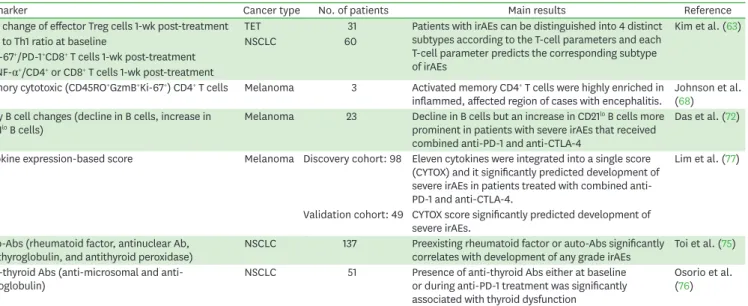
initiating anti-PD-1 therapy in TET and NSCLC patients (63). They found that the fold change in effector Treg cells post-treatment, the pre-treatment Th17 to Th1 ratio, post-treatment frequency of Ki-67+ cells among PD-1+CD8+ T cells, and post-treatment TNF-α+ cells among CD4+ or CD8+ T cells are associated with the development of irAEs. Interestingly, in clustering analysis, they found that patients with irAEs can be distinguished into 4 subtypes according to the T-cell parameters. The first subtype had a high frequency of post-treatment TNF-α+ cells among CD4+ or CD8+ T cells. The role of TNF-α in autoimmunity is well-described, and TNF-α blockers are currently used to treat autoimmune disorders (64). Moreover, infliximab is recommended for the management of severe irAEs in patients who do not respond to corticosteroids (65). The second subtype had a high pre-treatment Th17 to Th1 ratio (63).
Th17 cells secrete cytokines, including IL-17A, IL-17F, and IL-22; play protective roles against infection; and are involved in tissue inflammation in autoimmune disease (66). Interestingly, IL-17A blockade can successfully treat irAEs induced by anti-PD-1 therapy (67,68). The third and fourth subtypes involve a high post-treatment frequency of Ki-67+ cells among PD-1+CD8+ T cells, accompanied by either a high increase in effector Treg cells post-treatment or no increase in effector Treg cells (63). The authors referred to the third and fourth subtypes as Treg-compensated and Treg-uncompensated, respectively. Notably, the Treg-compensated subtype exhibited no severe (grade ≥3) irAEs, whereas the Treg-uncompensated subtype mostly consisted of severe irAEs. Abundant CD8+ T-cell infiltration of affected organs has been reported in patients with irAEs (69,70). This implies a central role of CD8+ T cells in the pathogenesis of certain types of irAEs, and a sufficient increase in effector Treg cells may compensate for the activity of pathogenic CD8+ T cells. The authors also investigated the predictive value of T-cell parameters used for clustering analysis. Each T-cell parameter significantly predicted the corresponding subtype of irAEs (63). For example, the frequency of post-treatment TNF-α+ cells among CD4+ T cells significantly predicted the first subtype, and the Th17 to Th1 ratio significantly predicted the second subtype. Overall, this study provides insight into the immunological heterogeneity underlying irAEs and suggests clinical implications of early evaluation of the immunological responses for predicting irAEs.
The role of memory cytotoxic (CD45RO+GzmB+Ki-67+) CD4+ T cells in irAEs has also been reported recently (71). In a fatal case of encephalitis in a patient with melanoma induced after anti-PD-1 treatment, the authors found perivascular lymphocytic infiltrate in the brain tissue with abundant CD4+ and CD8+ T cells (71). They spatially profiled the gene expression across several regions of interest, revealing an increase in CD45RO, GzmB, and Ki-67 in CD4+ T cells.
Furthermore, TCR sequencing revealed high clonality of CD4+ T cells, including a single clone comprising 19.6% of all infiltrating CD4+ T cells. Bioinformatics analysis revealed that the expanded TCR clones in the inflamed brain were EBV-specific. They also found evidence of EBV infection in the brain lesions, suggesting an interplay between viral infection and irAEs.
Early B-cell changes have also been suggested to predict irAEs following combined anti- PD-1 and anti-CTLA-4 therapy (72). Changes in peripheral blood B cells detectable after the first cycle of therapy are characterized by a decrease in B cells and an increase in CD21lo B cells and plasmablasts. The early B-cell changes are more prominent in patients with severe irAEs, and patients with B-cell changes have a greater risk of irAEs than those without B-cell changes. Another study demonstrated an association between B-cell lymphopenia and the development of myositis in patients with thymoma treated with anti-PD-L1 Abs (73).
Although the mechanisms underlying such B-cell changes are unknown, it is interesting that
the decline in circulating B cells and specific increase in circulating CD21lo B cells has been documented in humans with germline CTLA-4 deficiency (74).
Other peripheral blood-based factors associated with irAEs
Peripheral blood Ab profiles and cytokine levels have also been studied in patients with irAEs.
Pre-existing auto-Abs significantly correlate with the development of irAEs of any grade in NSCLC patients (75). Osorio et al. (76) demonstrated that the presence of anti-thyroid Abs at baseline or during anti-PD-1 treatment is significantly associated with thyroid dysfunction after treatment in NSCLC patients. Lim et al. (77) proposed a cytokine expression-based score to predict the development of irAEs following immune checkpoint blockade. They evaluated the expression of 65 cytokines in 98 patients with melanoma treated with anti-PD-1 therapy alone or in combination with anti-CTLA-4, and in an independent validation cohort of 49 patients treated with combination anti-PD-1 and anti-CTLA-4 therapy. Eleven cytokines (G-CSF, GM-CSF, fractalkine, fibroblast growth factor-2, IFN-α2, IL12p70, IL-1α, IL-1β, IL1Rα, IL-2, and IL-13) were significantly elevated at baseline in patients with severe irAEs and were integrated into a single score. The predictive power of this score was moderate, with an area under the curve value of 0.68 (77). We summarized the peripheral blood-based biomarkers that can predict irAEs in Table 2.
CONCLUSION AND FUTURE PERSPECTIVES
The use of peripheral blood in the discovery of potent biomarkers is actively being performed in the era of immune-oncology due to its safe and less invasive nature of acquisition. In addition, recent evidence indicates that tumor-specific immune responses can be detected in circulating adaptive immune cells. As blood sampling can easily be repeated multiple times over the course of treatment, it can facilitate on-treatment monitoring of the anti-tumor immune response following anti-PD-1/PD-L1 therapy. Considering the mode-of-action of anti-PD-1/PD-L1 treatment, monitoring the responses of the main target cells, PD-1+CD8+ and CD4+ T cells, can be a promising approach for predicting treatment outcomes. The
Table 2. Summary of relevant biomarker studies predicting irAEs
Biomarker Cancer type No. of patients Main results Reference
Fold change of effector Treg cells 1-wk post-treatment TET 31 Patients with irAEs can be distinguished into 4 distinct subtypes according to the T-cell parameters and each T-cell parameter predicts the corresponding subtype of irAEs
Kim et al. (63)
Th17 to Th1 ratio at baseline NSCLC 60
%Ki-67+/PD-1+CD8+ T cells 1-wk post-treatment
%TNF-α+/CD4+ or CD8+ T cells 1-wk post-treatment
Memory cytotoxic (CD45RO+GzmB+Ki-67+) CD4+ T cells Melanoma 3 Activated memory CD4+ T cells were highly enriched in
inflammed, affected region of cases with encephalitis. Johnson et al.
(68) Early B cell changes (decline in B cells, increase in
CD21lo B cells) Melanoma 23 Decline in B cells but an increase in CD21lo B cells more
prominent in patients with severe irAEs that received combined anti-PD-1 and anti-CTLA-4
Das et al. (72)
Cytokine expression-based score Melanoma Discovery cohort: 98 Eleven cytokines were integrated into a single score (CYTOX) and it significantly predicted development of severe irAEs in patients treated with combined anti- PD-1 and anti-CTLA-4.
Lim et al. (77)
Validation cohort: 49 CYTOX score significantly predicted development of severe irAEs.
Auto-Abs (rheumatoid factor, antinuclear Ab,
antithyroglobulin, and antithyroid peroxidase) NSCLC 137 Preexisting rheumatoid factor or auto-Abs significantly
correlates with development of any grade irAEs Toi et al. (75) Anti-thyroid Abs (anti-microsomal and anti-
thyroglobulin) NSCLC 51 Presence of anti-thyroid Abs either at baseline
or during anti-PD-1 treatment was significantly associated with thyroid dysfunction
Osorio et al.
(76) NSCLC, non-small-cell lung cancer; TET, thymic epithelial tumor.
magnitude of reinvigoration of the exhausted T cells in cancer patients would be influenced by the efficiency of Ag presentation, blockade of immune checkpoint receptors, trafficking and infiltration of T cells to tumors, inhibitory immune cells, and soluble inhibitors, and each of these factors is commonly suggested as a baseline biomarker. Unlike baseline biomarkers, early T-cell responses triggered by PD-1 blockade are a sum of these factors and reflect a much broader aspect of the interaction between tumors and the immune system. However, the accessibility, reproducibility, and cost-effectiveness of assessing peripheral blood immune cells by flow cytometry are issues that limit the use of such biomarkers in routine clinical practice. Moreover, most studies are limited in the number of patients and require further prospective validation including multiple types of cancers. Despite such limitations, peripheral blood immune cell-based biomarkers are still an attractive option, and recent data and advances in current immune cell profiling techniques indicate that this approach has potential for use in personalized clinical management of cancer patients receiving ICIs.
ACKNOWLEDGEMENTS
This study was supported by the National Research Foundation (grant NRF-2018M3A9D3079498), which is funded by the Ministry of Science and ICT.
REFERENCES
1. McLane LM, Abdel-Hakeem MS, Wherry EJ. CD8 T cell exhaustion during chronic viral infection and cancer. Annu Rev Immunol 2019;37:457-495.
PUBMED | CROSSREF
2. Ribas A, Wolchok JD. Cancer immunotherapy using checkpoint blockade. Science 2018;359:1350-1355.
PUBMED | CROSSREF
3. Zajac AJ, Blattman JN, Murali-Krishna K, Sourdive DJ, Suresh M, Altman JD, Ahmed R. Viral immune evasion due to persistence of activated T cells without effector function. J Exp Med 1998;188:2205-2213.
PUBMED | CROSSREF
4. Gallimore A, Glithero A, Godkin A, Tissot AC, Plückthun A, Elliott T, Hengartner H, Zinkernagel R.
Induction and exhaustion of lymphocytic choriomeningitis virus-specific cytotoxic T lymphocytes visualized using soluble tetrameric major histocompatibility complex class I-peptide complexes. J Exp Med 1998;187:1383-1393.
PUBMED | CROSSREF
5. Hashimoto M, Kamphorst AO, Im SJ, Kissick HT, Pillai RN, Ramalingam SS, Araki K, Ahmed R.
CD8 T cell exhaustion in chronic infection and cancer: opportunities for interventions. Annu Rev Med 2018;69:301-318.
PUBMED | CROSSREF
6. Blank CU, Haining WN, Held W, Hogan PG, Kallies A, Lugli E, Lynn RC, Philip M, Rao A, Restifo NP, et al. Defining 'T cell exhaustion'. Nat Rev Immunol 2019;19:665-674.
PUBMED | CROSSREF
7. Wherry EJ, Kurachi M. Molecular and cellular insights into T cell exhaustion. Nat Rev Immunol 2015;15:486-499.
PUBMED | CROSSREF
8. Hargadon KM, Johnson CE, Williams CJ. Immune checkpoint blockade therapy for cancer: an overview of FDA-approved immune checkpoint inhibitors. Int Immunopharmacol 2018;62:29-39.
PUBMED | CROSSREF
9. Cho JH. Immunotherapy for non-small-cell lung cancer: current status and future obstacles. Immune Netw 2017;17:378-391.
PUBMED | CROSSREF
10. Callahan MK, Postow MA, Wolchok JD. Targeting T cell co-receptors for cancer therapy. Immunity 2016;44:1069-1078.
PUBMED | CROSSREF
11. Sharma P, Hu-Lieskovan S, Wargo JA, Ribas A. Primary, adaptive, and acquired resistance to cancer immunotherapy. Cell 2017;168:707-723.
PUBMED | CROSSREF
12. Baxi S, Yang A, Gennarelli RL, Khan N, Wang Z, Boyce L, Korenstein D. Immune-related adverse events for anti-PD-1 and anti-PD-L1 drugs: systematic review and meta-analysis. BMJ 2018;360:k793.
PUBMED | CROSSREF
13. Wang DY, Salem JE, Cohen JV, Chandra S, Menzer C, Ye F, Zhao S, Das S, Beckermann KE, Ha L, et al.
Fatal toxic effects associated with immune checkpoint inhibitors: a systematic review and meta-analysis.
JAMA Oncol 2018;4:1721-1728.
PUBMED | CROSSREF
14. Champiat S, Ferrara R, Massard C, Besse B, Marabelle A, Soria JC, Ferté C. Hyperprogressive disease:
recognizing a novel pattern to improve patient management. Nat Rev Clin Oncol 2018;15:748-762.
PUBMED | CROSSREF
15. Pitt JM, Vétizou M, Daillère R, Roberti MP, Yamazaki T, Routy B, Lepage P, Boneca IG, Chamaillard M, Kroemer G, et al. Resistance mechanisms to immune-checkpoint blockade in cancer: tumor-intrinsic and -extrinsic factors. Immunity 2016;44:1255-1269.
PUBMED | CROSSREF
16. Patel SP, Kurzrock R. PD-L1 expression as a predictive biomarker in cancer immunotherapy. Mol Cancer Ther 2015;14:847-856.
PUBMED | CROSSREF
17. Ilie M, Long-Mira E, Bence C, Butori C, Lassalle S, Bouhlel L, Fazzalari L, Zahaf K, Lalvée S, Washetine K, et al. Comparative study of the PD-L1 status between surgically resected specimens and matched biopsies of NSCLC patients reveal major discordances: a potential issue for anti-PD-L1 therapeutic strategies. Ann Oncol 2016;27:147-153.
PUBMED | CROSSREF
18. Hofman P. PD-L1 immunohistochemistry for non-small cell lung carcinoma: which strategy should be adopted? Expert Rev Mol Diagn 2017;17:1097-1108.
PUBMED | CROSSREF
19. Liu Y, Dong Z, Jiang T, Hou L, Wu F, Gao G, He Y, Zhao J, Li X, Zhao C, et al. Heterogeneity of PD-L1 expression among the different histological components and metastatic lymph nodes in patients with resected lung adenosquamous carcinoma. Clin Lung Cancer 2018;19:e421-e430.
PUBMED | CROSSREF
20. Cristescu R, Mogg R, Ayers M, Albright A, Murphy E, Yearley J, Sher X, Liu XQ, Lu H, Nebozhyn M, et al. Pan- tumor genomic biomarkers for PD-1 checkpoint blockade-based immunotherapy. Science 2018;362:eaar3593.
PUBMED | CROSSREF
21. Lu S, Stein JE, Rimm DL, Wang DW, Bell JM, Johnson DB, Sosman JA, Schalper KA, Anders RA, Wang H, et al. Comparison of biomarker modalities for predicting response to PD-1/PD-L1 checkpoint blockade: a systematic review and meta-analysis. JAMA Oncol 2019;5:1195.
PUBMED | CROSSREF
22. Petitprez F, de Reyniès A, Keung EZ, Chen TW, Sun CM, Calderaro J, Jeng YM, Hsiao LP, Lacroix L, Bougoüin A, et al. B cells are associated with survival and immunotherapy response in sarcoma. Nature 2020;577:556-560.
PUBMED | CROSSREF
23. Cabrita R, Lauss M, Sanna A, Donia M, Skaarup Larsen M, Mitra S, Johansson I, Phung B, Harbst K, Vallon-Christersson J, et al. Tertiary lymphoid structures improve immunotherapy and survival in melanoma. Nature 2020;577:561-565.
PUBMED | CROSSREF
24. Helmink BA, Reddy SM, Gao J, Zhang S, Basar R, Thakur R, Yizhak K, Sade-Feldman M, Blando J, Han G, et al. B cells and tertiary lymphoid structures promote immunotherapy response. Nature 2020;577:549-555.
PUBMED | CROSSREF
25. Gerlinger M, Rowan AJ, Horswell S, Math M, Larkin J, Endesfelder D, Gronroos E, Martinez P, Matthews N, Stewart A, et al. Intratumor heterogeneity and branched evolution revealed by multiregion sequencing.
N Engl J Med 2012;366:883-892.
PUBMED | CROSSREF
26. Lesterhuis WJ, Bosco A, Millward MJ, Small M, Nowak AK, Lake RA. Dynamic versus static biomarkers in cancer immune checkpoint blockade: unravelling complexity. Nat Rev Drug Discov 2017;16:264-272.
PUBMED | CROSSREF
27. Tumeh PC, Harview CL, Yearley JH, Shintaku IP, Taylor EJ, Robert L, Chmielowski B, Spasic M, Henry G, Ciobanu V, et al. PD-1 blockade induces responses by inhibiting adaptive immune resistance. Nature 2014;515:568-571.
PUBMED | CROSSREF
28. Simoni Y, Becht E, Fehlings M, Loh CY, Koo SL, Teng KW, Yeong JP, Nahar R, Zhang T, Kared H, et al.
Bystander CD8+ T cells are abundant and phenotypically distinct in human tumour infiltrates. Nature 2018;557:575-579.
PUBMED | CROSSREF
29. Thommen DS, Koelzer VH, Herzig P, Roller A, Trefny M, Dimeloe S, Kiialainen A, Hanhart J, Schill C, Hess C, et al. A transcriptionally and functionally distinct PD-1+ CD8+ T cell pool with predictive potential in non-small-cell lung cancer treated with PD-1 blockade. Nat Med 2018;24:994-1004.
PUBMED | CROSSREF
30. Gros A, Parkhurst MR, Tran E, Pasetto A, Robbins PF, Ilyas S, Prickett TD, Gartner JJ, Crystal JS, Roberts IM, et al. Prospective identification of neoantigen-specific lymphocytes in the peripheral blood of melanoma patients. Nat Med 2016;22:433-438.
PUBMED | CROSSREF
31. Gros A, Tran E, Parkhurst MR, Ilyas S, Pasetto A, Groh EM, Robbins PF, Yossef R, Garcia-Garijo A, Fajardo CA, et al. Recognition of human gastrointestinal cancer neoantigens by circulating PD-1+
lymphocytes. J Clin Invest 2019;129:4992-5004.
PUBMED | CROSSREF
32. Kim KH, Cho J, Ku BM, Koh J, Sun JM, Lee SH, Ahn JS, Cheon J, Min YJ, Park SH, et al. The first-week proliferative response of peripheral blood PD-1+CD8+ T cells predicts the response to anti-PD-1 therapy in solid tumors. Clin Cancer Res 2019;25:2144-2154.
PUBMED | CROSSREF
33. Kamphorst AO, Pillai RN, Yang S, Nasti TH, Akondy RS, Wieland A, Sica GL, Yu K, Koenig L, Patel NT, et al. Proliferation of PD-1+ CD8 T cells in peripheral blood after PD-1-targeted therapy in lung cancer patients. Proc Natl Acad Sci U S A 2017;114:4993-4998.
PUBMED | CROSSREF
34. Huang AC, Postow MA, Orlowski RJ, Mick R, Bengsch B, Manne S, Xu W, Harmon S, Giles JR, Wenz B, et al. T-cell invigoration to tumour burden ratio associated with anti-PD-1 response. Nature 2017;545:60-65.
PUBMED | CROSSREF
35. Huang AC, Orlowski RJ, Xu X, Mick R, George SM, Yan PK, Manne S, Kraya AA, Wubbenhorst B, Dorfman L, et al. A single dose of neoadjuvant PD-1 blockade predicts clinical outcomes in resectable melanoma. Nat Med 2019;25:454-461.
PUBMED | CROSSREF
36. Zappasodi R, Budhu S, Hellmann MD, Postow MA, Senbabaoglu Y, Manne S, Gasmi B, Liu C, Zhong H, Li Y, et al. Non-conventional inhibitory CD4+Foxp3-PD-1hi T cells as a biomarker of immune checkpoint blockade activity. Cancer Cell 2018;33:1017-1032.e7.
PUBMED | CROSSREF
37. Juliá EP, Mandó P, Rizzo MM, Cueto GR, Tsou F, Luca R, Pupareli C, Bravo AI, Astorino W, Mordoh J, et al. Peripheral changes in immune cell populations and soluble mediators after anti-PD-1 therapy in non- small cell lung cancer and renal cell carcinoma patients. Cancer Immunol Immunother 2019;68:1585-1596.
PUBMED | CROSSREF
38. Hopkins AC, Yarchoan M, Durham JN, Yusko EC, Rytlewski JA, Robins HS, Laheru DA, Le DT, Lutz ER, Jaffee EM. T cell receptor repertoire features associated with survival in immunotherapy-treated pancreatic ductal adenocarcinoma. JCI Insight 2018;3;e122092.
PUBMED | CROSSREF
39. Han J, Duan J, Bai H, Wang Y, Wan R, Wang X, Chen S, Tian Y, Wang D, Fei K, et al. TCR repertoire diversity of peripheral PD-1+CD8+ T cells predicts clinical outcomes after immunotherapy in patients with non-small cell lung cancer. Cancer Immunol Res 2020;8:146-154.
PUBMED | CROSSREF
40. Chen DS, Mellman I. Oncology meets immunology: the cancer-immunity cycle. Immunity 2013;39:1-10.
PUBMED | CROSSREF
41. Blank CU, Haanen JB, Ribas A, Schumacher TN. Cancer immunology. The “cancer immunogram”. Science 2016;352:658-660.
PUBMED | CROSSREF
42. Zuazo M, Arasanz H, Fernández-Hinojal G, García-Granda MJ, Gato M, Bocanegra A, Martínez M, Hernández B, Teijeira L, Morilla I, et al. Functional systemic CD4 immunity is required for clinical responses to PD-L1/PD-1 blockade therapy. EMBO Mol Med 2019;11:e10293.
PUBMED | CROSSREF
43. Krieg C, Nowicka M, Guglietta S, Schindler S, Hartmann FJ, Weber LM, Dummer R, Robinson MD, Levesque MP, Becher B. High-dimensional single-cell analysis predicts response to anti-PD-1 immunotherapy. Nat Med 2018;24:144-153.
PUBMED | CROSSREF
44. Kim HR, Park SM, Seo SU, Jung I, Yoon HI, Gabrilovich DI, Cho BC, Seong SY, Ha SJ, Youn JI. The ratio of peripheral regulatory T cells to Lox-1+ polymorphonuclear myeloid-derived suppressor cells predicts the early response to anti-PD-1 therapy in patients with non-small cell lung cancer. Am J Respir Crit Care Med 2019;199:243-246.
PUBMED | CROSSREF
45. Borghaei H, Paz-Ares L, Horn L, Spigel DR, Steins M, Ready NE, Chow LQ, Vokes EE, Felip E, Holgado E, et al. Nivolumab versus docetaxel in advanced nonsquamous non-small-cell lung cancer. N Engl J Med 2015;373:1627-1639.
PUBMED | CROSSREF
46. Bellmunt J, de Wit R, Vaughn DJ, Fradet Y, Lee JL, Fong L, Vogelzang NJ, Climent MA, Petrylak DP, Choueiri TK, et al. Pembrolizumab as second-line therapy for advanced urothelial carcinoma. N Engl J Med 2017;376:1015-1026.
PUBMED | CROSSREF
47. Ferris RL, Blumenschein G Jr, Fayette J, Guigay J, Colevas AD, Licitra L, Harrington K, Kasper S, Vokes EE, Even C, et al. Nivolumab for recurrent squamous-cell carcinoma of the head and neck. N Engl J Med 2016;375:1856-1867.
PUBMED | CROSSREF
48. Powles T, Durán I, van der Heijden MS, Loriot Y, Vogelzang NJ, De Giorgi U, Oudard S, Retz MM, Castellano D, Bamias A, et al. Atezolizumab versus chemotherapy in patients with platinum-treated locally advanced or metastatic urothelial carcinoma (IMvigor211): a multicentre, open-label, phase 3 randomised controlled trial. Lancet 2018;391:748-757.
PUBMED | CROSSREF
49. Ferrara R, Mezquita L, Texier M, Lahmar J, Audigier-Valette C, Tessonnier L, Mazieres J, Zalcman G, Brosseau S, Le Moulec S, et al. Hyperprogressive disease in patients with advanced non-small cell lung cancer treated with PD-1/PD-L1 inhibitors or with single-agent chemotherapy. JAMA Oncol 2018;4:1543-1552.
PUBMED | CROSSREF
50. Champiat S, Dercle L, Ammari S, Massard C, Hollebecque A, Postel-Vinay S, Chaput N, Eggermont A, Marabelle A, Soria JC, et al. Hyperprogressive disease is a new pattern of progression in cancer patients treated by anti-PD-1/PD-L1. Clin Cancer Res 2017;23:1920-1928.
PUBMED | CROSSREF
51. Saâda-Bouzid E, Defaucheux C, Karabajakian A, Coloma VP, Servois V, Paoletti X, Even C, Fayette J, Guigay J, Loirat D, et al. Hyperprogression during anti-PD-1/PD-L1 therapy in patients with recurrent and/
or metastatic head and neck squamous cell carcinoma. Ann Oncol 2017;28:1605-1611.
PUBMED | CROSSREF
52. Kato S, Goodman A, Walavalkar V, Barkauskas DA, Sharabi A, Kurzrock R. Hyperprogressors after immunotherapy: analysis of genomic alterations associated with accelerated growth rate. Clin Cancer Res 2017;23:4242-4250.
PUBMED | CROSSREF
53. Kim CG, Kim KH, Pyo KH, Xin CF, Hong MH, Ahn BC, Kim Y, Choi SJ, Yoon HI, Lee JG, et al.
Hyperprogressive disease during PD-1/PD-L1 blockade in patients with non-small-cell lung cancer. Ann Oncol 2019;30:1104-1113.
CROSSREF
54. Kim Y, Kim CH, Lee HY, Lee SH, Kim HS, Lee S, Cha H, Hong S, Kim K, Seo SW, et al. Comprehensive clinical and genetic characterization of hyperprogression based on volumetry in advanced non-small cell lung cancer treated with immune checkpoint inhibitor. J Thorac Oncol 2019;14:1608-1618.
PUBMED | CROSSREF
55. Kamada T, Togashi Y, Tay C, Ha D, Sasaki A, Nakamura Y, Sato E, Fukuoka S, Tada Y, Tanaka A, et al. PD-1+ regulatory T cells amplified by PD-1 blockade promote hyperprogression of cancer. Proc Natl Acad Sci U S A 2019;116:9999-10008.
PUBMED | CROSSREF
56. Miyara M, Yoshioka Y, Kitoh A, Shima T, Wing K, Niwa A, Parizot C, Taflin C, Heike T, Valeyre D, et al. Functional delineation and differentiation dynamics of human CD4+ T cells expressing the FoxP3 transcription factor. Immunity 2009;30:899-911.
PUBMED | CROSSREF
57. Postow MA, Sidlow R, Hellmann MD. Immune-related adverse events associated with immune checkpoint blockade. N Engl J Med 2018;378:158-168.
PUBMED | CROSSREF
58. June CH, Warshauer JT, Bluestone JA. Is autoimmunity the Achilles' heel of cancer immunotherapy? Nat Med 2017;23:540-547.
PUBMED | CROSSREF
59. Wing K, Sakaguchi S. Regulatory T cells exert checks and balances on self tolerance and autoimmunity.
Nat Immunol 2010;11:7-13.
PUBMED | CROSSREF
60. Bettelli E, Oukka M, Kuchroo VK. T(H)-17 cells in the circle of immunity and autoimmunity. Nat Immunol 2007;8:345-350.
PUBMED | CROSSREF
61. Walter U, Santamaria P. CD8+ T cells in autoimmunity. Curr Opin Immunol 2005;17:624-631.
PUBMED | CROSSREF
62. Lipsky PE. Systemic lupus erythematosus: an autoimmune disease of B cell hyperactivity. Nat Immunol 2001;2:764-766.
PUBMED | CROSSREF
63. Kim KH, Hur JY, Cho J, Ku BM, Koh J, Koh JY, Sun JM, Lee SH, Ahn JS, Park K, et al. Immune-related adverse events are clustered into distinct subtypes by T-cell profiling before and early after anti-PD-1 treatment. OncoImmunology 2020;9:e1722023.
CROSSREF
64. Tracey D, Klareskog L, Sasso EH, Salfeld JG, Tak PP. Tumor necrosis factor antagonist mechanisms of action: a comprehensive review. Pharmacol Ther 2008;117:244-279.
PUBMED | CROSSREF
65. Haanen JBAG, Carbonnel F, Robert C, Kerr KM, Peters S, Larkin J, Jordan K; ESMO Guidelines Committee. Management of toxicities from immunotherapy: ESMO clinical practice guidelines for diagnosis, treatment and follow-up. Ann Oncol 2017;28:iv119-iv142.
PUBMED | CROSSREF
66. Korn T, Bettelli E, Oukka M, Kuchroo VK. IL-17 and Th17 cells. Annu Rev Immunol 2009;27:485-517.
PUBMED | CROSSREF
67. Esfahani K, Miller WH Jr. Reversal of autoimmune toxicity and loss of tumor response by interleukin-17 blockade. N Engl J Med 2017;376:1989-1991.
PUBMED | CROSSREF
68. Johnson D, Patel AB, Uemura MI, Trinh VA, Jackson N, Zobniw CM, Tetzlaff MT, Hwu P, Curry JL, Diab A. IL17A blockade successfully treated psoriasiform dermatologic toxicity from immunotherapy. Cancer Immunol Res 2019;7:860-865.
PUBMED | CROSSREF
69. Johnson DB, Balko JM, Compton ML, Chalkias S, Gorham J, Xu Y, Hicks M, Puzanov I, Alexander MR, Bloomer TL, et al. Fulminant myocarditis with combination immune checkpoint blockade. N Engl J Med 2016;375:1749-1755.
PUBMED | CROSSREF
70. Martinez-Calle N, Rodriguez-Otero P, Villar S, Mejías L, Melero I, Prosper F, Marinello P, Paiva B, Idoate M, San-Miguel J. Anti-PD1 associated fulminant myocarditis after a single pembrolizumab dose: the role of occult pre-existing autoimmunity. Haematologica 2018;103:e318-e321.
PUBMED | CROSSREF
71. Johnson DB, McDonnell WJ, Gonzalez-Ericsson PI, Al-Rohil RN, Mobley BC, Salem JE, Wang DY, Sanchez V, Wang Y, Chastain CA, et al. A case report of clonal EBV-like memory CD4+ T cell activation in fatal checkpoint inhibitor-induced encephalitis. Nat Med 2019;25:1243-1250.
PUBMED | CROSSREF
72. Das R, Bar N, Ferreira M, Newman AM, Zhang L, Bailur JK, Bacchiocchi A, Kluger H, Wei W, Halaban R, et al. Early B cell changes predict autoimmunity following combination immune checkpoint blockade.
J Clin Invest 2018;128:715-720.
PUBMED | CROSSREF
73. Mammen AL, Rajan A, Pak K, Lehky T, Casciola-Rosen L, Donahue RN, Lepone LM, Zekeridou A, Pittock SJ, Hassan R, et al. Pre-existing antiacetylcholine receptor autoantibodies and B cell lymphopaenia are associated with the development of myositis in patients with thymoma treated with avelumab, an immune checkpoint inhibitor targeting programmed death-ligand 1. Ann Rheum Dis 2019;78:150-152.
PUBMED | CROSSREF
74. Kuehn HS, Ouyang W, Lo B, Deenick EK, Niemela JE, Avery DT, Schickel JN, Tran DQ, Stoddard J, Zhang Y, et al. Immune dysregulation in human subjects with heterozygous germline mutations in CTLA4.
Science 2014;345:1623-1627.
PUBMED | CROSSREF
75. Toi Y, Sugawara S, Sugisaka J, Ono H, Kawashima Y, Aiba T, Kawana S, Saito R, Aso M, Tsurumi K, et al.
Profiling preexisting antibodies in patients treated with anti-PD-1 therapy for advanced non-small cell lung cancer. JAMA Oncol 2019;5:376-383.
PUBMED | CROSSREF
76. Osorio JC, Ni A, Chaft JE, Pollina R, Kasler MK, Stephens D, Rodriguez C, Cambridge L, Rizvi H, Wolchok JD, et al. Antibody-mediated thyroid dysfunction during T-cell checkpoint blockade in patients with non- small-cell lung cancer. Ann Oncol 2017;28:583-589.
PUBMED | CROSSREF
77. Lim SY, Lee JH, Gide TN, Menzies AM, Guminski A, Carlino MS, Breen EJ, Yang JY, Ghazanfar S, Kefford RF, et al. Circulating cytokines predict immune-related toxicity in melanoma patients receiving anti-PD-1- based immunotherapy. Clin Cancer Res 2019;25:1557-1563.
PUBMED | CROSSREF