HIGHLIGHTS
• Repetitive transcranial magnetic stimulation (rTMS) can improve upper extremity motor function in post-stroke patients with severe upper-limb motor impairment.
• Low- and high-frequency rTMS may have similar effects in improving upper extremity motor function.
Original Article
Received: Sep 11, 2019 Revised: Oct 4, 2019 Accepted: Oct 6, 2019 Correspondence to Deog Young Kim
Department of Rehabilitation Medicine and Research Institute of Rehabilitation Medicine, Yonsei University College of Medicine, 50 Yonsei-ro, Seodaemun-gu, Seoul 03722, Korea.
E-mail: kimdy@yuhs.ac
Ju Sun Kim, Dae Hyun Kim, Hyun Jung Kim, Kang Jae Jung, Juntaek Hong, Deog Young Kim
Effect of Repetitive Transcranial
Magnetic Stimulation in Post-stroke
Patients with Severe Upper-Limb
Motor Impairment
ABSTRACT
Repetitive transcranial magnetic stimulation (rTMS) has been known to improve the motor function through modulation of excitability in the cerebral cortex. However, most studies with rTMS were limited to post-stroke patients with mild to moderate motor impairments.
The effect of rTMS on severe upper-limb motor impairment remains unclear. Therefore, this study investigated the effects of rTMS on the upper extremity function in post-stroke patients with severe upper-limb motor impairment. Subjects were divided into 3 groups, low-, high-frequency rTMS and control group were received stimulation 10 times for 2 weeks. The motor scale of Fugl-Meyer Assessment (FMA) and cortical excitability on the unaffected hemisphere were measured before and after performing 10 rTMS sessions. The motor scale of upper extremity FMA (UE-FMA) and shoulder component of the UE-FMA were significantly improved in both low- and high-frequency rTMS groups. However, no significant improvement was observed in the wrist and hand components. No significant differences were noted in low- and high-frequency rTMS groups. The amplitude of motor evoked potential on the unaffected hemisphere showed a significant decrease in the low- and high-frequency stimulation groups. rTMS may be helpful in improving upper extremity motor function even in post-stroke patients with severe upper-limb motor impairment.
Keywords: Transcranial magnetic stimulation; Recovery of function; Upper extremity;
Cortical excitability
INTRODUCTION
Stroke is a major cause of serious long-term disability in adults, and post-stroke motor impairment is the primary cause of disability [1]. Hence, stroke burden is likely to increase due to its disabling impact. However, effective therapies for patients with stroke have been limited especially in upper limb motor impairment.
Original Article
Received: Sep 11, 2019 Revised: Oct 4, 2019 Accepted: Oct 6, 2019 Correspondence to Deog Young Kim
Department of Rehabilitation Medicine and Research Institute of Rehabilitation Medicine, Yonsei University College of Medicine, 50 Yonsei-ro, Seodaemun-gu, Seoul 03722, Korea.
E-mail: kimdy@yuhs.ac
Copyright © 2020. Korea Society for Neurorehabilitation
This is an Open Access article distributed under the terms of the Creative Commons Attribution Non-Commercial License (https://
creativecommons.org/licenses/by-nc/4.0) which permits unrestricted non-commercial use, distribution, and reproduction in any medium, provided the original work is properly cited.
ORCID iDs Ju Sun Kim
https://orcid.org/0000-0003-1178-7884 Dae Hyun Kim
https://orcid.org/0000-0002-5065-4286 Hyun Jung Kim
https://orcid.org/0000-0002-3802-7829 Kang Jae Jung
https://orcid.org/0000-0003-2515-4850 Juntaek Hong
https://orcid.org/0000-0002-1597-4137 Deog Young Kim
https://orcid.org/0000-0001-7622-6311
Ju Sun Kim ,1 Dae Hyun Kim ,2 Hyun Jung Kim ,1 Kang Jae Jung ,3 Juntaek Hong ,1 Deog Young Kim 1
1 Department of Rehabilitation Medicine and Research Institute of Rehabilitation Medicine, Yonsei University College of Medicine, Seoul, Korea
2Department of Physical Medicine and Rehabilitation, Veterans Health Service Medical Center, Seoul, Korea
3 Department of Physical Medicine and Rehabilitation, Eulji University Hospital, Eulji University School of Medicine, Daejeon, Korea
Effect of Repetitive Transcranial
Magnetic Stimulation in Post-stroke
Patients with Severe Upper-Limb
Motor Impairment
Funding
This research was supported by Basic Science Research Program through the National Research Foundation of Korea (NRF) funded by the Ministry of Education, Science and Technology E00455.
Conflict of Interest
The authors have no potential conflicts of interest to disclose.
Repetitive transcranial magnetic stimulation (rTMS) has been known as an effective therapy for motor impairment by modulating cortical excitability and enhancing neural plasticity [2].
Previous research has shown that rTMS can improve the motor function in patients with acute and chronic stroke [3-8]. However, these studies only investigated patients with mild or moderate motor deficits who were able to move fingers individually [3], tap the fingers [5,7,8], and grip the hands [6] or those with Motricity index range 72–76 [4]. It is still unclear whether rTMS can be an effective treatment for post-stroke patients with severe upper-limb motor impairment. Boggio et al. [9] reported only one patient with stroke and severe motor deficits who had improved motor function after low-frequency rTMS. Moreover, it remains unclear which stimulation method is more effective either high-frequency stimulation on lesional primary motor cortex (M1) or low-frequency stimulation on contralesional M1 for patients with stroke. Khedr et al. [5] have suggested that low-frequency rTMS was more effective than high-frequency rTMS. In contrast, Sasaki et al. [10] observed that high- frequency rTMS was more effective than low-frequency rTMS in improving the hand grip and finger tapping frequency of the affected hand in patients with early-stage stroke.
This study aimed to investigate whether rTMS improved the motor function in post-stroke hemiplegic patients with severe upper-limb motor impairment and to compare the effect of high-frequency stimulation on lesional M1 and low-frequency stimulation on contralesional M1.
MATERIALS AND METHODS
Subjects
Patients were included based on the following criteria: 1) first-ever stroke confirmed by magnetic resonance imaging or computed tomography, 2) the location of the stroke lesion is within the supratentorial area, 3) severely impaired hemiplegic patients who scored below 25 in the upper extremity motor scale of the Fugl-Meyer Assessment (FMA) [11], and 4) ability to maintain a sitting posture and proper cognitive function to cooperate during the procedure. Patients were excluded if they had 1) a seizure history or ictal waveform on electroencephalogram, 2) an intracranial metallic implantation, 3) a cardiac pacemaker, 4) impaired upper or lower extremity motor function before a stroke attack, 5) evidence of peripheral neuropathy by diabetic mellitus or focal peripheral neural injury, and 6) coexisting neurological and psychiatric diseases. Sixty patients who were admitted in the department of rehabilitation medicine in our hospital were enrolled in the study. However, 2 patients in low-frequency stimulation group (premature discharge: 1, transferred to another department: 1) were excluded. Thus, a total of 58 post-stroke patients with severe upper- limb motor impairment (38 men, 20 women) with a mean age of 62.9 years were analyzed in our study. Fifty patients had infarction while 8 patients had hemorrhage. The duration from stroke onset was 8.2 ± 5.9 weeks. The patients' characteristics are summarized in Table 1. No statistically significant differences were observed in the age, sex, duration from stroke onset, and other characteristics among the 3 groups.
The study was approved by the local ethics committee. Informed consent was obtained from each patient prior to participation in the study.
Study design
This was a single-blind randomized controlled study. The subjects were randomly assigned into 3 groups: patients with 1 Hz rTMS (low-frequency stimulation group), those with 20 Hz rTMS (high-frequency stimulation group), and those with sham stimulation (control group). All groups received 10 stimulation sessions 5 days per week for 2 weeks. In the low-frequency rTMS group, 1 Hz rTMS was applied on the contralesional M1. In the high-frequency rTMS group, 20 Hz rTMS was applied on the lesional M1. The control group received sham stimulation on the primary motor hotspot of the contralesional hemisphere of the cerebral cortex. The FMA of the involved side and cortical excitability on the unaffected hemisphere were measured before and after the rTMS in all groups. Clinical assessment was conducted by an experienced physician blinded to patients' group allocation and rTMS was performed by another investigator who was not involved in clinical assessment, patient follow up, or data analysis.
rTMS
The subjects were comfortably seated in the chair with both hands supinated on the pillow. TMS of the motor cortex was performed with a figure-of-eight coil connected to a Magstim rapid stimulator (Magstim Co., Wales, UK). Prior to the rTMS intervention, the motor hotspot of the abductor digiti minimi (ADM) muscle was evaluated according to the recommendations of the International Federation of Clinical Neurophysiology [12]. The electromyography (EMG) was recorded from the ADM muscle of the hand via silver-silver chloride surface electrodes using a muscle belly-tendon set-up via a Synergy EMG system (Medelec Co., Oxford, UK). The hotspot was defined as the location where stimulation of a slightly suprathreshold intensity elicited the largest motor evoked potential (MEP) in the ADM muscle. If no lesional MEP response was observed, the hotspot was obtained on the contralesional hemisphere. Once motor hotspot was identified, the resting motor threshold (RMT) was evaluated. RMT was defined as the lowest stimulus intensity to elicit MEPs ≥50 uV peak-to-peak amplitude in 5 of 10 consecutive trials in a relaxed muscle [3]. The EMG activity was amplified in every rTMS trial. Each motor hotspot and RMT were reevaluated just before the stimulation; then, rTMS was delivered. One session of rTMS per day was performed on each subject for a total of 10 sessions for 2 weeks (5 days/week).
Patients in the low-frequency stimulation group received rTMS as follow; 1 Hz, total of 1,500 pulses and 90% RMT were applied on the motor hotspot of the ADM muscle in the contralesional hemisphere. For the high-frequency stimulation group, patients received rTMS as follows; 20 Hz, 5 seconds, intertrain interval 55 seconds, 15 trains, total 1,500 pulses and 90% RMT were applied on the motor hotspot of the ADM muscle in the lesional hemisphere. If no lesional MEP response was observed, the location for stimulation was decided to the equal point of scalp in the lesional hemisphere from the contralesional motor hotspot and 90% RMT of contralesional M1 was applied. In the sham stimulation group, the stimulation was delivered to the contra-lesional motor hotspot area with the coil Table 1. Baseline characteristics of subjects
Group Low frequency group (n = 18) High frequency group (n = 20) Control group (n = 20)
Sex (M:F) 13:5 15:5 10:10
Age (yr) 61.8 ± 12.4 62.7 ± 14.2 64.0 ± 13.4
Duration (wk) 8.7 ± 7.1 8.2 ± 5.3 7.9 ± 5.5
Location of lesion (C:S) 13:5 13:7 15:5
Side of stroke (R:L) 10:8 12:8 13:7
Values are mean ± standard deviations. Duration from stroke onset to experimental day (week).
M, male; F, female; C, cortical; S, subcortical; R, right; L, left.
held perpendicular to the scalp with 1 Hz and 1,500 pulses. All patients received the same conventional physical and occupational therapy to maintain optimal passive and active range of motion of all affected upper extremity joints for 30 minutes respectively after each rTMS session, which were conducted by well-trained therapists.
Measurement of motor function and cortical excitability
The FMA was used to assess the changes in motor function on the involved side. It is a well- known assessment tool to measure post-stroke physical impairments related to functional recovery. Furthermore, it is a reliable and valid measurement tool to assess the ability to isolate movements at each joint and the influence of unwanted synergies on movement [11].
Of its 5 domains (motor, sensory, balance, range of motion, joint pain), the motor scale of upper extremity FMA (UE-FMA) and lower extremity FMA (LE-FMA) are used to measure voluntary movement and include 33 items (score range, 0–66) and 17 items (score range, 0–34), respectively. Additionally, motor scale of UE-FMA has been subscaled into shoulder/
elbow/forearm (score range, 0–36), wrist (score range, 0–10), and hand (score range, 0–14).
The MEP on the contralesional hemisphere was measured by single pulse TMS with 120%
RMT on the primary motor hotspot of hand ADM area. Ten successive stimulations with more than 5 seconds of interval were performed, and MEP was calculated as the average of peak-to-peak amplitude of collected response. To assess whether the rTMS treatments cause changes in intrinsic cortical excitability, intercortical inhibition (ICI) and intercortical facilitation (ICF) were evaluated at baseline and after 10 sessions of rTMS treatment.
Additionally, the ICI and ICF were examined in the fully relaxed ADM muscle using a paired conditioning-test pulse paradigm [13]. The intensity of the test stimulus was 120% RMT and that of the conditioned stimulus was 80% RMT. The interstimulus interval (ISI) were 3 ms and 5 ms for ICI and 10 ms and 15 ms for ICF. For each ISI, 5 trials were performed and the average value was calculated and divided by the mean MEP amplitude. Therefore, the amplitudes of the ICI and ICF were expressed as a ratio of the amplitude. All measurements were performed before and after 10 stimulation sessions by the examiner who was blinded to the stimulation methods used.
Statistical analysis
Statistical analysis was performed using the SPSS 20.0 software (SPSS Inc., Chicago, IL, USA). Sex, laterality, and location of the lesion among the different groups were compared using the χ2 test. Meanwhile, age and duration from stroke onset to experimental day were compared using a one-way analysis of variance (ANOVA) at baseline. Repeated measures ANOVA was used to evaluate the effects of TIME before, after and GROUP low, high, control to compare the FMA and cortical excitability among the 3 groups. Comparison of the changes of cortical excitability and FMA among the 3 groups were performed using a one-way ANOVA. A post hoc analysis was performed with Bonferroni's correction. The level of significance was set at p < 0.05. All data are expressed as mean ± standard deviations.
RESULTS
Motor function
No significant difference was noted in the UE-FMA measured at baseline among the 3 groups.
Repeated measures ANOVA revealed a significant interaction between TIME before, after and GROUP low, high, control on the total UE-FMA (F = 8.36; p = 0.001). On the contrary, no significant
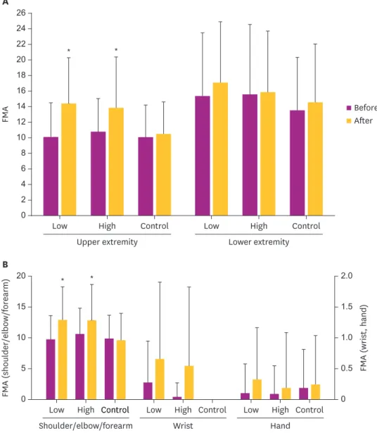
difference was found in the total LE-FMA (F = 2.58; p = 0.085). UE-FMA significantly improved in both high- and low-frequency rTMS groups following the intervention (p < 0.05), but not in the control group (Fig. 1A). Changes in the UE-FMA after stimulation were significantly more improved in both low- and high-frequency rTMS groups compared to the control group (p < 0.05). However, no significant difference was noted between low- and high-frequency rTMS groups (p > 0.05, Table 2).
Moreover, repeated measures ANOVA revealed a significant interaction between TIME before, after
and GROUP low, high, control on the shoulder/elbow/forearm subscore of the UE-FMA (F = 5.29;
* *
14 26 24
10
6
2 4 16
12
8 22 20 18
0
FMA
Low High
Upper extremity
Control Low High Control
A
Lower extremity
15
5 10 20
0
1.5
0.5 1.0 2.0
FMA (shoulder/elbow/forearm) 0 FMA (wrist, hand)
Low
*
High
Shoulder/elbow/forearm
*
Control
Control Low High Control Low High Control B
Wrist Hand
Before After
Fig. 1. Comparison of changes of FMA after stimulation according to stimulation methods. (A) UE-FMA scores were improved by low and high frequency rTMS. Repeated measures ANOVA showed a significant TIME × GROUP factor interaction (p < 0.05). (B) UE-FMA scores of shoulder/elbow/forearm were improved by low and high frequency rTMS. ANOVA showed a significant TIME × GROUP factor interaction (p < 0.05).
FMA, Fugl-Meyer Assessment; UE-FMA, upper extremity Fugl-Meyer Assessment; rTMS, repetitive transcranial magnetic stimulation; ANOVA, analysis of variance; Low, low frequency group; High, high frequency group;
Control, control group; Before, before stimulation; After, After stimulation.
*Significant at p < 0.05 vs. before stimulation.
p = 0.008), but not on the wrist and hand subscore of the FMA (F = 1.83; p = 0.171, F = 0.82;
p = 0.446). The shoulder/elbow/forearm subscale of the UE-FMA significantly increased in both high- and low-frequency rTMS groups (p < 0.05), but not in the control group (Fig. 1B).
Changes after the stimulation were significantly more improved in both low- and high- frequency rTMS groups compared to the control group (p < 0.05), but we did not find any significant difference between low- and high-frequency rTMS groups (p > 0.05). Detailed information of FMA after the stimulation are summarized in Table 2.
Cortical excitability
No lesional MEP response was observed in 54 of 58 patients in this study. Of these, 16 were in low-frequency stimulation group, 19 were in high-frequency stimulation group and 19 were in control group. There were no significant differences in response of lesional MEP among the 3 groups. No significant differences were found in the amplitude of MEP and MEP with ISI (3 ms, 5 ms, 10 ms, 15 ms) measured at baseline when stimulating the contralesional hemisphere among the 3 groups. Repeated measures ANOVA revealed a significant
interaction between TIME before, after and GROUP low, high, control on the amplitude of MEP (F = 6.15;
p = 0.004). However, no significant difference was observed in the amplitude of MEP with ISI (3 ms, 5 ms, 10 ms, 15 ms) (F = 0.95; p = 0.392, F = 0.87; p = 0.426, F = 2.45; p = 0.096, F = 1.12; p = 0.334, respectively). The amplitude of MEP significantly decreased in both high- and low-frequency rTMS groups (p < 0.05). A significant difference was noted in the changes after the stimulation in both low- and high-frequency rTMS groups compared to the control group (p < 0.05); however, no significant difference was observed between low- and high-frequency rTMS groups (p > 0.05). Detailed information on cortical excitability after stimulation are summarized in Table 3.
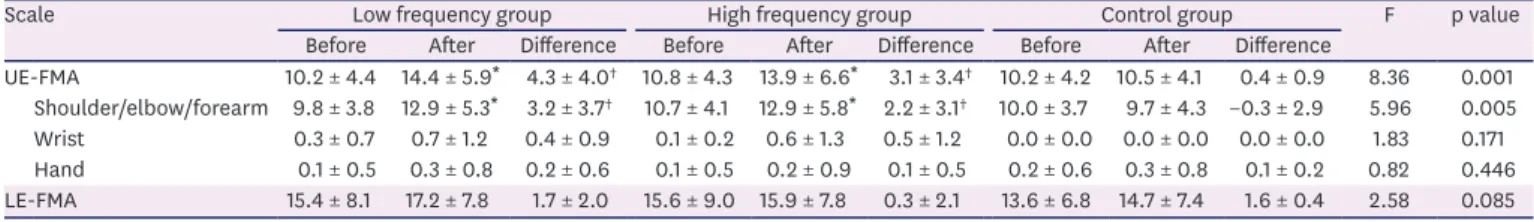
Table 2. Comparison of changes of FMA after stimulation according to stimulation methods
Scale Low frequency group High frequency group Control group F p value
Before After Difference Before After Difference Before After Difference
UE-FMA 10.2 ± 4.4 14.4 ± 5.9* 4.3 ± 4.0† 10.8 ± 4.3 13.9 ± 6.6* 3.1 ± 3.4† 10.2 ± 4.2 10.5 ± 4.1 0.4 ± 0.9 8.36 0.001 Shoulder/elbow/forearm 9.8 ± 3.8 12.9 ± 5.3* 3.2 ± 3.7† 10.7 ± 4.1 12.9 ± 5.8* 2.2 ± 3.1† 10.0 ± 3.7 9.7 ± 4.3 −0.3 ± 2.9 5.96 0.005 Wrist 0.3 ± 0.7 0.7 ± 1.2 0.4 ± 0.9 0.1 ± 0.2 0.6 ± 1.3 0.5 ± 1.2 0.0 ± 0.0 0.0 ± 0.0 0.0 ± 0.0 1.83 0.171 Hand 0.1 ± 0.5 0.3 ± 0.8 0.2 ± 0.6 0.1 ± 0.5 0.2 ± 0.9 0.1 ± 0.5 0.2 ± 0.6 0.3 ± 0.8 0.1 ± 0.2 0.82 0.446 LE-FMA 15.4 ± 8.1 17.2 ± 7.8 1.7 ± 2.0 15.6 ± 9.0 15.9 ± 7.8 0.3 ± 2.1 13.6 ± 6.8 14.7 ± 7.4 1.6 ± 0.4 2.58 0.085 Values are mean ± standard deviations.
FMA, Fugl-Meyer Assessment; UE-FMA, upper extremity Fugl-Meyer Assessment; LE-FMA, lower extremity Fugl-Meyer Assessment; Before, before stimulation;
After, after stimulation.
*p < 0.05 vs. before stimulation; †p < 0.05 vs. control group.
Table 3. Comparison of changes of contralesional cortical excitability after stimulation according to stimulation methods
Variables Low frequency group High frequency group Control group F p value
Before After Difference Before After Difference Before After Difference
MEP (mV) 1.07 ± 0.47 0.92 ± 0.41* −0.15 ± 0.21† 1.03 ± 0.41 0.93 ± 0.39*−0.10 ± 0.15† 0.94 ± 0.39 0.98 ± 0.39 0.04 ± 0.15 6.15 0.004 ISI 3 ms 0.50 ± 0.24 0.41 ± 0.16 −0.10 ± 0.15 0.50 ± 0.23 0.42 ± 0.20 −0.08 ± 0.19 0.37 ± 0.19 0.33 ± 0.18 −0.03 ± 0.08 0.95 0.392 ISI 5 ms 0.60 ± 0.20 0.47 ± 0.17 −0.13 ± 0.19 0.59 ± 0.25 0.49 ± 0.25 −0.10 ± 0.11 0.47 ± 0.22 0.40 ± 0.22 −0.07 ± 0.12 0.87 0.426 ISI 10 ms 1.24 ± 0.41 1.22 ± 0.37 −0.02 ± 0.20 1.17 ± 0.34 0.99 ± 0.35 −0.18 ± 0.23 0.99 ± 0.42 0.95 ± 0.33 −0.03 ± 0.31 2.45 0.096 ISI 15 ms 1.49 ± 0.59 1.41 ± 0.47 −0.08 ± 0.22 1.30 ± 0.30 1.07 ± 0.37 −0.23 ± 0.25 1.24 ± 0.57 1.10 ± 0.39 −0.15 ± 0.38 1.12 0.334 Values are mean ± standard deviations.
MEP, motor evoked potential; ISI, interstimulus interval; Before, before stimulation; After, after stimulation.
*p < 0.05 vs before stimulation; †p < 0.05 vs control group.
DISCUSSION
The findings in this study indicated that low- and high-frequency rTMS improved the upper extremity motor function, especially in the proximal upper limb, compared with the sham stimulation in post-stroke patients with severe upper-limb motor impairment. Additionally, the improvement was not statistically significant between low- and high-frequency rTMS.
These positive findings after rTMS were consistent with most randomized controlled trials documenting the effects of rTMS on the upper limb function in post-stroke patients [6,14- 21]. However, the upper limb function of their subjects was heterogeneous or mild to moderate. Khedr et al. [5] reported a significant positive correlation between pre rTMS MEP amplitude of the affected hemisphere and percent improvement in National Institutes of Health Stroke Scale and modified Rankin Scale. Carey et al. [22] reported that the responders showed a significantly greater baseline paretic hand function and preservation volume of the ipsilesional posterior limb of the internal capsule than the non-responders. Futhermore, Lee et al. [23] reported that the presence of an MEP response and high Motricity index arm score were significantly associated with a response to rTMS. Therefore, few studies have been reported on the effects of rTMS in post-stroke patients with severe upper-limb motor impairment. Boggio et al. [9] reported only one patient with stroke with a severe motor deficit whose motor function improved following low-frequency rTMS. Demirtas-Tatlidede et al. [24] reported motor improvement in post-stroke patients with severe upper-limb motor impairment after 1 Hz rTMS on the contralesional M1. However, this study was a single open- label trial. To our knowledge, this is the first randomized controlled study on post-stroke patients with severe upper-limb motor impairment.
This study revealed that rTMS was also effective even in post-stroke patients with severe upper-limb motor impairment. Interestingly, this study showed improvement of the FMA in the shoulder/elbow/forearm subscore rather than the wrist or hand subscore, although the hotspot of the ADM muscle was targeted. It is possible to explain that the rTMS induced rapid changes in the distorted cortical representation after extensive neuronal damage and improved the motor function. Although it was not clearly understood that the improvement of motor function mechanism was associated with rTMS, it has been thought that rTMS modulates cortical excitability [25]. After stroke, the abnormal interhemispheric inhibition is increased and may impede motor function [4,21,26]. Modulation of cortical excitability using rTMS would result in a decrease in the transcallosal inhibition to the affected hemisphere and improve motor function [4,21]. Animal studies showed that one strategy for recovery after stroke is for the surrounding surviving cortex to regain control over the function of the damaged motor area through somatotopic reorganization, a phenomenon called vicariation [27-29]. However, when extensive neuronal damage has occurred, there may not be enough surviving cortical neurons for representation in the perilesional area [30]. Extensive neuronal damage induces reorganization of perilesional and secondary motor area in functional recovery after stroke [30]. Nudo et al. [31] demonstrated that focal neuroplastic changes occurred following intracortical stimulation to the motor region representing a rat forelimb.
This kind of reorganization, placed in the motor region, modifies the motor cortical representational map, and this process can be induced with rTMS [32]. Another hypothesis is that rTMS stimulate the extrapyramidal tract as well as pyramidal tract. Extrapyramidal system has been known to influence the proximal (midline) more than the distal (peripheral) muscles and plays a key role in maintaining proper tone and posture. Zappulla et al. [33]
reported the components of the spinal MEP in rats arise from pathways outside the motor
cortex, such as the subcortical and brain stem structures, reflecting extrapyramidal system function. Kamida et al. [34] also reported that magnetic MEPs recorded from the limb muscles mainly reflect extrapyramidal activities. Therefore, proximal muscles such as shoulders and forearms may have shown a greater degree of recovery than the distal counterparts.
The excitability of the contralesional motor cortex shows physiological plasticity during functional recovery after stroke, and hyperactivity in the contralesional motor cortex occurs early after stroke [8]. Decreased contralesional M1 activity is correlated with functional recovery [35]. Cortical activity is well reflected by the amplitude of the MEP. Low-frequency rTMS produces a decreasing cortical activity, whereas high-frequency rTMS induces an increasing cortical excitability. This study showed that the amplitude of the MEP in the contralesional hemisphere significantly decreased after both contralesional low-frequency and lesional high-frequency rTMS. Matsuura et al. [35] reported that the reduction of contralesional M1 hyperactivity was related to the functional recovery after stroke. Veldema et al. [36] demonstrated that the greater the increase of ipsilesional corticospinal excitability, the better hand motor recovery. It may be hypothesized that the contralesional hemisphere activity was inhibited through transcallosal inhibition by increasing lesional cortical excitability by high-frequency stimulation on the affected M1 and directly by decreasing contralesional cortical excitability by low-frequency stimulation on the unaffected M1.
Nonetheless, it remains unclear what mechanism has reduced the amplitude of the
contralesional MEP in lesional high-frequency rTMS and contralesional low-frequency rTMS in this study. Future study on this matter will be necessary.
It is still controversial which stimulation method is more effective either high-frequency stimulation on lesional M1 or low-frequency stimulation on contralesional M1 in mild to moderate stroke [5,37,38]. Khedr et al. [5] previously revealed that 1 Hz on the contralesional hemisphere showed better improvement (hand grip, pegboard task, and keyboard tapping) than 3 Hz on the lesional hemisphere. Emara et al. [37] reported the similarity of 1 Hz and 5 Hz rTMS in improving the finger tapping test. In this study, both low-frequency rTMS on the contralesional hemisphere and high-frequency rTMS on the lesional hemisphere showed no significant difference in improving the motor function. This study was only a clinical trial comparing both stimulation methods in post-stroke patients with severe upper-limb motor impairment and was not designed to explain the possible mechanism for these findings.
Thus, several further clinical studies with a study design to reveal the mechanism in post- stroke patients with severe upper-limb motor impairment will be also necessary.
This study had several limitations. First, subjects with a relatively broad duration from stroke onset were enrolled from 1 week to 21 weeks. Second, the reliability of ICI and ICF measurements may be lower than expected because it was obtained by repeating 5 times, lower than minimum number of pulses needed to achieve reliable ICI, ICF suggested by Chang et al. [39]. Third, we could not reveal if the effect is sustained for a long time because of the short follow-up duration. Therefore, we hope to address this issue in future studies.
Moreover, the study was not designed to explain the possible mechanisms, but to reveal the clinical outcomes. Therefore, future large-scaled well-designed randomized controlled clinical study and basic research will be needed.
In conclusion, this study showed that rTMS may be helpful in improving the upper extremity motor function even in post-stroke patients with severe upper-limb motor impairment
and that the effects between contralesional low-frequency and lesional high-frequency stimulation may be similar.
REFERENCES
1. Kim DY, Ohn SH, Yang EJ, Park CI, Jung KJ. Enhancing motor performance by anodal transcranial direct current stimulation in subacute stroke patients. Am J Phys Med Rehabil 2009;88:829-836.
PUBMED | CROSSREF
2. Bütefisch CM, Khurana V, Kopylev L, Cohen LG. Enhancing encoding of a motor memory in the primary motor cortex by cortical stimulation. J Neurophysiol 2004;91:2110-2116.
PUBMED | CROSSREF
3. Ko MH, Jeong YC, Seo JH, Kim YH. The after-effect of sub-threshold 10 Hz repetitive transcranial magnetic stimulation on motor cortical excitability. J Korean Acad Rehabil Med 2006;30:436-440.
4. Avenanti A, Coccia M, Ladavas E, Provinciali L, Ceravolo MG. Low-frequency rTMS promotes use- dependent motor plasticity in chronic stroke: a randomized trial. Neurology 2012;78:256-264.
PUBMED | CROSSREF
5. Khedr EM, Abdel-Fadeil MR, Farghali A, Qaid M. Role of 1 and 3 Hz repetitive transcranial magnetic stimulation on motor function recovery after acute ischaemic stroke. Eur J Neurol 2009;16:1323-1330.
PUBMED | CROSSREF
6. Khedr EM, Etraby AE, Hemeda M, Nasef AM, Razek AA. Long-term effect of repetitive transcranial magnetic stimulation on motor function recovery after acute ischemic stroke. Acta Neurol Scand 2010;121:30-37.
PUBMED | CROSSREF
7. Mansur CG, Fregni F, Boggio PS, Riberto M, Gallucci-Neto J, Santos CM, Wagner T, Rigonatti SP, Marcolin MA, Pascual-Leone A. A sham stimulation-controlled trial of rTMS of the unaffected hemisphere in stroke patients. Neurology 2005;64:1802-1804.
PUBMED | CROSSREF
8. Nowak DA, Grefkes C, Dafotakis M, Eickhoff S, Küst J, Karbe H, Fink GR. Effects of low-frequency repetitive transcranial magnetic stimulation of the contralesional primary motor cortex on movement kinematics and neural activity in subcortical stroke. Arch Neurol 2008;65:741-747.
PUBMED | CROSSREF
9. Boggio PS, Alonso-Alonso M, Mansur CG, Rigonatti SP, Schlaug G, Pascual-Leone A, Fregni F. Hand function improvement with low-frequency repetitive transcranial magnetic stimulation of the unaffected hemisphere in a severe case of stroke. Am J Phys Med Rehabil 2006;85:927-930.
PUBMED | CROSSREF
10. Sasaki N, Mizutani S, Kakuda W, Abo M. Comparison of the effects of high- and low-frequency repetitive transcranial magnetic stimulation on upper limb hemiparesis in the early phase of stroke. J Stroke Cerebrovasc Dis 2013;22:413-418.
PUBMED | CROSSREF
11. Luft AR, McCombe-Waller S, Whitall J, Forrester LW, Macko R, Sorkin JD, Schulz JB, Goldberg AP, Hanley DF. Repetitive bilateral arm training and motor cortex activation in chronic stroke: a randomized controlled trial. JAMA 2004;292:1853-1861.
PUBMED | CROSSREF
12. Groppa S, Oliviero A, Eisen A, Quartarone A, Cohen LG, Mall V, Kaelin-Lang A, Mima T, Rossi S, Thickbroom GW, Rossini PM, Ziemann U, Valls-Solé J, Siebner HR. A practical guide to diagnostic transcranial magnetic stimulation: report of an IFCN committee. Clin Neurophysiol 2012;123:858-882.
PUBMED | CROSSREF
13. Chistyakov AV, Kaplan B, Rubichek O, Kreinin I, Koren D, Hafner H, Feinsod M, Klein E. Effect of electroconvulsive therapy on cortical excitability in patients with major depression: a transcranial magnetic stimulation study. Clin Neurophysiol 2005;116:386-392.
PUBMED | CROSSREF
14. Watanabe K, Kudo Y, Sugawara E, Nakamizo T, Amari K, Takahashi K, Tanaka O, Endo M, Hayakawa Y, Johkura K. Comparative study of ipsilesional and contralesional repetitive transcranial magnetic stimulations for acute infarction. J Neurol Sci 2018;384:10-14.
PUBMED | CROSSREF
15. Lüdemann-Podubecká J, Bösl K, Nowak DA. Inhibition of the contralesional dorsal premotor cortex improves motor function of the affected hand following stroke. Eur J Neurol 2016;23:823-830.
PUBMED | CROSSREF
16. Cha HG, Kim MK. Effects of repetitive transcranial magnetic stimulation on arm function and decreasing unilateral spatial neglect in subacute stroke: a randomized controlled trial. Clin Rehabil 2016;30:649-656.
PUBMED | CROSSREF
17. Zheng CJ, Liao WJ, Xia WG. Effect of combined low-frequency repetitive transcranial magnetic stimulation and virtual reality training on upper limb function in subacute stroke: a double-blind randomized controlled trail. J Huazhong Univ Sci Technolog Med Sci 2015;35:248-254.
PUBMED | CROSSREF
18. Matsuura A, Onoda K, Oguro H, Yamaguchi S. Magnetic stimulation and movement-related cortical activity for acute stroke with hemiparesis. Eur J Neurol 2015;22:1526-1532.
PUBMED | CROSSREF
19. Sung WH, Wang CP, Chou CL, Chen YC, Chang YC, Tsai PY. Efficacy of coupling inhibitory and facilitatory repetitive transcranial magnetic stimulation to enhance motor recovery in hemiplegic stroke patients. Stroke 2013;44:1375-1382.
PUBMED | CROSSREF
20. Chang WH, Kim YH, Bang OY, Kim ST, Park YH, Lee PK. Long-term effects of rTMS on motor recovery in patients after subacute stroke. J Rehabil Med 2010;42:758-764.
PUBMED | CROSSREF
21. Fregni F, Boggio PS, Valle AC, Rocha RR, Duarte J, Ferreira MJ, Wagner T, Fecteau S, Rigonatti SP, Riberto M, Freedman SD, Pascual-Leone A. A sham-controlled trial of a 5-day course of repetitive transcranial magnetic stimulation of the unaffected hemisphere in stroke patients. Stroke 2006;37:2115-2122.
PUBMED | CROSSREF
22. Carey JR, Deng H, Gillick BT, Cassidy JM, Anderson DC, Zhang L, Thomas W. Serial treatments of primed low-frequency rTMS in stroke: characteristics of responders vs. nonresponders. Restor Neurol Neurosci 2014;32:323-335.
PUBMED
23. Lee JH, Kim SB, Lee KW, Kim MA, Lee SJ, Choi SJ. Factors associated with upper extremity motor recovery after repetitive transcranial magnetic stimulation in stroke patients. Ann Rehabil Med 2015;39:268-276.
PUBMED | CROSSREF
24. Demirtas-Tatlidede A, Alonso-Alonso M, Shetty RP, Ronen I, Pascual-Leone A, Fregni F. Long-term effects of contralesional rTMS in severe stroke: safety, cortical excitability, and relationship with transcallosal motor fibers. NeuroRehabilitation 2015;36:51-59.
PUBMED
25. Siebner HR, Rothwell J. Transcranial magnetic stimulation: new insights into representational cortical plasticity. Exp Brain Res 2003;148:1-16.
PUBMED | CROSSREF
26. Murase N, Duque J, Mazzocchio R, Cohen LG. Influence of interhemispheric interactions on motor function in chronic stroke. Ann Neurol 2004;55:400-409.
PUBMED | CROSSREF
27. Merzenich MM, Nelson RJ, Stryker MP, Cynader MS, Schoppmann A, Zook JM. Somatosensory cortical map changes following digit amputation in adult monkeys. J Comp Neurol 1984;224:591-605.
PUBMED | CROSSREF
28. Sanes JN, Suner S, Donoghue JP. Dynamic organization of primary motor cortex output to target muscles in adult rats. I. Long-term patterns of reorganization following motor or mixed peripheral nerve lesions.
Exp Brain Res 1990;79:479-491.
PUBMED | CROSSREF
29. Donoghue JP, Suner S, Sanes JN. Dynamic organization of primary motor cortex output to target muscles in adult rats. II. Rapid reorganization following motor nerve lesions. Exp Brain Res 1990;79:492-503.
PUBMED | CROSSREF
30. Hoyer EH, Celnik PA. Understanding and enhancing motor recovery after stroke using transcranial magnetic stimulation. Restor Neurol Neurosci 2011;29:395-409.
PUBMED
31. Nudo RJ, Jenkins WM, Merzenieh MM. Repetitive microstimulation alters the cortical representation of movements in adult rats. Somatosens Mot Res 1990;7:463-483.
PUBMED | CROSSREF
32. Harvey RL, Nudo RJ. Cortical brain stimulation: a potential therapeutic agent for upper limb motor recovery following stroke. Top Stroke Rehabil 2007;14:54-67.
PUBMED | CROSSREF
33. Zappulla RA, Hollis P, Ryder J, Moore FM, Adamson J, Moustakis W, Malis LI. Noncortical origins of the spinal motor evoked potential in rats. Neurosurgery 1988;22:846-852.
PUBMED | CROSSREF
34. Kamida T, Fujiki M, Hori S, Isono M. Conduction pathways of motor evoked potentials following transcranial magnetic stimulation: a rodent study using a “figure-8” coil. Muscle Nerve 1998;21:722-731.
PUBMED | CROSSREF
35. Matsuura A, Karita T, Nakada N, Fukushima S, Mori F. Correlation between changes of contralesional cortical activity and motor function recovery in patients with hemiparetic stroke. Phys Ther Rev 2017;20:28-35.
CROSSREF
36. Veldema J, Bösl K, Nowak DA. Cortico-spinal excitability and hand motor recovery in stroke: a longitudinal study. J Neurol 2018;265:1071-1078.
PUBMED | CROSSREF
37. Emara TH, Moustafa RR, Elnahas NM, Elganzoury AM, Abdo TA, Mohamed SA, Eletribi MA. Repetitive transcranial magnetic stimulation at 1Hz and 5Hz produces sustained improvement in motor function and disability after ischaemic stroke. Eur J Neurol 2010;17:1203-1209.
PUBMED | CROSSREF
38. Takeuchi N, Tada T, Toshima M, Matsuo Y, Ikoma K. Repetitive transcranial magnetic stimulation over bilateral hemispheres enhances motor function and training effect of paretic hand in patients after stroke. J Rehabil Med 2009;41:1049-1054.
PUBMED | CROSSREF
39. Chang WH, Fried PJ, Saxena S, Jannati A, Gomes-Osman J, Kim YH, Pascual-Leone A. Optimal number of pulses as outcome measures of neuronavigated transcranial magnetic stimulation. Clin Neurophysiol 2016;127:2892-2897.
PUBMED | CROSSREF