217
Immune Network
Lines, Scid.adh, R1.1 and EL-4
Jong Seok Chae1, Hae-jung Kim1, Weon Seo Park1, Youngmee Bae1 and Kyeong Cheon Jung2
1Department of Pathology, Kangwon National University College of Medicine, Chunchon, Korea;
2Department of Pathology, Hallym University College of Medicine, Chuncheon, Korea
ABSTRACT
Background: Scid.adh is a recently developed murine thymic lymphoma cell line, which has been used as in vitro model for the study of double negative stage III thymocytes.
In this study, we compared the expression profile of a number of genes and proteins, which are tightly related to T cell development and apoptosis, in thymic lymphoma cell lines, R1.1, EL-4, and Scid.adh for the developmental staging. Methods: We examined the expression of development marker genes and proteins in three lymphoma cell lines by flow cytometry and RT-PCR. In addition, the expression of apoptosis-related molecules including bcl-2, bax and Fas was also investigated. Results: As previously reported, Scid.adh cell line expressed CD8 and CD25 but not TCR α chain, while R1.1 cells expressed TCR α chain and both CD4 and CD8 transcripts. These suggest that R1.1 might be in double positive stage, and low level of CD44 expression and the absence of CD25 support this suggestion. In contrast, EL-4 cells showed high level of TCR α chain transcript, and low-level of CD4 expression, suggesting that EL-4 is in more mature stage than R1.1. Further, this suggestion was supported by the lack of mT-20 in EL-4 cells, which is expressed in the immature thymocytes, and Scid.adh and R1.1 cell lines, but not in the terminally differentiated thymocytes and peripheral T cells. Among the apoptosis-related gene, transcripts of bcl-2 gene were detected in both R1.1 and EL-4 but not in Scid.adh cells, while bax was expressed in all cell lines.
Fas expression was the highest in EL-4 cells and low in Scid.adh cell line. Conclusion:
R1.1 cell may represent double positive stage, and EL-4 is more differentiated cell line.
In addition, Scid.adh and EL-4 cell lines are suspected to be useful for the study of function of bcl-2 family and Fas during the thymocyte development, respectively.
(Immune Network 2002;2(4):217-222)
Key Words: Thymocyte development, lymphoma cell line
Correspondence to: Kyeong Cheon Jung, Department of Pathol- ogy, Hallym University College of Medicine, 1 Okchon-dong, Chuncheon 200-702, Korea. (TEL) +82-33-240-1631, (FAX) +82-33-241-8250, (E-mail) jungkc@hallym.ac.kr
This work was supported by the fund from Hallym University (2000).
Introduction
Intrathymic T cell development is marked by the ordered changes in expression of the CD4 and CD8 differentiation antigens (1). The immature thymocytes are characterized by germline configuration of TCR gene and the absence of both CD4 and CD8 cor- eceptors (termed double negative, DN, stage). Further, progression of the immature thymocytes is defined by the sequential expression of CD44 and CD25 pro- teins. For instance, CD44+CD25- (DN I) cells are immediately followed by CD25 expression (the CD44+
CD25+ DN II), and then lose CD44 expression and continue the TCR-β chain gene rearrangement (CD44- CD25+ DN III). At DN III stage, thymocytes, that express functional TCR-β chains associated with the pre-TCR-α chain as a pre-TCR complex on the sur- face, are known to downregulate CD25 expression, and differentiate into CD44-CD25- DN IV cells (2, 3). At the next developmental stage, thymocytes coexpress the CD4 and CD8 proteins, and become CD4+CD8+ DP cells. During this process, TCR-α genes are now to allow undergoing rearrangement.
Subsequently, only the thymocytes that have success- fully rearranged the TCR-α genes and expressed α/β TCR complexes undergo positive and negative se- lection (4,5). Upon interaction of TCR and MHC- peptide complex, DP cells that express TCRs of intermediate affinity are positively selected, differen- tiate into CD4 or CD8 single positive (SP) cells, and
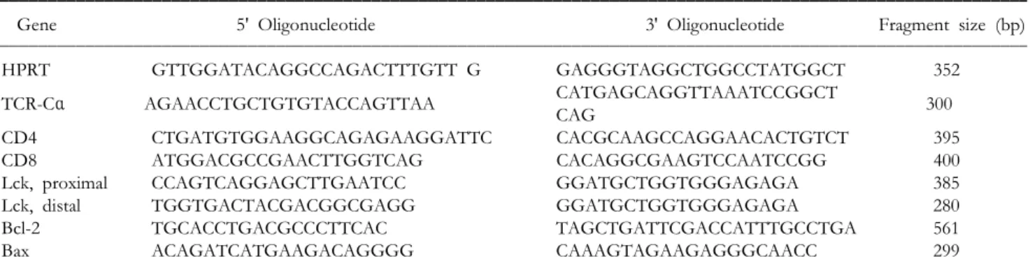
Table I. Oligonucleotides used to amplify genes from Scid.adh, R1.1 and EL-4 cDNA
Gene 5' Oligonucleotide 3' Oligonucleotide Fragment size (bp)
HPRT GTTGGATACAGGCCAGACTTTGTT G GAGGGTAGGCTGGCCTATGGCT 352
CATGAGCAGGTTAAATCCGGCT
TCR-Cα AGAACCTGCTGTGTACCAGTTAA 300
CAG
CD4 CTGATGTGGAAGGCAGAGAAGGATTC CACGCAAGCCAGGAACACTGTCT 395
CD8 ATGGACGCCGAACTTGGTCAG CACAGGCGAAGTCCAATCCGG 400
Lck, proximal CCAGTCAGGAGCTTGAATCC GGATGCTGGTGGGAGAGA 385
Lck, distal TGGTGACTACGACGGCGAGG GGATGCTGGTGGGAGAGA 280
Bcl-2 TGCACCTGACGCCCTTCAC TAGCTGATTCGACCATTTGCCTGA 561
Bax ACAGATCATGAAGACAGGGG CAAAGTAGAAGAGGGCAACC 299
allow exiting to the peripheral lymphatic organs.
In order to understand the mechanisms involved in thymocyte maturation, several in vitro culture systems have been established (6). For example, the commonly used system to study negative selection involves stimulation of DP thymocytes with anti-CD3 antibody or antigenic peptides-pulsed antigen pres- enting cells (7). In contrast, fetal thymic organ culture system is used to provide an experimental system in which thymocytes development proceeds in a manner comparable to that seen in vivo (8). In addition, thymic lymphoma cell lines might be useful. Especially Scid.adh cell line, which was derived from scid mouse, was established as a model cell line that mimics the DN III stage thymocytes (9,10). EL-4 and R1.1 are other thymic lymphoma cell lines available, although these are not fully characterized. Therefore, we compared the expression of developmentally reg- ulated genes and proteins in R1.1 and EL-4 cell lines with Scid.adh to assess the developmental stages of these cell lines. In addition, the expression of apoptosis- related genes were analyzed.
Materials and Methods
Cell line. EL-4 and R1.1 cell lines were purchased from American Type Culture Collection (Manassas, VA), and Scid.adh cell line was obtained from Dr.
David L. Wiest (Fox Chase Cancer, Philadelphia, PA) with permission. EL-4 and R1.1 cell lines were cultured in DMEM media (Gibco BRL, Rockville, MD) supplemented with 10% hoarse serum, while Scid.adh cell line was maintained in IMEM media supplemented, as previously described (11).
Flow cytometry and antibodies. For each sample, 106 cells were washed in staining buffer (Phosphate buffered saline with 0.1% sodium azide), stained for 30 min at 4oC with the indicated fluorochrome-conjugated monoclonal antibodies (mAbs), washed twice in staining buffer, and analyzed by flow cytometry using
a FACS Calibur cytometer (Becton Dickinson, San
Jose, CA) and CELLQUEST software (Becton Dick- inson). Dead cells were excluded using the vital dye 7-aminoactinomcyin D (Sigma, St. Louis, Mo). The following fluorochrome-conjugated mAbs were used in flow cytometry: anti-CD3ε-CyChrome (145-2C11), anti-CD5-biotin (53-7.3), anti-CD24-PE (M1/69), anti- CD25-FITC (7D4), anti-CD44-PE (IM7), anti-CD69- FITC (H1.2F3), anti-Fas-PE (Jo2), anti-TCR-β-FITC (H57-597) [purchased from PharMingen, San Diego, CA], anti-CD4-CyChrome (GK1.5), and anti-CD8α- PE (53-6.7) [purchased from DiNonA, Seoul, Korea].
One anti-mouse thymocytes antibody, named as mT- 20, was developed in our laboratory and conjugated with FITC in DiNonA for flow cytometry.
RT-PCR. Total RNA was isolated from a total of 1×10⁶ thymic lymphoma cells into a final volume of 10μl of RNase-free H2O, using RNA extraction kit (Bioneer, Taejon, Korea) and cDNA was first pre- pared by reverse transcription of 10μl RNA in a 50μl reaction volume containing 1 mM DTT, 200μM dNTPs, and 200 pmol oligo dT18. 2.5μl cDNA were amplified in PCR master mix composed of 10 mM Tris-HCl, pH 9.0, 1 U Taq DNA polymerase, 250μM dNTP, 40 mM KCl, 1.5 mM MgCl2 (Bioneer, Taejon, Korea), using primers specific for: HPRT, TCR-Cα, CD4, CD8, proximal lck, distal lck, Bcl-2, and Bax (see Table I). The PCR products were resolved by 2% agarose gel electrophoresis and stained with ethidium bromide.
Results
Differential expression of development marker antigens in three cell lines. As the CD4 and CD8 proteins are usually accepted makers for the identification of three de- velopmental stages of intrathymic T cells, we first com- pared the expression of these markers in three thymic lymphoma cell lines by flow cytometry and RT-PCR (Fig. 1, Table II). As known previously (9), Scid.adh cells expressed CD8 but not CD4, and this was also
confirmed by RT-PCR. While neither CD4 nor CD8
Figure 1. Expression of TCR, CD3, CD4, and CD8 in thymic lymphoma cell lines. A. Tumor cells were stained with indicated antibodies and analyzed by flow cytometry. The positive cell percentages are marked. B. RNAs were extracted from cultured tumor cells, followed by RT-PCR. The PCR products were resolved by agarose gel electrophoresis and stained with ethidium bromide.
Figure 2. Expression of CD25, CD44, and mT-20 in thymic lymphoma cell lines. Tumor cells were stained with indicated antibodies and analyzed by flow cytometry. The positive cell percentages are marked.
Table II. Summary of surface antigen phenotype of R1.1, EL-4 and Scid.adh
R1.1 EL-4 Scid.adh
TCR-β 0.38* 38.44 0.58
CD3 3.79 15.14 4.85
CD4 1.23 7.54 3.73
CD8 2.13 1.58 46.33
CD25 0.53 0.28 96.25
CD44 12.68 99.36 1.83
mT-20 98.52 1.67 97.2
CD5 37.16 99.8 99.4
CD24 45.3 83.18 49.59
CD69 2.99 0.52 7.2
CD95 (Fas) 59.02 81.03 18.27
*The percentages of positive cell populations are indicated
Figure 3. Expression of lck gene by proximal and distal pro- moters. The products of RT-PCR using the primers specific for transcripts of lck by proximal or distal promoter were resolved by agarose gel electrophoresis and stained with ethidium bromide.
was expressed on the surface of R1.1 cells, transcripts of both CD4 and CD8 were detected in this cell line.
EL-4 cells expressed only low-level of CD4 molecules (Fig. 1, Table II).
Rearrangement of TCR gene and surface expres- sion of TCR and CD3 molecules are also critical in intrathymic T cell development. As a consequence, we compared surface expression of TCR and CD3 molecules in three cell lines by flow cytometry and rearrangement status of TCR α gene was assessed by RT-PCR (Fig. 1, Table II). The TCR protein was not expressed on the surface of Scid.adh cells and these cells showed scanty expression of CD3 mol- ecules on the surface. In addition, transcripts of TCR Cα gene were not detected in Scid.adh cell line (Fig.
1), as previously reported (9). In contrast, transcripts of TCR Cα gene were detected in R1.1 cell line, although this cell line did not express TCR protein on the surface. In addition, EL-4 cell line showed low-level of TCR expression on the surface and abundant transcripts of TCR Cα gene (Fig. 1). These suggest that Scid.adh represents DN stage before
TCR α gene rearrangement, while R1.1 and EL-4 cell lines represent more mature stage.
Surface expression of CD25, CD44 and mT-20. In addi- tion to CD4 and CD8 antigens, DN thymocytes are further classified into four stage based on the surface expression of CD25 and CD44 antigens. CD25 protein is expressed exclusively in subsets of DN thymocytes, whereas subsets of DN and DP thymocytes express CD44. Especially, β selection in DN stage induces the differentiation of CD25+CD44- (DN III) thy- mocytes to the CD25-CD44- (DN IV) stage and the rearrangement of TCR α gene. In this study, the expression pattern of CD25 and CD44 was also examined in three cell lines (Fig. 2, Table II). As previously reported, Scid.adh cell line was CD25+
CD44-, while subset of R1.1 cells and most of EL-4 cells expressed CD44 antigen but not CD25 mol- ecule.
We recently developed a monoclonal antibody, named mT-20, that reacts with mouse thymocytes.
Although the antigen recognized by mT-20 mAb was
Figure 4. Expression of apoptosis-related gene. A. The expres- sion of Fas on the surface of lymphoma cells were analyzed by flow cytometry. B. The transcripts of bax and bcl-2 genes were detected by RT-PCR, followed by staining with ethidium bromide.
not fully identified, studies in our laboratory showed that the expression patterns of mT-20 in lymphoid cells were similar to those of CD24 (heat stable antigen). Especially, this antibody showed the bright- est staining in DN thymocytes and intermediate staining in DP thymocytes, while it's expression was downregulated in SP thymocytes and peripheral T cells were not reactive to the mT-20 mAb (unpub- lished data). These suggested that the phenotypic analysis with mT-20 mAb might be useful for the classification of thymocytes differentiation stages. We next analyzed the mT-20 reactivity in thymic lym- phoma cell lines, and both Scid.adh and R1.1 cell lines were stained by mT-20 mAb, with higher expression in Scid.adh as compared to R1.1 (Fig. 2, Table II). In contrast, interestingly, EL-4 cell line did not express mT-20 antigen at all.
All these results suggest that Scid.adh cell line might represent DN III stage and R1.1 represents DP stage. In addition, EL-4 represents more mature stage than R1.1 in the respects of higher expression of TCR α chain and the absence of mT-20.
Additionally, expression pattern of other surface antigens is summarized in Table II.
Expression of p56lck. Lck gene expression is regulated in T lymphocytes through the coordinated activities of two independent promoter elements (12,13). The promoter lying directly upstream of the lck structural exons (proximal) is active throughout thymopoiesis (14,15). In contrast, the activity of the distal lck promoter (residing approximately 20 kB upstream of the lck proximal promoter) is developmentally de- layed as compared to that of the proximal promoter, coinciding with the acquisition of surface TCR expres- sion in developing thymocytes and higher in mature DP thymocytes and peripheral T cells (16). We ex- amined the expression of lck gene regulated by two independent promoters in three thymic lymphoma cell lines. As shown in Fig. 3, only the proximal transcripts were detected in all cell lines (Fig. 3).
These results suggest that all three lymphoma cell lines are in intrathymic stage.
Expression of apoptosis-associated gene. Apoptosis is a critical process for the elimination of both non- functioning and self-reactive thymocytes during thy- mic selection and thymic lymphoma cell lines could be useful for the study of thymocyte apoptosis. In this study, we measured the expression of bcl-2, bax and Fas genes in three thymic lymphoma cell lines.
Flow cytometric analysis showed the highest and intermediate expression of Fas on the surface of EL-4 and R1.1 cell lines, respectively, whereas Fas expression on the surface of Scid.adh was very low (Fig. 4A, Table II). Transcripts of bcl-2, one of anti-apoptotic genes, were abundant in R1.1 and also
detected in EL-4 cell line, but hardly detectable in Scid.adh cells (Fig. 4B). Transcripts of bax were abundant in all three cell lines (Fig. 4B).
Discussion
For the study of functions of several genes during thymic education, thymic lymphoma cell lines could be useful prior to the generation of genetically ma- nipulated mouse. For example, several studies using Scid.adh cell line revealed that CD3ε-signaling and the expression of egr gene are important during β selection of DN thymocytes (9,10). In this study, we compared the expression of several marker genes in R1.1 and EL-4 cell line with that in Scid.adh cell line and showed that R1.1 is in DP stage, and EL-4 represents the more mature stages than R1.1.
During thymic development, intrathymic T cells are classified into three populations based on the surface expression pattern of CD4 and CD8 proteins.
Flow cytometric analysis showed that CD8 and CD4 expression were present on the surface of Scid.adh and EL-4 cell lines, respectively, while either protein were not expressed on the surface of R1.1. However, Scid.adh cell line was known to represent DN III stage in spite of CD8 expression, because TCR α gene was not rearranged. Therefore, we assessed the rearrangement status of TCR gene in three cell lines.
During thymic education, TCR β gene is initially rearranged and expressed for β selection in DN stage, followed by differentiation into DP stage and rearrangement and transcription of TCR α gene.
Therefore, transcripts of TCR α gene are not detected in DN thymocytes and Scid.adh cell line (Fig. 1B; 10).
However, transcripts of TCR α gene were detected in both R1.1 and EL-4 cell lines and more abundant in EL-4 than in R1.1. In addition, transcripts of both CD4 and CD8 were detected in R1.1 cell line.
Therefore, these results suggest that R1.1 cell line represents the DP stage, while EL-4 represents the more differentiated DP or SP stage.
The differentiation stage of three lymphoma cell lines was further assessed by the expression of CD25, mT-20 and p56lck. CD25 is expressed in DN II and III thymocytes. Upon β selection, these cells undergo proliferative expansion, down-regulate CD25, pro- gress to the immature single positive (ISP) stage by up-regulating CD8, and finally develop into DP thymocytes that do not express CD25 (9). The mT- 20, which was recently developed anti-mouse thy- mocyte mAb in our laboratory, shows strongest reactivity against DN thymocyte and intermediate reactivity in DP thymocytes (unpublished data). Its immunoreactivity was further decreased in SP thy- mocyte and not detected in terminally differentiated SP thymocytes and peripheral T cells (unpublished data). Therefore, the expression pattern of CD25 and mT-20 might be useful for the evaluation of thy- mocyte differentiation stage and the expression of these molecules was assessed in three lymphoma cell lines. As know previously (9), Scid.adh cell line express CD25 antigen and also showed mT-20- immunoreativity. In contrast, CD25 was not expres- sed on the surface of R1.1 cell line and R1.1 cells showed lower intensity against mT-20. EL-4 cell line did not express either CD25 or mT-20. These results support the suggestion that R1.1 line is in DP stage and EL-4 cell line is more differentiated than R1.1 Lck is another differentiation marker of thymo- cytes and T cells, as the activity of proximal lck promoter is high only in immature thymocytes, while activity of distal lck promoter is strong in mature thymocyte and peripheral T cells (16). As expected, only the proximal transcripts of lck were detected in Scid.adh and R1.1. In addition, EL-4 cell line also showed proximal transcripts only. These results indicate that EL-4 cell line is not fully maturated.
During thymic education, non-functioning and self-reactive thymocytes are eliminated by apoptosis (17). Since apoptosis plays such an important role in T cell development, a number of studies have ex- amined the function of apoptosis regulatory mol- ecules in this process. In particular, a bcl-2 transgene protect thymocyte against dexamethasone-induced apoptosis, while a bax transgene accelerates thy- mocyte apoptosis (18-20). In this study, the expres- sion of bcl-2 and bax genes was evaluated in the lymphoma cell lines by RT-PCR. Characteristically, abundant transcripts of bcl-2 gene were detected in
R1.1 and EL-4 cell line, but not in Scid.adh. In contrast, transcripts of bax gene were detected in all cell lines.
In addition, Fas is one of apoptosis-inducing proteins in thymocytes. Further, recent work suggests that Fas is involved in thymic T cell development as a costimulatory molecules (21). Flow cytometric analysis showed that highest expression of Fas on the surface of EL-4 cell line, and intermediate expression in R1.1 cell line. Expression of Fas in Scid.adh cells was very low (Fig. 4).
These results imply that Scid.adh cell line might be useful for the study of bcl-2-associated apoptosis during thymic selection, while EL-4 could be useful to evaluate the Fas-mediated function in thymic ed- ucation.
Conclusively, this work suggests that R1.1 cells represent DP stage and EL-4 cell line is more dif- ferentiated than R1.1. In addition, Scid.adh and EL-4 could be useful for the study of function of bcl-2 family and Fas, respectively.
Acknowledgment
We thank Dr. David L. Wiest for Scid.adh cell line.
We thank Jae Nam Seo, Jin Sil Choi, Seung Hee Lee, and Ju Hyun Kim (Departement of Patholgoy, Hallym University College of Medicine) for their kind technical assistance.
References
1. Shortman K, Wu L: Early T lymphocyte progenitors. Annu Rev Immunol 14;29-47, 1996
2. Dudley EC, Petrie HT, Shah LM, Owen MJ, Hayday AC:
T cell receptor b chain gene rearrangement and selection during thymocyte development in adult mice. Immunity 1;83-93, 1994
3. Levelt CN, Eichmann K: Receptors and signals in early thymic selection. Immunity 3;667-672, 1995
4. Nossal GJ: Negative selection of lymphocytes. Cell 76;229- 239, 1994
5. Von Boehmer H: Positive selection of lymphocytes. Cell 76;219-228, 1994
6. Hare KJ, Jenkinson EJ, Anderson G: In vitro models of T cell development. Semin Immunol 11;3-12, 1999
7. Swat W, Ignatowicz L, von Boehmer H, Kisielow P: Clonal deletion of immature CD4+8+ thymocytes in suspension culture by extrathymic antigen-presenting cells. Nature 351;
150-153, 1991
8. Jenkinson EJ, Anderson G: Fetal thymic organ cultures. Curr Opin Immunol 6;293-297, 1994
9. Carleton M, Ruetsch NR, Berger MA, Rhodes M, Kaptik S, Wiest DL: Signals transduced by CD3e, but not by surface pre-TCR complexes, are able to induce maturation of an early thymic lymphoma in vitro. J Immunol 163;2576-2585, 1999
10. Carleton M, Haks MC, Smeele SA, Jones A, Belkowski SM, Berger MA, Linsley P, Kruisbeek AM, Wiest DL: Early growth response transcription factors are required for de- velopment of CD4-CD8- thymocytes to the CD4+CD8+ stage. J Immunol 168;1649-1658, 2002
11. Wiest DL, Burgess WH, McKean D, Kearse KP, Singer A:
The molecular chaperone calnexin is expressed on the surface of immature thymocytes in association with clonotype- independent CD3 complexes. EMBO J 14;3425-33, 1995 12. Adler HT, Reynolds PJ, Kelley CM, Sefton BM: Tran-
scriptional activation of lck by retrovirus promoter insertion between two lymphoid-specific promoters. J Virol 62;4113- 4122, 1988
13. Garvin AM, Pawar S, Marth JD, Perlmutter RM: Structure
of the murine lck gene and its rearrangement in a murine lymphoma cell line. Mol Cell Biol 8;3058-3064, 1988 14. Reynolds PJ, Lesley J, Trotter J, Schulte R, Hyman R, Sefton
BM: Changes in the relative abundance of type I and type II lck mRNA transcripts suggest differential promoter usage during T-cell development. Mol Cell Biol 10;4266-4270, 1990 15. Wildin RS, Garvin AM, Pawar S, Lewis DB, Abraham KM, Forbush KA, Ziegler SF, Allen JM, Perlmutter RM: Devel- opmental regulation of lck gene expression in T lymphocytes.
J Exp Med 173;383-393, 1991
16. Wildin RS, Wang HU, Forbush KA, Perlmutter RM: Func- tional dissection of the murine lck distal promoter. J Immunol 155;1286-1295, 1995
17. Sebzda E, Mariathasan S, Ohteki T, Jones R, Bachmann MF, Ohashi PS: Selection of the T cell repertoire. Annu Rev Immunol. 17;829-874, 1999
18. Sentman CL, Shutter JR, Hockenbery D, Kanagawa O, Korsmeyer SJ: bcl-2 inhibits multiple forms of apoptosis but not negative selection in thymocytes. Cell 67;879-888, 1991 19. Strasser A, Harris AW, Cory S: bcl-2 transgene inhibits T cell
death and perturbs thymic self-censorship. Cell 67;889-899, 1991
20. Brady HJ, Salomons GS, Bobeldijk RC, Berns AJ: T cells from baxalpha transgenic mice show accelerated apoptosis in response to stimuli but do not show restored DNA damage- induced cell death in the absence of p53 gene product in.
EMBO J 15;1221-1230, 1996
21. Kurasawa K, Hashimoto Y, Kasai M, Iwamoto I: The fas antigen is involved in thymic T-cell development as a costimulatory molecule, but not in the deletion of neglected thymocytes. J Allergy Clin Immunol 106;S19-S31, 2000