Differences of Chest and Waist Circumferences in Spastic Diplegic and Hemiplegic Cerebral Palsy
Purpose: Circumference of the chest and waist can be one of clinical indicator to reflect respiratory function in children with cerebral palsy. In this study, we compared to differences in the chest/waist circumference and maximal phonation time between children with spastic diplegia and hemiplegia.
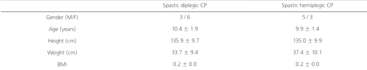
Methods: Seventeen children with spastic diplegic and hemiplegic cerebral palsy were recruited, who were matched to gender, age, height, weight, and body mass index for control of the known factors affected to respiratory function. The chest/waist circumference and were measured in each group, when children took a breath at rest and at maximal voluntary inspiration/
expiration.
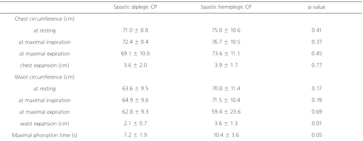
Results: No significant differences were found in the chest and waist circumference and expansion between the two groups.
However, only in the waist expansion, children with diplegic CP were significantly lower extensibility of lung, compared to the other group. In comparison of the maximal phonation time, a significant lower score was shown in children with spastic diplegic CP, compared to children with hemiplegic CP.
Conclusion: Our results indicated that children with spastic diplegic CP had smaller chest wall and waist, compared to children with spastic hemiplegic CP. In addition, they showed a shorter time for sustaining phonation than spastic hemiplegic CP did.
Therefore, spastic diplegic CP will be required for careful monitor regarding respiratory function in rehabilitation settings.
Key Words: Cerebral palsy, Chest circumference, Waist circumference, Maximal phonation time.
Ki Seok Nam
1, Hye Young Lee
21
Department of Physical Therapy, Yeungnam College of Science and Technology,
2Department of Rehabilitation Science, Graduate School, Daegu University
The Journal of Korean Society of Physical Therapy Original Article
Ⅰ. Introduction
Cerebral palsy (CP) is one of common disease in pediatric physical therapy, which is defined as a non-progressive neurological disorder that causes sensoriomotor dysfunction in physical development.
1-3Children with CP suffer fromvarious symptoms in terms of cognitive, intelligent, and respiratory problems, aside from sensoriomotor disability. In particular, high incidence of respiratory dysfunctionin CP is observed in rehabilitation clinical settings, which consists of
Copylight ⓒ 2013 The Korea Society of Physical Therapy
This is an Open Access article distribute under the terms of the Creative Commons Attribution Non-commercial License (Http:// creativecommons.org/license/by-nc/3.0.) which permits unrestricted non-commercial use, distribution,and reproduction in any medium, provided the original work is properly cited.
Recived June 13, 2013 Revised June 14, 2013 Accepted June 14, 2013
Corresponding author Hye Young Lee, happypt@hanmail.net
pneumonia, atelectasis, bronchiectasis, and so forth.
4,5observed in rehabilitation clinical settings, which consists of pneumonia, atelectasis, bronchiectasis, and so forth.
4,5These clinical symptoms are assumed to result from poor cough/airway clearance, respiratory muscle weakness, kyphoscoliosis, and brachial hyperactivity. In addition, according to Plioplys et al’ study,
6a half percentile of postnatal survival is expired by respiratory problems. So, respiratory function plays an important role in performing functional activities and sustaining life quality.
Normal respiratory function is composed of contraction of
the diaphragm, external/internal intercostal and abdominal
muscles, which results in elevation and depression of the rib
case as well as expansion of abdomen. In numerous cases
of CP, inefficiency of these respiratory muscles caused by
neuromuscular impairment leads to impede bio-mechanic
mechanism of normal respiratory, although no directly impairment of parenchymal lung structure and airway pathway existed.
7, 8The respiratory dysfunction, such as swallow and low volumetric breathing could lead to decrease of parenchymal lung distensibility.
9In addition, these problems would be affected to phonation mechanism required for normal development of language function.
According to previous studies,
4, 10-12the chest/waist circumference and maximal phonation time have been used as simple clinical assessment tools for respiratory function of children with CP.
CP is clinically classified into several kinds of major typical types according to region of involved limb, such as diplegic, hemiplegic, quadriplec CP. A variety of clinical features and symptoms have been observed depending on the types. Many children with CP belong to spastic diplegic and hemiplegic CP. To our knowledge, there are a few evidences regarding differences of chest and waist circumferences between spastic diplegic and hemiplegic CP, although circumference of the chest and waist are used as a valuable evaluation tool of respiratory function. Therefore, we investigated whether differences of chest/waist circumference and maximal phonation time were observed between spastic diplegic and hemiplegic CP.
II. Methods
1. Subjects
Seventeen children with spastic diplegic and hemiplegic cerebral palsy were recruited from a university hospital or rehabilitation facilities in local community in this study.
Inclusive criterions were included as following: ⑴ children with spastic diplegic and hemiplegic cerebral palsy diagnosed by a pediatric medical doctor, ⑵ no language and cognitive problem capable of measuring respiratory function test,
⑶ medical clearance such as psychiatric and neurological disease except cerebral palsy, ⑷ below III level of the Gross Motor Function Classification System, ⑸ their parents’
agreement for participation of this experiment. Nine spastic diplegic cerebral palsy group (SD-CP group; boys: 3, age:
9.9±1.4) and eight spastic hemiplegic cerebral palsy group (SH-CP group; boys: 6, age: 10.4±1.9) were matched to gender,
age, height, weight, and body mass index for control of the known factors affected to respiratory function. All of their parents had written the informed consent before participation of this experiment.
2. Measurement of the chest/waist circumference and maximal phonation time
1) Measurement of chest and waist circumference and expansion
All children were lying in comfortable position on a bed in silent room for pediatric physical therapy, and their head and trunk was straightly positioned with the legs extended.
The circumference of chest and waist was measured at the
three respiratory points, when children breathed in and
out within tidal volume without extra effort (circumference
at resting), when they breathed in (circumference at
maximal voluntary inspiration) and out (circumference at
maximal voluntary expiration) with maximal voluntary
effort. The measurement of circumference at resting in the
chest and waist was performed, when their breath was
hold by examiner, at the end point of a relaxed expiration
within tidal volume. They were asked to breathe in and
out as much as possible, and then hold their breath, when
the circumference at maximal voluntary inspiration and
expiration were assessed. Measurement of the chest and
waist circumference was performed by the examiner who
had over 5-year clinical experience with pediatric physical
therapy, using a tape marked in 0.1cm increments. The
chest circumference was horizontally measured at the
level of articulated junction between the xiphoid process
and sternum. The waist circumference was horizontally
measured through the narrowest region of the trunk, which
was between the lowest rib and the iliac crest of pelvis. In
addition, expansion of chest and waist was calculated as
the difference between maximal voluntary inspiration and
maximal voluntary expiration. These methods were used
by several previous studies.
10, 11, 13Before assessment of the
circumference, they were instructed to breathe in and out
comfortably without extra effort for several times. During
the three different points of respiratory execution, rest
time was provided for three minutes, in order to prevent
hyperventilation phenomenon.
2) Measurement of maximal phonation time
All children were in a sitting position on a chair without back support. They were asked to sustain ‘ah” vowel sounds at a relatively comfortable vocal tone and loudness as long as possible after taking a deep breath, when examiner produced ‘start sign’. At this time, examiner measured total time (second) of phonation using a stopwatch. The best value of three trials was used as individual MPT.
3. Statistical analysis
For comparison of demographic data (i.e., gender, age, weight, height, and body mass index) and dependent variables between the two groups, chi-square and independent t-test were performed in terms of the chest and waist circumference during three different conditions, and the maximal phonation time. In addition, Pearson correlation analysis was conducted among thechest and waist expansion and the maximal phonation time. All data were analyzed using statistical software, PAWS 18.0 (SPSS, Chicago, IL, USA). A level of p<0.05 was used as a cut-off level for statistical significance.
III. Results
Table 1 shows demographic information of children with spastic hemiplegic CP and spastic diplegic CP, in terms of gender, age, height, weight, and body mass index. In general, children with spastic hemiplegic CP was older, and had higher physical indices on height, Weight, and body mass index, compared to children with spastic diplegic CP. However, an independent t-test indicated that no significant differences between the two groups existed in all of demographic variables. Table 2 indicates the chest and waistcircumference in the two groups, when they breathed
in/out forcefully, and took a rest without extra effort. In addition, the chest/waist expansion and maximal phonation time were compared between the two groups. There were no significant difference between the two groups in chest circumference and expansion, although children with spastic diplegic CP had lower scores in all variables. In waist circumference, all variables were lower in children with spastic diplegic CP, compared to the other group. However, only significant difference was observed in the waist expansion between the two groups. In comparison of the maximal phonation time, children with spastic diplegic CP showed a significant shorter time than the other group did.
In addition, the phonation time was significant correlated with only the waist expansion.
IV. Discussion
In the current study, we attempted to investigate differences of the chest and waist circumference between children with spastic diplegia and hemiplegia, while breathing in and out without extra effort, or taking maximal voluntary inspiration and expiration. As our first finding, regarding the chest and waist circumference at three measurement conditions, children with diplegic CP had smaller appearance and extensibility of the chest, compared to the other group, although no significant differences were statistically observed in the two groups. In our opinion, this result might be attributed that children with spastic hemiplegia showed relatively higher body mass index and distribution of boys, even if these variables were not significant between the two groups. In addition, according to previous studies,
9,14-16