• 검색 결과가 없습니다.

Use of an Extraoral Transfer Jig and a Handheld Face Scanner App for Integrating Face Scan Data into Prosthesis Design

N/A
N/A
Protected

Academic year: 2021

Share "Use of an Extraoral Transfer Jig and a Handheld Face Scanner App for Integrating Face Scan Data into Prosthesis Design"

Copied!
5
0
0

로드 중.... (전체 텍스트 보기)

전체 글

(1)

pISSN 1229-5418 eISSN 2671-6623

Implantology 2019; 23(2): 106-110 https://doi.org/10.32542/implantology.2019009

Received: June 13, 2019 Revised: June 28, 2019 Accepted: June 28, 2019 ORCID

Ji-Man Park

https://orcid.org/0000-0003-0018-1166 Copyright © 2019. The Korean Academy of Oral &

Maxillofacial Implantology

This is an Open Access article distributed under the terms of the Creative Commons Attribution Non-Commercial License (http://creativecommons.

org/licenses/by-nc/4.0/) which permits unrestricted non-commercial use, distribution, and reproduction in any medium, provided the original work is properly cited.

OPEN ACCESS

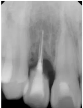
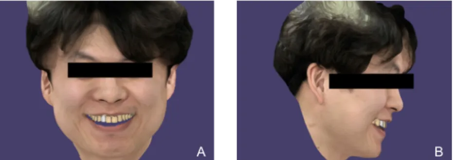
This article demonstrates a case that successfully rehabilitated the maxillary anterior missing tooth area with a single-tooth implant prosthesis by integrating three-dimensional face scan data into the computer-aided design software. An extraoral transfer jig was self-invented to achieve this purpose, and a handheld face scanner app was used to accomplish economic benefits. Not only was the protocol convenient and efficient for both the patient and clinician, but it also resulted in a satisfactory outcome.

Keywords: Extraoral transfer jig, Computer-Aided design, Three-dimensional face scan, Aesthetics

Abstract

*Corresponding author: Ji-Man Park, [email protected]

Face Scanner App for Integrating Face Scan Data into Prosthesis Design

Kyung Chul Oh and Ji-Man Park

*

Department of Prosthodontics, Yonsei University College of Dentistry, Seoul, Republic of Korea

Ⅰ. Introduction

Consideration of facial aesthetics is of significant importance for the rehabilitation of the anterior tooth area

1

. Harmony of facial and dental midlines, amount of tooth exposure while smiling and when in physiologic rest position, and lip support are some of the factors that need to be considered

2, 3

. In the past, it was difficult to incorporate facial information into a single workflow. However, with the development of dental computer-aided design (CAD) software and the widespread use of digital single-lens reflex cameras, it is possible to utilize two-dimensional facial photographs for the design of dental prostheses

4

. Despite the successful application of this technique, special attention is required so that the photographs are taken without distortion or from multiple angles

5

.

Three-dimensional (3D) facial scanning has partially solved these difficulties. It has

become possible to examine the design of the prosthesis along with the corresponding

facial appearance in 3D view

6

. However the integration of the scan data of the intraoral

condition into the face scan data still poses a challenge with regard to accurate

superimposition. One method to resolve this issue is to use a highly accurate face scanner

that visualizes the teeth portion as well as the facial area. However, these face scanners, in

(2)

most cases, take up a lot of space. In contrast, portable handheld face scanners are advantageous because the scan data can be readily obtained and they do not require additional storage place. However, their accuracy may be relatively inferior when compared to the fixed-type face scanners. To achieve more accurate outcomes from the integration, an extraoral transfer jig can be utilized. The present case described herein reports a successful application of a self-invented extraoral transfer jig to integrate the scan data obtained from the handheld face scanner with those of the intraoral condition in the rehabilitation of the anterior missing tooth area.

Ⅱ. Case Report

A 33-year-old man presented to the Department of Conservative Dentistry at Yonsei University Dental Hospital to receive a treatment for the mobile maxillary right lateral incisor. The tooth was diagnosed as an external root resorption with hopeless prognosis (Fig. 1). Hence, it was decided that the

Fig. 1. Periapical radiograph exhibiting an external root resorption of the maxillary right lateral incisor.

Kyung Chul Oh and Ji-Man Park : Use of an Extraoral Transfer Jig and a Handheld Face Scanner App for Integrating Face Scan Data into Prosthesis Design. Implantology 2019

Fig. 2. Placement of a single-implant (NR line 3.6x11 mm; Dentium) followed by the guided bone regeneration procedures. A: Surgical view in occlusal aspect. B: Periapical radiograph after implant placement.

Kyung Chul Oh and Ji-Man Park : Use of an Extraoral Transfer Jig and a Handheld Face Scanner App for Integrating Face Scan Data into

Prosthesis Design. Implantology 2019

(3)

tooth be extracted and rehabilitated with a single-tooth implant restoration. Three months after the extraction, an implant (NR line 3.6×11 mm; Dentium) was placed, and guided bone regeneration procedures were performed in the buccal dehiscence defect area using synthetic bone substitutes (Osteon III; Genoss), collagen membrane (Collagen Graft; Genoss), and metal pins (Membrane pin; Dentium) (Fig. 2). The implant was submerged for 6 months, after which a second surgery was performed to engage the healing abutment (5.5M; Dentium).

The final impression was obtained with a polyether impression material (Impregum Penta; 3M ESPE) and the definitive cast was fabricated with a type IV dental stone (Snowrock; DK Mungyo). The 3D face scans were taken with a handheld face scanner app (Bellus3D; Bellus3D) under three conditions: with closed mouth, in smile view, and with an extraoral transfer jig hold state (Fig. 3). Prior to taking the face scan with the transfer jig, the horizontal part of the jig was filled with bite registration material (Blu- Mousse; Parkell Inc.) and the interocclusal relationship was obtained. These data were transferred to the CAD software (exocad DentalCAD; exocad GmbH) and were accurately integrated by means of the

Fig. 3. Three-dimensional (3D) face scans taken with a handheld face scanner app (Bellus3D; Bellus3D).

A: With closed mouth. B: In smile view. C: Design of an extraoral transfer jig.

Kyung Chul Oh and Ji-Man Park : Use of an Extraoral Transfer Jig and a Handheld Face Scanner App for Integrating Face Scan Data into Prosthesis Design. Implantology 2019

Fig. 4. Design of a definitive zirconia crown in the computer-aided design software (exocad DentalCAD;

exocad GmbH). A: Frontal smile view. B: Lateral smile view.

Kyung Chul Oh and Ji-Man Park : Use of an Extraoral Transfer Jig and a Handheld Face Scanner App for Integrating Face Scan Data into

Prosthesis Design. Implantology 2019

(4)

transfer jig. The definitive zirconia crown was designed using the same software, and evaluated in both frontal and lateral smile views (Fig. 4). The definitive prosthesis was evaluated for its connection with the implant fixture, occlusion, and aesthetics, and was placed in the patient’s mouth (Fig. 5). The patient expressed satisfaction with the outcome with regard to both aesthetics and function.

Ⅲ. Discussion

Integration of the face scan data with the prosthesis-designing workflow is particularly effective for communication with patients because it visualizes the expected outcomes and enables the immediate design modification of the prosthesis in accordance with patients’ reasonable preferences in the CAD stage. The extraoral transfer jig that consists of a horizontal part to contain bite registration material and a vertical part with a number of target markers was invented. All acquired digital data can be successfully merged by additionally scanning this transfer jig with the desktop scanner and scanning the face in the jig hold state. The presented protocol does not require patients’ movement to other locations because the face scan software program is embedded in a mobile app. This may be advantageous when treating elderly patients.

This approach can broaden its application range to more complicated clinical situations in which multiple anterior teeth require rehabilitation. Furthermore, studies assessing the accuracy of this self- invented extraoral transfer jig in comparison with the commercial ones may be worth investigating.

Ⅳ. Conclusion

A maxillary anterior missing tooth area was rehabilitated with a single-tooth implant by integrating Fig. 5. Frontal view after the placement of the definitive zirconia crown.

Kyung Chul Oh and Ji-Man Park : Use of an Extraoral Transfer Jig and a Handheld Face Scanner App for Integrating Face Scan Data into

Prosthesis Design. Implantology 2019

(5)

3D face scan data into the CAD software in the process of designing the prosthesis. A self-invented extraoral jig was used to achieve this incorporation. The patient was satisfied in terms of both aesthetic and functional outcomes. Further studies assessing the jig’s accuracy and its applicability to clinical situations in which the rehabilitation of multiple anterior teeth are required.

Acknowledgements

This study was supported by a grant of the Korea Health Technology R&D Project through the Korea Health Industry Development Institute (KHIDI), funded by the Ministry of Health & Welfare (HI18C0435). The authors thank Pf. Ui-Won Jung for conducting implant surgery in this case.

References

1. Hasanreisoglu U, Berksun S, Aras K, et al. An analysis of maxillary anterior teeth: facial and dental proportions. J Prosthet Dent. 2005; 94: 530-538.

2. Greenberg JR, Bogert MC. A dental esthetic checklist for treatment planning in esthetic dentistry.

Compend Contin Educ Dent. 2010; 31: 630-634, 6, 8.

3. Koidou VP, Chatzopoulos GS, Rosenstiel SF. Quantification of facial and smile esthetics. J Prosthet Dent. 2018; 119: 270-277.

4. Coachman C, Calamita M, Ricci A. Digital Smile Design: A Digital Tool for Esthetic Evaluation, Team Communication, and Patient Management. Ronald E. Goldstein's Esthetics in Dentistry. 2018:

84-111.

5. Stanley M, Paz AG, Miguel I, et al. Fully digital workflow, integrating dental scan, smile design and CAD-CAM: case report. BMC Oral Health. 2018; 18: 134.

6. Pozzi A, Arcuri L, Moy PK. The smiling scan technique: Facially driven guided surgery and

prosthetics. J Prosthodont Res. 2018; 62: 514-517.

수치

Fig. 1. Periapical radiograph exhibiting an external root resorption of the maxillary right lateral incisor.
Fig. 4. Design of a definitive zirconia crown in the computer-aided design software (exocad DentalCAD;

참조

관련 문서