병변(white matter lesion)과 열공경색(lacunar infarct)으로 구분된 다[3, 4]. 이들의 발병기전은 혈관 내경의 협착과 뇌혈류의 자동조 절의 이상으로 뇌의 백질과 대뇌피질하 회백질의 허혈성 손상에 의한다고 알려져 있다[5, 6]. 그러나 Wardlaw 등[7]은 뇌백질병변이 뇌혈관의 죽상경화반 형성과 혈관내피의 손상으로 혈관 내의 혈 장 독성 성분이 뇌실질로 유출되어 발생한다고 하였으며, Bamford 등[8]은 뇌의 열공경색은 뇌의 소동맥 벽의 유리지질증과 섬유 괴 사와 관련있다고 하였다. 또한 Fernando 등[9]은, 열공경색에서는 뇌의 소동맥의 폐쇄가 관찰되나, 뇌백질병변에서는 뇌의 소동맥의 협착이나 폐쇄는 관찰되지 않는다고 하였다.
한편, 심혈관계질환의 위험인자들이 뇌졸증이나 무증상 뇌경색 과 관련이 있고[10], 심혈관계질환 위험인자를 갖는 경우에 뇌백질 병변이 더 흔하다는 연구결과들이 보고되고 있다[11]. 그러나 뇌백 질병변과 열공경색들의 대사적 차이나 심혈관계질환 위험인자와 의 연관성 차이를 연구한 결과들은 드물다.
이에 본 연구에서는 신경학적 증상이 없는 건강검진자에서 뇌백
서 론
뇌의 작은혈관질환(cerebral small vessel disease)은 신경학적 이 상이 없는 사람들의 뇌 MRI 검사에서 드물지 않게 발견된다. 이 질 환은 향후 뇌졸증이나 인지기능장애로 발전될 수 있음이 보고되 고 있다[1, 2]. 뇌의 작은혈관질환은 뇌 MRI의 소견에 따라 뇌백질
뇌백질병변과 열공경색의 심혈관계질환 위험인자와의 관계
Relationship of White-Matter Lesions and Lacunar Infarcts with Cardiovascular Risk Factors
나은희·조한익
Eun Hee Nah, M.D., Han-Ik Cho, M.D.
한국건강관리협회
Korea Association of Health Promotion, Seoul, Korea http://dx.doi.org/10.3343/lmo.2012.2.2.95
Corresponding author: Eun Hee Nah, M.D.
Korea Association of Health Promotion, 335 Hwagok-ro, Gangseo-gu, Seoul 157-704, Korea
Tel: +82-2-2600-2000, Fax: +82-2-2696-4500, E-mail: [email protected] Received: September 6, 2011
Revision received: October 18, 2011 Accepted: October 31, 2011
This article is available from http://www.labmedonline.org 2012, Laboratory Medicine Online
This is an Open Access article distributed under the terms of the Creative Commons Attribution Non-Commercial License (http://creativecommons.org/licenses/by-nc/3.0/) which permits unrestricted non-commercial use, distribution, and reproduction in any medium, provided the original work is properly cited.
Background: Magnetic resonance imaging (MRI) findings of white-matter lesions are different from those of lacunar infarcts; however, both these conditions are related to cardiovascular risk factors. This study was performed to investigate the differences in the relationships of white- matter lesions and lacunar infarcts with cardiovascular risk factors and differences between the metabolic characteristics of patients with these conditions.
Methods: We included 4,255 patients who showed neurological deficits during health checkups. These individuals were classified into the follow- ing 3 groups on the basis of MRI findings: normal, white-matter lesion, and lacunar infarct. The groups were compared for age; weights; prevalence of metabolic syndrome; and levels of blood pressure, blood glucose, lipid, high sensitivity C-reactive protein, and HbA1c.
Results: Age, body mass index (BMI); waist circumference; levels of blood pressure, blood glucose, triglycerides and HbA1c; and prevalence of metabolic syndrome and its components were the highest in lacunar infarct group, followed by white matter lesion group, and normal group. Age and diastolic blood pressure level were related to white matter lesions, and age, systolic blood pressure level, and blood glucose level were relat- ed to lacunar infarcts. Further, the prevalence of the above-mentioned lesions increased with increase of the number of the components of meta- bolic syndrome.
Conclusions: This study suggests that lacunar infarct is more advanced lesion than white matter lesion. Among all the cardiovascular risk factors, high blood pressure and impaired fasting blood glucose levels were significantly related to white-matter lesions and lacunar infarct.
Key Words: White matter lesion, Lacunar infarct, Cardiovascular risk factors, Metabolic syndrome
질병변 또는 열공경색과 심혈관계질환의 위험요소와의 연관성 및 대사적 특징을 밝히고, 이들 뇌의 작은혈관질환 사이에 어떤 차이 가 있는지 알아보고자 하였다.
대상 및 방법
1. 대상
2009년 4월부터 2010년 12월까지 건강증진센터에서 신경학적 질환이 없이 건강검진을 목적으로 뇌 MRI 검사를 받은 20세 이상 성인들 중 뇌의 작은혈관질환 이외의 병변을 갖는 경우를 제외한 4,255명(남, 1,788명; 여, 2,467명)을 대상으로 하였다. 이들을 뇌 MRI 소견에 따라, 뇌의 병변이 없는 정상군(2,618명), 뇌백질병변 군(1,007명), 열공경색군(630명)으로 구분하였다.
2. 뇌의 작은혈관질환 측정
검진에 사용된 MRI는 1.5 T MRI (Sigma HDe, GE Yokogawa Med- ical Systems, Tokyo, Japan)였으며, axial T2-, T1-weighted spin echo, FLAIR, T2-weighted GRE 등으로 검사하였다. 열공경색의 진단기 준은, T2-weighted 영상에서 직경이 3-15 mm 정도의 고밀도 음영 이면서 T1-weighted 영상에서 저밀도 음영인 병변으로 하였다. 뇌 백질병변의 진단기준은, T2-weighted 영상에서 고밀도 음영이 보 이지만 T1-weighted 영상에서는 뚜렷한 저밀도 음영이 관찰되지 않는 병변으로 하였다.
3. 심혈관계질환 위험인자 측정
혈압 측정은 최소 5분 이상 안정을 취한 후 표준혈압계를 사용 하여 상완에서 수동으로 측정하였다. 정상혈압이 아닌 경우는 좀 더 안정을 취한 후 재측정하였고, 만약 재측정값이 처음 값과 10 mmHg 이상 차이가 날 때는 한 번 더 측정하여 근접한 2회 측정값 의 평균치로 정하였다. 체질량지수(body mass index, BMI)는 체 중(kg)/신장2(m2)의 공식에 의해 계산하였다.
혈액화학검사는 10시간 공복 후 채혈한 혈액의 혈청에서 공복혈 당, 중성지방, 총콜레스테롤, HDL콜레스테롤, LDL콜레스테롤, 고 감도 C-반응성 단백질(high sensitivity C-reactive protein, hsCRP) 을 Hitachi 7600 (Hitachi, Nakai, Japan)으로 측정하였다. 총콜레스 테롤, HDL콜레스테롤, 중성지방, 혈당은 아산시약(Asan Inc., Hwaseong, Korea)을 사용하여 효소법으로, LDL콜레스테롤은 Daiichi 시약(Daiichi Pure Chemicals Co., Tokyo, Japan)을 사용하 여 효소법으로, hsCRP는 Daiichi 시약을 사용하여 혼탁면역법으 로 측정하였다. 총콜레스테롤, HDL콜레스테롤, 중성지방 측정의 보정은 로슈사의 보정물질(Roche Diagnostics, Mannheim, Ger- many)을, LDL콜레스테롤 측정의 보정은 Daiichi 의 보정물질(Dai-
ichi Pure Chemicals Co., Tokyo, Japan)을 사용하였다. 지질검사의 정밀도와 정확도는 정도관리물질인 Lychocheck levels I, II (Bio- Rad Lab., Irvine, USA)를 사용하였고, 대한임상검사정도관리협회 의 외부정도관리사업에 참여하여 관리하였다. C-반응성 단백질 (hsCRP)의 측정범위는 0.03-30 mg/dL이었고, 참고범위는 0.3 mg/
dL 미만으로 하였다. 당화혈색소(HbA1c)는 고성능액체크로마토그 래피법을 이용한 HLC-723 G7 (Tosoh Corporation, Tokyo, Japan) 으로 측정하였다. 당화혈색소의 참고범위는 6% 이하, 정밀도는 검 사 내 및 총 변이계수가 모두 2.5% 이내였다.
대사증후군의 판정은 개정된 National Cholesterol Education Panel (NCEP) 기준[12]에 따랐다. 즉, 공복혈당이 100 mg/dL 이상, 혈압이 130/85 mmHg 이상, 중성지방이 150 mg/dL 이상, HDL콜 레스테롤은 남자는 40 mg/dL 미만, 여자는 50 mg/dL 미만인 경우, 허리둘레는 WHO의 서태평양지역의 기준[13]에 따라 남자는 90 cm, 여자는 80 cm를 초과하는 경우로 하였으며, 이들 중 3가지 이 상에 해당할 때 대사증후군으로 판정하였다.
4. 통계 분석
정상군, 뇌백질병변군, 열공경색군 사이의 임상 및 대사적 차이 를 밝히기 위해 일원분산분석(one-way ANOVA)을 하였으며, 각 군 간의 차이를 알기 위해 사후분석으로 다중비교를 하였다. 뇌의 작 은혈관질환과 심혈관계질환의 위험인자와의 연관성을 알기 위해 다중회귀분석을 하였다. 뇌백질병변군과 열공경색군에서 대사증 후군 및 대사증후군 요소의 유병률 차이를 알기 위해 교차분석을 하였다. 통계 프로그램은 SPSS version 12.0 (SPSS Inc., Chicago, IL, USA)을 이용하였고, P<0.05를 통계적으로 유의한 것으로 하였다.
결 과
1. 연령대에 따른 뇌의 작은혈관질환의 유병률
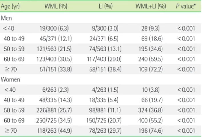
대상자의 평균 연령은 54.6±11.9세였으며, 남녀 모두에서 연령 이 증가할수록 뇌의 작은혈관질환의 유병률은 통계적으로 유의하 게 증가하였다(P<0.01). 즉, 남성의 유병률은 40대에서 18.6%, 50 대에서 34.6%로, 60대에서 59.5%, 70세 이상에서 72.2%였고, 여성 의 유병률은 40대에서 19.7%, 50대에서 36.8%, 60대에서 55.2%, 70 세 이상에서 74.6%였다(Table 1).
2. 뇌의 작은혈관질환의 임상적 및 대사적 특징
정상군, 뇌백질병변군 및 열공경색군의 평균 연령은 각각 50.3± 12.0세, 60.0±9.6세, 62.2±9.6세로 의미있게 차이가 있었다(P<
0.01). 체질량지수, 허리둘레, 혈압, 공복혈당은 정상군, 뇌백질병변 군, 열공경색군 순으로 의의있게 높았다(P<0.01). 중성지방도 정상
군, 뇌백질병변군, 열공경색군 순으로 점차 증가하였다(P=0.045).
HbA1c는 뇌백질병변군 5.87±0.73%, 열공경색군 5.93±0.79%로 정상군의 5.65±0.71%보다 높았다(P<0.01) (Table 2).
3. 뇌의 작은혈관질환과 심혈관계질환 위험인자와의 연관성 뇌백질병변군의 발생은 연령과 관련이 있었다. 즉, 뇌백질병변 연 관성은 40세 미만에 비해 50대에서 10.45배 더 높았고(교차비: 10.45, 95% 신뢰구간: 1.38-79.38), 60대에서는 18.69배 더 높았으며(교차 비: 18.69, 95% 신뢰구간: 2.42-144.58), 70세 이상에서는 69.30배 더 높았다(교차비: 69.3, 95% 신뢰구간: 8.21-585.31). 이완기 혈압도 뇌 백질병변과 연관성이 있었는데, 이완기 혈압이 85 mmHg 미만에 비해 85 mmHg 이상인 경우에 연관성이 2.30배 더 높았다(교차비:
2.30, 95% 신뢰구간: 1.28-4.13). 그러나 성별, 흡연상태, 허리둘레, 체질량지수, 수축기 혈압, 공복혈당, 총콜레스테롤, 저밀도 콜레스 테롤, 중성지방, hsCRP, HbA1c와 뇌백질병변 발생과는 연관성이 없 었다.
한편, 열공경색 발생도 연령과 연관성이 있었다. 40세 미만에 비 해 60대에서 4.95배 연관성이 더 높았고(교차비: 4.95, 95% 신뢰구 간: 1.08-22.74), 70세 이상에서 13.11배 더 높았다(교차비: 13.11, 95% 신뢰구간: 2.46-69.80). 수축기 혈압과 열공경색과의 연관성 은, 수축기 혈압 130 mmHg 미만에 비해 130 mmHg 이상인 경우 에 1.92배 더 높았다(교차비: 1.92, 95% 신뢰구간: 1.14-3.23). 공복 혈당과의 연관성은 공복혈당 100 mg/dL 미만에 비해 공복혈당 100-125 mg/dL인 경우에 2.23배 더 높았다(교차비: 2.23, 95% 신뢰 Table 1. Prevalence of cerebral small vessel diseases
Age (yr) WML (%) LI (%) WML+LI (%) P value*
Men
<40 19/300 (6.3) 9/300 (3.0) 28 (9.3) <0.001 40 to 49 45/371 (12.1) 24/371 (6.5) 69 (18.6) <0.001 50 to 59 121/563 (21.5) 74/563 (13.1) 195 (34.6) <0.001 60 to 69 123/403 (30.5) 117/403 (29.0) 240 (59.5) <0.001 ≥70 51/151 (33.8) 58/151 (38.4) 109 (72.2) <0.001 Women
<40 6/263 (2.3) 4/263 (1.5) 10 (3.8) <0.001 40 to 49 48/335 (14.3) 18/335 (5.4) 66 (19.7) <0.001 50 to 59 226/881 (25.7) 98/881 (11.1) 324 (36.8) <0.001 60 to 69 250/725 (34.5) 150/725 (20.7) 400 (55.2) <0.001 ≥70 118/263 (44.9) 78/263 (29.7) 196 (74.6) <0.001
*P values were determined by using X2 test for comparing the prevalence of WML and LI in individuals of different age groups.
Abbreviations: WML, white-matter lesion; LI, lacunar infarct.
Table 2. Intergroup differences in patient characteristics and variables Normal
(N=2,618) WML
(N=1,007) LI
(N=630) P value Age (yr) 50.3±12.0*† 60.0±9.6‡ 62.2±9.6 <0.01
Men (%) 43.8 35.7 44.8 0.213
Smoking 0.056
Never 62.2 (%) 68.5 67.3
Past 20.2 (%) 17.8 21.5
Current 17.6 (%) 13.7 11.2
BMI (kg/m2) 24.0±3.2† 24.2±3.0‡ 24.6±3.0 <0.01 WC (cm) 80.7±9.1*† 82.0±8.3‡ 83.6±8.4 <0.01 SBP level (mmHg) 119.8±14.5*† 124.7±16.1‡ 128.3±15.8 <0.01 DBP level (mmHg) 73.9±10.2*† 75.7±10.5‡ 77.1±10.4 <0.01 FBS level (mg/dL) 98.1±21.2*† 100.7±20.6‡ 105.3±24.0 <0.01 TC level (mg/dL) 191.5±35.6 195.0±35.1 194.1±35.4 0.056 TG level (mg/dL) 112.2±76.6 119.0±79.3 119.7±64.3 0.045 HDL-C level (mg/dL) 55.8±9.3 56.0±9.0 55.3±9.4 0.418 LDL-C level (mg/dL) 113.3±32.2 115.6±32.0 115.3±32.3 0.225 hsCRP level (mg/dL) 0.15±0.39 0.19±0.52 0.20±0.42 0.108 HbA1C level (%) 5.65±0.71*† 5.87±0.73 5.93±0.79 <0.01 P value derived from one-way analysis of variance test used for intergroup com- parison.
*P<0.05 derived from posthoc comparisons (Dunnett’s test) between the normal group and white-matter lesion group; †P<0.05 derived from the comparison of variables between the normal gruop and the lacunar infarct group; ‡P<0.05 de- rived from the comparison of variables between white-matter lesion group and the lacunar infarct group.
Abbreviations: WML, white-matter lesion; LI, lacunar infarct; BMI, body mass in- dex; WC, waist circumference; SBP, systolic blood pressure; DBP, diastolic blood pressure; FBS, fasting blood sugar; TC, total cholesterol; TG, triglyceride; LDL-C, LDL-cholesterol; HDL-C, HDL-cholesterol; hsCRP, high sensitivity C-reactive pro- tein; HbA1c, hemoglobin A1c.
Table 3. Odds ratio (95% confidence interval) of multiple regression analysis for cardiovascular risk factors in WML and LI groups*
Characteristics WML LI
OR (95% CI) P value OR (95% CI) P value Age (yr)
<40 1 1
40 to 49 4.78 (0.59–39.05) 0.140 1.21 (0.23-6.28) 0.824 50 to 59 10.45 (1.38-79.38) 0.023 2.39 (0.53-10.75) 0.258 60 to 69 18.69 (2.42-144.58) 0.005 4.95 (1.08-22.74) 0.040 ≥70 69.30 (8.21-585.31) <0.001 13.11 (2.46-69.80) 0.003 SBP level
<130 mmHg 1 1
≥130 mmHg 0.73 (0.44-1.21) 0.226 1.92 (1.14-3.23) 0.014 DBP level
<85 mmHg 1 1
≥85 mmHg 2.30 (1.28-4.13) 0.005 1.26 (0.68-2.35) 0.465 FBS level (mg/dL)
<100 1 1
100 to 125 1.04 (0.61-1.75) 0.893 2.23 (1.28-3.91) 0.005 ≥126 0.75 (0.29-1.94) 0.547 2.27 (0.83-6.23) 0.111
*Multiple regression analysis performed for the WML and LI groups included the following variables: age, sex, smoking, waist circumference, body mass index, sys- tolic and diastolic blood pressure levels, fasting blood glucose levels, total choles- terol levels, triglyceride levels, HDL-C levels, LDL-C levels, hsCRP levels and HbA1c levels.
Abbreviations: WML, white-matter lesion; LI, lacunar infarct; SBP, systolic blood pres- sure; DBP, diastolic blood pressure; FBS, fasting blood sugar.
구간: 1.28-3.91). 그러나 열공경색과 성별, 흡연상태, 허리둘레, 체 질량지수, 이완기 혈압, 총콜레스테롤, 저밀도 콜레스테롤, 중성지 방, hsCRP, HbA1c와의 연관성은 없었다(Table 3).
4. 뇌의 작은 혈관질환과 대사증후군과의 관계
대사증후군의 유병률은 정상군에서 15.6%, 뇌백질병변군에서 23.7%, 열공경색군에서 30.3%로, 정상군에 비해 뇌백질병변군, 열 공경색군으로 갈수록 높았다(P<0.001). 대사증후군 요소 중 복부 비만 유병률은 정상군에서 30.6%, 뇌백질병변군에서 43.2%, 열공 경색군에서 46.3%로 정상군에 비해 뇌백질병변군, 열공경색군으 로 갈수록 높았다(P<0.001). 고혈압은 정상군에서 30.7%, 뇌백질 병변군에서 42.3%, 열공경색군에서 52.0%로 정상군에 비해 뇌백 질병변군, 열공경색군으로 갈수록 높았다(P<0.001). 고혈당은 정 상군에서 28.6%, 뇌백질병변군에서 35.7%, 열공경색군에서 45.0%
로 정상군에 비해 뇌백질병변군, 열공경색군으로 갈수록 높았다 (P<0.001) (Table 4). 뇌의 작은혈관질환의 유병률은 대사증후군 요소 수와도 관련이 있었다. 즉, 요소 수가 1개일 때 36.0%, 2개일 때 41.8%, 3개일 때 48.6%, 4개일 때 52.4%로 대사증후군 요소 수 가 증가할수록 의미있게 증가하였다(P<0.001) (Fig. 1).
고 찰
뇌백질병변은 뇌에서 정보처리속도와 수행능력의 장애와 연관 되어 있으며[14, 15], 열공경색은 인지장애나 치매의 위험성을 증가 시키는 병변이다[16]. 본 연구결과 뇌백질병변군과 열공경색군 사 이에 연령, 체질량지수, 허리둘레, 혈압, 공복혈당, 중성지방 및
HbA1c에 차이가 있음을 알 수 있었다. 즉, 정상군, 뇌백질병변, 열공 경색군으로 갈수록 이들의 평균값은 높아졌다. 대사증후군의 유 병률 또한 정상군, 뇌백질병변군, 열공경색군으로 갈수록 더 높았 다. Atwood 등[17, 18]은 뇌의 작은혈관질환은 여러 위험인자들과 유전적 요인들이 복합적으로 혈관벽의 손상을 축적시켜 발병한다 고 하였다. 한편, 대사증후군은 심혈관계질환 위험인자들의 조합 으로서, 대사증후군을 갖는 경우 심혈관계질환 발생의 위험이 증 가하며, 대사증후군 구성 요소에 해당하는 요인이 많을수록 심혈 관계질환 위험이 증가한다고 보고되고 있다[19, 20]. 또한 뇌의 작 은혈관질환 발생은 심혈관계질환과 밀접한 관계가 있다는 연구들 [10, 11]로 미루어 볼 때, 뇌백질병변보다는 열공경색이 좀 더 진행 된 뇌의 작은혈관질환으로 볼 수 있을 것 같다.
뇌의 열공경색은 연령과 연관성이 있었다. 연령대가 증가할수록 연관성이 높았으며, 특히 70세 이상에서는 높은 교차비를 보여 큰 연관성을 보였다. 본 연구에서 열공경색의 전체 유병률은 14.8%
로, Kwon 등[21]의 무증상 뇌경색의 유병률 5.5%에 비해 더 높았 다. 연령대별 유병률에서도 본 연구에서의 유병률은 Kwon 등[21]
에 비해 높은 경향을 보였다. Kwon 등[21]의 연구와 본 연구와의 이같은 차이의 원인으로 두 가지를 들 수 있다. 첫째는 Kwon 등 [21]의 연구에서는 전체 뇌 MRI 검사자 중 무증상 뇌경색의 유병률 을 기술한 반면, 본 연구에서는 신경학적 질환이 없는 검진자의 뇌 MRI 검사 결과 뇌의 병변이 없는 정상, 뇌백질병변, 열공경색의 소 견을 보이는 사람들 중의 열공경색의 비율을 보는 것으로 연구 대 상자의 차이로 볼 수 있다. 둘째는 Kwon 등의 연구에서는 뇌의 작 은혈관질환의 유형을 구분하지 않고 뇌경색의 증상은 보이지 않으 면서 뇌 MRI 소견상 뇌경색 소견을 보이는 무증상 뇌경색 집단을 대상으로 하였기 때문으로 생각되었다. 뇌백질병변 또한 연령과 연 관성이 있었으며, 열공경색에서보다 연관성이 더 컸다. 뇌백질병변 Table 4. Prevalence of the components of metabolic syndrome in pa-
tients with cerebral small vessel disease
Normal WML LI P value
Visceral obesity 665/2,171 342/791 227/490 <0.001
(30.6%) (43.2%) (46.3%)
Hypertension 598/1,946 293/693 233/448 <0.001
(30.7%) (42.3%) (52.0%)
Low HDL-C 214/1,943 91/697 59/439 0.189
(11.0%) (13.1%) (13.4%)
High TG 414/1,945 162/697 105/439 0.346
(21.3%) (23.2%) (23.9%)
Hyperglycemia 554/1,940 247/692 197/438 <0.001
(28.6%) (35.7%) (45.0%)
Metabolic 301/1,924 162/683 131/432 <0.001
syndrome (15.6%) (23.7%) (30.3%)
P values were derived from the X2 test used for comparing the prevalence of com- ponents of metabolic syndrome between the patients with white-matter lesions and those with lacunar infarcts.
Abbreviations: WML, white-matter lesion; LI, lacunar infarct; TG, triglyceride; HDL- C, HDL-cholesterol.
60 50 40 30 20 10 Prevalence of WML and LI (%) 0
0 1 2 3 4 No. of component of metabolic syndrome
WMLLI WML+LI
Fig. 1. Prevalence of white-matter lesions and lacunar infarct accord- ing to the number of the components of metabolic syndrome.
P<0.01 derived from X2 tests used to comparison the prevalence of white-matter lesion and lacunar infarct with reference to the number of components of the metabolic syndrome.
Abbreviations: WML, white-matter lesion; LI, lacunar infarct.
의 유병률은 연령에 따라 차이가 있게 보고되었는데, 64세 정도의 인구집단의 11-21%부터 82세의 유병률 94%까지 넓은 범위로 보고 되고 있다[22]. 본 연구에서 뇌백질병변의 유병률은 40세 이하에서 4.4%, 70세 이상에서 40.8%로, 평균 약 23.7%의 유병률을 보였고, 여성에서 유병률이 남성보다 의의있게 높았으며, 특히 50세 이상 의 여성에서 뇌백질병변 유병률이 남성보다 높았다. Manolio 등 [23]은 뇌백질병변의 유병률이 여성에서 더 높고, 이러한 유병률의 차이는 여성에서 폐경이 심혈관계질환 위험인자에 영향을 줌으로 써 생기는 결과라고 설명하였다. 그러나 열공경색의 유병률에 대 한 성별의 차이는 연구에 따라 상이하였으며[24, 25], 본 연구에서 도 성별에 따른 열공경색 유병률의 차이는 없었다.
본 연구에서, 조절 가능한 심혈관계 위험인자들 중 혈압과 공복 혈당장애가 열공경색과 연관성이 있었다. 이는 다른 위험인자들의 교란효과를 제거한 결과이다. Khan 등[26]은 뇌의 큰혈관질환과 작은혈관질환의 위험인자 비교연구에서, 고혈압이 뇌의 작은혈관 질환의 위험인자이며, 뇌의 작은혈관질환의 아형에 따른 비교연구 에서는, 작고 다수의 허혈성병변들은 고혈압과 연관이 있는 반면, 열공경색은 당뇨병, 고콜레스테롤혈증과 연관이 있어, 이는 죽상 경화가 열공경색의 병인일 수 있다는 것을 시사한다 하였다. Park 등[27]의 연구에서는 혈압과 공복혈당장애 이외에도 고중성지방혈 증과 복부비만도 무증상 열공경색과 연관이 있다고 하였다. 이는 Park 등의 연구가 40-59세의 중년을 대상으로 한 연구로 본 연구대 상과는 차이가 있으며, 본 연구에서도 복부비만이 열공경색 및 뇌 백질병변과 연관성이 있었으나 연령과 성의 인자를 제거한 후에는 연관성이 없어졌다. 한편, 본 연구에서 혈당 126 mg/dL 이상인 당 뇨병과 열공경색은 연관성이 없었다. 이는 본 연구에서는 무증상 열공경색만을 대상으로 하였으므로, 이미 당뇨병인 경우에는 좀 더 진행된 뇌혈관질환이 발생하여 본 연구대상에서 제외되었을 가능성을 시사한다. 뇌백질병변은 이완기 혈압과 연관성이 있었 다. 다른 연구들[28, 29]에서도 연령과 고혈압이 뇌백질병변의 주요 한 예견인자라 하였고, 흡연, 당뇨병, 다른 혈관질환의 위험인자들 이 뇌백질병변과 복합적으로 연관되어 있다고 하였다.
대사증후군 요소 수에 따른 뇌의 작은혈관질환 유병률을 볼 때, 대사증후군 요소 수가 많을수록 유병률은 증가하였으며, 비록 대 사증후군 요소 수가 1개인 경우에서도 0개인 경우에 비해 뇌의 작 은혈관질환 유병률은 증가하였다. 이는 대사증후군 요소가 1개라 도 있으면 뇌의 작은혈관질환 발생 위험성은 높아짐을 의미한다.
본 연구에서는 몇 가지 제한점이 있다. 첫째는 단면연구였으므 로 뇌백질병변 및 열공경색과 심혈관계질환 위험인자와의 인과관 계를 증명할 수 없었다. 둘째, 뇌 MRI 검사를 선택한 자발적 검진 자들을 대상으로 한 연구이므로 일반 집단을 대표한다고 할 수는 없었다. 셋째, 약물 복용 유무를 확인하거나 인지기능검사를 시행
할 수 없었던 점이다.
본 연구에서 뇌백질병변과 열공경색의 병인의 차이를 증명할 수 는 없었으나, 두 군 사이의 관련위험인자들의 정도와 대사증후군 의 유병률의 차이로 미루어, 뇌백질병변보다 열공경색이 더 진행 된 병변이며, 고혈압과 공복혈당장애가 뇌의 작은혈관질환과 연관 이 있음을 알 수 있었다. 또한, 대사증후군 요소의 갯수에 따라 뇌 의 작은혈관질환 유병률이 증가하기 때문에 이외의 대사증후군 요소들도 조절하는 것이 뇌의 작은혈관질환 예방에 도움이 되리 라 생각되었다.
요 약
배경: 뇌백질병변과 열공경색은 뇌 MRI상 다른 소견을 보이며, 심 혈관계질환 위험인자와 관련이 있다. 본 연구에서는 무증상 뇌백 질병변 또는 열공경색과 심혈관계질환 위험요소와의 연관성 및 대 사적 특징을 밝히고, 이들 사이에 어떤 차이가 있는지 알아보고자 하였다.
방법: 신경학적 증상이 없는 건강검진자 4,255명을 뇌 MRI 소견상 병변이 없는 정상군, 뇌백질병변군, 열공경색군으로 나누어, 연령, 비만도, 혈압, 혈당, 혈중지질, hsCRP, HbA1c들을 비교해 보았고, 대 사증후군 발생빈도를 알아보았다.
결과: 연령, 체질량지수, 허리둘레, 혈압, 혈당, 중성지방, HbA1c가 정상군, 뇌백질병변군, 열공경색군으로 갈수록 증가하였고, 대사 증후군 및 대사증후군 요소의 유병률이 위와 같은 순으로 증가하 였다. 뇌백질병변은 연령, 이완기 혈압과, 열공경색은 연령, 수축기 혈압, 공복혈당과 연관성이 있었고, 이 병변들의 유병률은 대사증 후군 요소 수가 증가함에 따라 증가하였다.
결론: 뇌백질병변보다 열공경색이 더 진행된 병변이며, 심혈관계질 환 위험인자 중, 고혈압, 공복혈당장애가 뇌백질병변 및 열공경색 과 연관성이 있었다.
참고문헌
1. Vermeer SE, Prins ND, den Heijer T, Hofman A, Koudstaal PJ, Breteler MM. Silent brain infarcts and the risk of dementia and cognitive de- cline. N Engl J Med 2003;348:1215-22.
2. Vermeer SE, Hollander M, van Dijk EJ, Hofman A, Koudstaal PJ, Bre- teler MM. Silent brain infarcts and white matter lesions increase stroke risk in the general population: the Rotterdam Scan Study. Stroke 2003;
34:1126-9.
3. Vermeer SE, Longstreth WT Jr, Koudstaal PJ. Silent brain infarcts: a sys- tematic review. Lancet Neurol 2007;6:611-9.
4. Kobayashi S, Okada K, Koide H, Bokura H, Yamaguchi S. Subcortical silent brain infarction as a risk factor for clinical stroke. Stroke 1997;28:
1932-9.
5. Pantoni L and Garcia JH. Pathogenesis of leukoaraiosis: a review. Stroke 1997;28:652-9.
6. de Leeuw FE, de Groot JC, Oudkerk M, Witteman JC, Hofman A, van Gijn J, et al. A follow-up study of blood pressure and cerebral white matter lesions. Ann Neurol 1999;46:827-33.
7. Wardlaw JM, Sandercock PA, Dennis MS, Starr J. Is breakdown of the blood-brain barrier responsible for lacunar stroke, leukoaraiosis, and dementia. Stroke 2003;34:806-12.
8. Bamford JM and Warlow CP. Evolution and testing of the lacunar hy- pothesis. Stroke 1988;19:1074-82.
9. Fernando MS, Simpson JE, Mathews F, Brayne C, Lewis CE, Barber R, et al. White matter lesions in an unselected cohort of the elderly: mo- lecular pathology suggests origin from chronic hypoperfusion injury.
Stroke 2006;37:1391-8.
10. Gállego J and Martínez-Vila E. Asymptomatic cerebrovascular disease and systemic diagnosis in stroke, atherothrombosis as a disease of the vascular tree. Cerebrovasc Dis 2005;20(S2):S1-10.
11. Launer LJ. Epidemiology of white matter lesions. Top Magn Reson Im- aging 2004;15:365-7.
12. Grundy SM, Cleeman JI, Daniels SR, Donato KA, Eckel RH, Franklin BA, et al. Diagnosis and management of the metabolic syndrome: an American Heart Association/National Heart, Lung, and Blood Institute Scientific Statement. Circulation 2005;112:2735-52.
13. WHO West Pacific Region. The Asia-pacific perspective: refining obe- sity and its treatment. London: International Obesity Taskforce, 2000.
14. Prins ND, van Dijk EJ, den Heijer T, Vermeer SE, Jolles J, Koudstaal PJ, et al. Cerebral small-vessel disease and decline in information process- ing speed, executive function and memory. Brain 2005;128:2034-41.
15. Sonohara K, Kozaki K, Akishita M, Nagai K, Hasegawa H, Kuzuya M, et al. White matter lesions as a feature of cognitive impairment, low vi- tality and other symptoms of geriatric syndrome in the elderly. Geriatr Gerontol Int 2008;8:93-100.
16. Bernick C, Kuller L, Dulberg C, Longstreth WT Jr, Manolio T, Beauchamp N, et al. Silent MRI infarcts and the risk of future stroke: the cardiovas- cular health study. Neurology 2001;57:1222-9.
17. Pantoni L. Cerebral small vessel disease: from pathogenesis and clinical characteristics to therapeutic challenges. Lancet Neurol 2010;9:689-701.
18. Atwood LD, Wolf PA, Heard-Costa NL, Massaro JM, Beiser A, D’Agos- tino RB, et al. Genetic variation in white matter hyperintensity volume in the Framingham Study. Stroke 2004;35:1609-13.
19. McNeill AM, Rosamond WD, Girman CJ, Golden SH, Schmidt MI, East HE, et al. The metabolic syndrome and 11-year risk of incident cardio- vascular disease in the atherosclerosis risk in communities study. Dia- betes Care 2005;28:385-90.
20. Klein BE, Klein R, Lee KE. Components of the metabolic syndrome and risk of cardiovascular disease and diabetes in Beaver Dam. Diabe- tes Care 2002;25:1790-4.
21. Kwon HM, Kim BJ, Lee SH, Choi SH, Oh BH, Yoon BW. Metabolic syndrome as an independent risk factor of silent brain infarction in healthy people. Stroke 2006;37:466-70.
22. American Psychiatric Association. Diagnostic and statistical manual of mental disorders. 4th ed. Washington: American Psychiatric Associa- tion, 1994.
23. Manolio TA, Furberg CD, Shemanski L, Psaty BM, O’Leary DH, Tracy RP, et al. Associations of postmenopausal estrogen use with cardiovas- cular disease and its risk factors in older women. The CHS Collabora- tive Research Group. Circulation 1993;88:2163-71.
24. Das RR, Seshadri S, Beiser AS, Kelly-Hayes M, Au R, Himali JJ, et al.
Prevalence and correlates of silent cerebral infarcts in the Framingham offspring study. Stroke 2008;39:2929-35.
25. Vermeer SE, Koudstaal PJ, Oudkerk M, Hofman A, Breteler MM. Prev- alence and risk factors of silent brain infarcts in the population-based Rotterdam scan study. Stroke 2002;33:21-5.
26. Khan U, Porteous L, Hassan A, Markus HS. Risk factor profile of cere- bral small vessel disease and its subtypes. J Neurol Neurosurg Psychia- try 2007;78:702-6.
27. Park K, Yasuda N, Toyonaga S, Tsubosaki E, Nakabayashi H, Shimizu K. Significant associations of metabolic syndrome and its components with silent lacunar infarction in middle aged subjects. J Neurol Neuro- surg Psychiatry 2008;79:719-21.
28. Longstreth WT Jr, Manolio TA, Amold A, Burke GL, Bryan N, Jungreis CA, et al. Clinical correlates of white matter findings on cranial mag- netic resonance imaging of 3301 elderly people. The Cardiovascular Health Study. Stroke 1996;27:1274-82.
29. Dufouil C, de Kersaint-Gilly A, Besancon V, Levy C, Auffray E, Brun- nereau L, et al. Longitudinal study of blood pressure and white matter hyperintensities: the EVA MRI Cohort. Neurology 2001;56:921-6.