뇌졸중 환자의 머리회전 각도가 내ㆍ외측 균형에 미치는 영향
이관섭1, 김중선2
1강병원 물리치료학과, 2대구대학교 재활과학대학 물리치료학과
The Effect of Medio-lateral Balance to Head Rotation in Stroke Patient
Kwan-Sub Lee1, Chung-Sun Kim2
1
Department of Physical Therapy, Kang Hospital,
2Department of Physical Therapy, College of Rehabilitation Science, Daegu University Purpose: This study was conducted in order to compare the ability to control postural sway during perturbation when stroke patients received postural sway induced by head rotation.
Methods: This study included 15 stroke patients and 15 healthy adults. Each group was measured by 3D motion analysis for determination of the angle of the neck in static position and by balance performance monitor for estimation of swaying angle in both neutral posture and head rotation position. These results were then analyzed in order to compare the healthy control group and the stroke patients group.
Results: In both static posture (60.7±4.81) and dynamic posture (51.46±6.87, 70.8±6.55), significant decreases were observed in the angle of head rotation of the patient group, compared to the healthy group (p<0.05), and significant decreases were observed in the sway angle of the patient group when in the neutral position (3.62±7, 24±0.60) and head rotation (3.04±0.80, 51.46±6.87), compared to the healthy group (p<0.05).
Conclusion: According to these findings, patients with stroke tend to restrict the ROM of head rotation and swaying angle in dynamic posture and maintain their posture instability using limitation of head movement relative to the trunk and sway angle of area which is larger than that of affected side in unaffected side.
Keywords: Head rotation, Swaying angle, Stroke patients
I. 서론
균형은 수의 동작 시 자세를 제어하면서, 외부 동요에 적절하 게 반응하여 자세를 유지하는 복합적인 과정이며,1 머리의 움 직임은 시각과 청각적 환경을 정상적으로 탐구하는 행동으 로,2 전신 활동 동안 머리 안정성의 중요성은 많은 연구에 의
해 지지된다.3,4 머리와 목 부분의 기능은 주변 환경에 대해 신 체의 기준을 설정해주며, 자세제어 동안 머리와 시각계 그리 고 전정계를 위한 안정된 지지 기저면(base of support)을 생산 하며,5 자세제어와 바로서기 반응은 머리제어의 근간이 된다.6 뇌졸중 후에 가장 일반적으로 나타나는 증상 중 하나가 마 비측의 근력약화이며, 이로 인한 근력 불균형에 의해 편마비 환자들은 비대칭적인 자세를 갖게 된다. 또한 자세 이미지나 신체 인식력의 문제로 인해 체간의 운동제어 능력이 저하되므 로,7 뇌졸중 환자는 저하된 균형제어 능력을 보상하기 위해 대 체전략을 사용하게 된다.
뇌졸중 환자는 종종 머리와 체간의 동작을 최소화함으로 써 불안정성을 보상하려 한다. 그 결과 목과 상체에 근긴장의
Received September 17, 2012 Revised October 17, 2012
Accepted October 17, 2012
Corresponding author Chung-Sun Kim, [email protected]
Copyright © 2012 by The Korean Society of Physical Therapy
This is an Open Access article distributed under the terms of the Creative Commons Attribution Non-Commercial License (http://creativecommons.org/licenses/by-nc/3.0/) which permits unrestricted non-commercial use, distribution, and reproduction in any medium, provided the original work is properly cited.
The J ournal of K orean S ociety of P hysical T herapy Original Article
증가, 피로, 통증과 같은 이차적인 근골격계 증상을 유발시킨 다.8 뇌졸중 환자의 마비측은 주로 목과 상체에 굽힘근 긴장도 가 증가되어 있으며, 굽힘근이 짧아져 원심성 근 활동 능력에 문제가 생기며 폄근과 같은 다른 근육은 생역학적으로 불리한 상태에 놓이게 되고,6 신체정렬의 변화와 관절가동범위의 제 한을 가진다.9 뇌졸중 환자는 머리회전 동안 균형을 유지하기 위해서 보다 큰 안정성 한계를 사용하여 변화된 자세제어를 보여주게 된다.10 근 활동과 안정성 사이의 불균형으로 인해 머리와 목이 비대칭적인 자세를 취하게 되며,11 이로 인해 시 각 정보가 왜곡되고 전정 기관의 기능에 영향을 주어 균형 제 어에 추가적인 문제를 야기할 수 있다.12
정적 기립상황에서 압력중심의 내ㆍ외측 이동은 일차적으 로 엉덩관절의 외전근에 의해 제어되며,13 머리의 회전 움직임 은 엉덩관절과 발관절을 반대방향으로 이동시키며, 이들 생역 학적 변화는 외측 흔들림을 제어하기 위한 특수 근육 반응과 연관이 있다.
자세 동요검사를 통한 압력중심의 측정은 지금까지 여러 연구에서 자세제어의 척도로써 사용되어 오고 있으며,14-16 신 체질량중심과 압력중심의 이동궤적을 반영하므로,17 본 연구 에서 뇌졸중 환자의 압력 중심 이동을 분석하는 데 이용하였 다.
뇌졸중 환자는 머리와 목의 비대칭적인 자세로 인해 관절 가동범위의 제한을 가지며 변화된 자세제어를 보여 주게 되므 로, 본 연구의 목적은 자세동요 검사를 통하여 정적ㆍ동적 상 황에서 뇌졸중 환자의 머리회전 각도가 내ㆍ외측 균형에 미치 는 영향을 알아보고자 한다.
II. 연구방법
1. 연구대상
본 연구는 대구 소재 K 병원에 입원하여 물리치료를 받고 있 는 환자 중 연구에 참여하기로 동의하고 연구 조건을 충족할 수 있는 뇌졸중으로 인한 편마비 환자 15명과 나이가 비슷한 정상인 15명을 대상으로 한다. 연구 대상자의 선정 조건은 다 음과 같다. 첫째, 뇌경색, 뇌출혈로 인하여 편마비가 된 환자.
둘째, 타인의 도움 없이 10미터 이상 독립보행이 가능한 환자.
셋째, 골반 및 양하지에 정형외과적 질환이 없는 환자. 넷째, 양발을 모으고 15초 이상 독립적으로 기립균형을 유지할 수 있는 환자. 다섯째, 연구자가 지시하는 내용을 이해하고 따를 수 있는 환자. 여섯째, 전정 장애(vestibular disorder)가 없는 환 자. 일곱째, 환측무시(neglect)가 없는 환자.
2. 실험방법 1) 측정도구
⑴ Balance performance monitor (BPM)
본 연구에서 균형능력 측정을 위하여 타당도와 신뢰도가 검증 된 BPM을 사용하였다. BPM은 다양한 시각 및 청각 피드백을 제공하는 균형 훈련과 균형을 측정하기 위한 도구로서 기립 자세에서 발판의 감지기가 움직임을 감지하여 신체 중심의 분 포와 동요각, 동요거리, 동요속도, 동요주기 등을 컴퓨터 스크 린 상에 수치화 및 그래프화 하여 나타나게 한다.18
(2) 3차원 동작 분석 시스템(3D-motion analysis measurement system)
목의 가동 범위를 측정하기 위해 ZEBRIS (MOTION LAB System Inc., LA, USA)를 사용하였다. 선택적으로 신체부위를 선택하여 입력단자를 부착한 후 삼차원적인 자세를 자료화하 는 운동학적 분석 장비로 모자와 같이 머리에 착용한 후, 상체 에 고정된 단자에 대해 머리의 회전만을 측정할 수 있게 된다.
이러한 분석 장비의 자료는 숫자와 그래프로 표시되어 객관적 인 자료를 얻을 수 있다.
2) 검사절차
선정기준에 적합한 15명의 대상자는 검사 전 치료사를 통하 여 유병 기간, 마비측, 발병원인, 진단 등의 특성을 기록하고 일반적 특성은 성별, 나이, 체중, 신장으로 구성하였고 검사 대상자에게 검사 목적과 과제에 대한 설명을 하였으며 검사 에 대한 동의를 얻도록 하였다. 머리에 3차원 동작 분석기를 착용하고 바닥에 놓여진 BPM 위에 올라서서 선 자세를 유지 하도록 하였다. 먼저 정적 선 자세 측정은 두발을 18 cm 거리 를 두고 시선은 눈높이 전방을 주시한다. 동적 선 자세는 두 발이 두 발이 BPM 위에서 떨어지지 않을 정도로 내ㆍ외측으 로 체중이동을 수행하며 시선은 전방을 주시한다. 정적 선 자 세에서 3차원 동작 분석기를 이용하여 대상자가 능동적으로 수행할 수 있는 머리회전 각도를 측정하였다. 머리회전 방향 은 검사자가 구두명령으로 지시하였으며, 머리회전 속도는 스스로 편안한 속도로 가능한 자연스럽게 수행하도록 하였 다.
동적 선 자세에서 검사 대상자는 머리를 중립 위에 유지하 면서 동요각을 측정하고, 마지막으로 검사자의 신호에 따라 실험 대상자는 능동적으로 소리가 나는 쪽으로 머리를 회전한 다. 동시에 머리회전 각도와 동요각을 각각 측정하였다.
3. 자료분석
본 연구에서 얻은 자료는 SPSS ver. 14.0 통계 프로그램(SPSS Inc., Chicago, IL, USA)을 이용하여 통계 처리하였다. 연구대상 자의 일반적 특성은 기술통계로 분석하였으며, 편마비군과 대 조군의 머리회전 그리고 동요각의 차이는 t-test를 사용하였으 며, 정적 및 동적 선 자세의 비교는 대응 t-test로 하였다. 통계 학적 유의수준 α는 0.05로 하였다.
III. 결과
1. 연구 대상자의 일반적 특성
본 연구에 참여한 전체 대상자는 총 30명이었으며, 각각 대조 군 15명, 편마비군 15명으로 구성되었다. 대상자의 일반적 특 성은 Table 1과 같다.
2. 대조군과 편마비군에서 정적 및 동적 선 자세의 머리회전 각도 비교
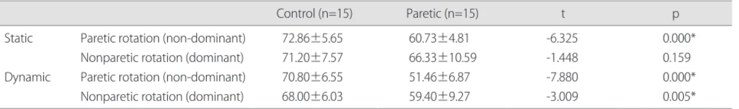
대조군과 편마비군에서 정적 및 동적 선 자세의 머리회전 각 도의 차이를 비교하였다. 대조군과 편마비군의 마비측 비교에 서 정적 및 동적 선 자세에서 통계학적으로 유의한 목각도의 감소가 있었다(p<0.05) (Table 2). 편마비군에서 정적 및 동적 선 자세의 머리회전 각도 비교에서 마비측 및 비마비측 모두 통계학적으로 유의한 목각도의 감소가 있었다(p<0.05) (Table 3).
3. 대조군과 편마비군의 동적 선 자세에서 머리 회전에 따른 동요각 비교
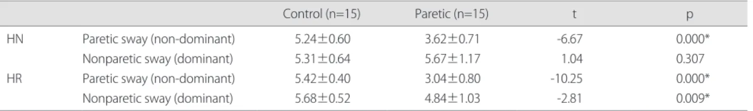
BPM을 이용하여 동적 선 자세에서 머리회전에 따른 집단 내 동요각을 측정하였다. 대조군과 편마비군의 마비측 동요각 비 교에서 머리 중립위와 머리회전 자세에서 통계학적으로 유의 한 감소가 있었다(p<0.05) (Table 4). 동적 선 자세에서 편마비 군의 동요각은 마비측 및 비마비측 모두 통계학적으로 유의한 감소가 있었다(p<0.05) (Table 5).
IV. 고찰
본 연구는 뇌졸중 환자의 일상생활에 많은 영향을 미치는 동 적 균형유지 능력에 방향을 바꾸거나 주위 환경의 탐구를 위 해 필요한 머리의 회전 움직임을 결합하여 더 큰 자세동요를 유발하여 뇌졸중 환자의 동요에 대항해 신체를 유지하는 자세
Table 1. General characteristics of the subjects
Division Control Paretic
Male/Female Weight (kg) Age (yr) Height (cm)
8/7 62.80±6.44 53.80±8.46 167.10±7.57
10/5 63.00±12.11 55.70±6.56 164.90±4.15 Values are presented as number or mean±standard deviation.
Table 2. The comparison of head rotation angle between static and dynamic in each group
(unit: degree)
Control (n=15) Paretic (n=15) t p
Static Dynamic
Paretic rotation (non-dominant) Nonparetic rotation (dominant) Paretic rotation (non-dominant) Nonparetic rotation (dominant)
72.86±5.65 71.20±7.57 70.80±6.55 68.00±6.03
60.73±4.81 66.33±10.59 51.46±6.87 59.40±9.27
-6.325 -1.448 -7.880 -3.009
0.000*
0.159 0.000*
0.005*
Values are presented as mean±standard deviation.
*p<0.05.
Table 3. The comparison of head rotation angle between static and dynamic in paretic group
(unit: degree) Paretic (n=15)
Static Dynamic t p
Paretic rotation Nonparetic rotation
60.73±4.81 66.33±10.59
51.46±6.87 59.40±9.27
6.577 4.045
0.000*
0.001*
Values are presented as mean±standard deviation.
*p<0.05.
제어 능력을 비교하는 데 목적이 있었다.
본 연구에서는 측정 시 오차를 줄이기 위해 MOTION LAB System사의 3차원 동작 분석기(ZEBRIS)를 이용하여 머리회전 각도를 측정하였으며, 나이에 따라 각도 차이가 존재하므로 대조군을 동일 연령대로 선정하였다. 본 연구에서는 뇌졸중 환자의 머리회전 각도를 측정하여 마비측과 비마비측의 차이 를 비교하고 대조군과 비교하여 관절 가동 범위의 제한을 검 사하였다. 그 결과 대조군의 평균 머리회전 각도는 좌우 회전 의 합이 평균 144.06o, 편마비군은 평균 127.06o로 편마비 그룹 의 머리회전 각도가 정상가동 범위에 미치지 못함을 나타내었 다. 뇌졸중 환자의 마비측은 주로 목과 상부 체간에 굽힘근 긴 장도가 증가되어 원심성 근 활동 능력에 문제가 생겨 대조군 과의 비교에서 차이를 보인 것으로 여겨진다.
Keshner와 Dhaher5는 뇌졸중 환자의 선 자세에서 주위를 탐색하는 자연스런 움직임 시 체간의 신체 동요는 증가되지만 머리 움직임은 감소된다고 하였고, Gjelsvik6은 동요 시 머리와 체간의 동작을 최소화함으로써 신체정렬의 변화와 관절가동 범위의 제한을 가진다고 하였다. 본 연구에서 내ㆍ외측 동요 동안 머리회전 각도는 정상군에서 138.8o, 편마비군은 110.86o 로 동요 동안 머리회전 각도는 유의한 감소를 보여 선행연구 와 유사함을 보였다. 뇌졸중 환자는 동요 동안 자세제어의 문 제와 시각의 자유도 감소로 시각 의존적 움직임을 하게 되고,
자세제어나 균형 유지에 시각의 참여가 증가되어 신체를 고정 하거나 관절가동범위를 제한시키는 것으로 여겨진다. 전후 방 향보다 내ㆍ외측 방향으로의 압력중심 움직임은 낙상을 예견 할 수 있는 균형 지수로서 중요한 요소라 하였고,19-21 Prieto 등22 과 Choi 등23은 자세 균형 수행 평가에 정적 선 자세와 발판 위 에서 움직임을 통한 동적 선 자세를 사용하였으며, 본 연구에 서는 동적 선 자세 균형은 BPM의 발판 위에서 30초간 수의적 으로 내ㆍ외측 방향으로 자연스런 움직임을 수행하도록 하였 다. 본 연구결과 대조군에서는 머리 중립 위와 회전 시 동요각 은 각각 10.55o, 11.1o로 약간의 증가가 있었고, 편마비군에서 는 각각 9.29o, 7.88o로 동요각의 감소가 있었다. 본 연구에서 동적 선 자세에서 편마비군의 머리 중립 위보다 회전 시 동요 각이 감소하였다는 사실은 머리회전이 동적 균형에 중요한 요 인이 될 수 있음을 나타낸다고 할 수 있다.
본 연구결과 동적 선 자세에서 편마비군 마비측과 비마비 측의 동요각 비교는 머리 중립 위 상태의 동요각은 각각 3.62o, 5.67o로 차이를 보였고, 머리 회전 시는 각각 3.04o, 4.84o로 차 이를 보였다. 편마비군에서는 머리 중립 위와 비교해 회전 시 마비측과 비마비측 모두 동요각의 감소를 보였으며, 마비되지 않은 하지 아래에서 더 넓은 범위를 보여 주었다. Geiger 등24의 연구에서 동요각은 마비된 쪽과 비교해 마비되지 않은 하지 아래에서 넒은 범위를 드러내며 안정성 한계도 감소한다고 하
Table 4.
The comparison of sway angle between neutral head and head rotation in each group (unit: degree)
Control (n=15) Paretic (n=15) t p
HN HR
Paretic sway (non-dominant) Nonparetic sway (dominant) Paretic sway (non-dominant) Nonparetic sway (dominant)
5.24±0.60 5.31±0.64 5.42±0.40 5.68±0.52
3.62±0.71 5.67±1.17 3.04±0.80 4.84±1.03
-6.67 1.04 -10.25 -2.81
0.000*
0.307 0.000*
0.009*
Values are presented as mean±standard deviation.
HN: head neutral, HR: head rotation.
*p<0.05.
Table 5. The comparison of sway angle between neutral head and head rotation in paretic group
(unit: degree) Paretic (n=15)
HN HR t p
Paretic sway Nonparetic sway
3.62±0.71 5.67±1.17
3.04±0.80 4.84±1.03
4.263 3.166
0.001*
0.007*
Values are presented as mean±standard deviation.
HN: head neutral, HR: head rotation.
*p<0.05.
여 본 연구를 지지한다. 이는 머리회전을 통한 시각적 보상으 로 신체의 근 활성을 증가시켜 동요에 대한 불안정성을 감소 시키려는 비효율적인 자세 움직임으로 보여지며, 시각은 자세 동요에 크게 영향을 미치며, 증가된 동요에 있어 시각의 보상 기능이 중요하게 작용하는 것을 알 수 있다.
적절한 자세 제어는 기립자세를 유지하고 모든 일상생활 동작에 필요한 자발적인 사지와 머리의 움직임을 수행하고 신 체를 안정화시키기 위해 필요한데, 편마비 환자는 중력에 대 항하는 움직임 부족과 협조성 패턴 장애 그리고 자세 적응이 잘 이루어지지 않기 때문에 정상적인 자세제어 수행의 어려움 을 겪는 것으로 보여진다. 동요가 뇌졸중 환자의 머리회전과 균형능력에 영향을 미치고 불안정성을 감소시키려는 변화된 자세제어 양상을 보임으로 임상에서 다양한 시각적 환경의 적 응과 과제 수행을 위한 뇌졸중 환자의 동적 평가에서 도움이 될 수 있는 자료가 될 것으로 사료된다. 본 연구에서 뇌졸중 환 자의 좌우 동요에 대한 동요각만 측정하였으며, 전후 방향의 동요에 대한 측정을 하지 못한 점과 시각적 보상에 대한 양적 자료를 측정하지 못한 제한점이 있다. 추후 연구에서 뇌졸중 환자의 전후 방향의 동요에 대한 자세제어 평가와 함께 시각 적 보상에 대한 측정이 필요할 것으로 여겨진다.
본 연구결과 뇌졸중 환자는 동요 동안 머리회전의 관절 가 동 범위를 제한하며, 체간에 대한 머리 움직임의 제한을 통해 뇌졸중 환자의 자세 불안정성을 유지하려는 경향을 나타냈다.
이는 자연스런 시각적 보상으로 신체의 근 활성을 증가시켜 동요에 대한 불안정성을 감소시키려는 비효율적인 자세 움직 임으로 정상군과 비교해 변화된 자세제어 양상을 나타내었다.
따라서 뇌졸중 환자의 머리회전 움직임은 다양한 시각적 환경 의 적응과 과제 수행을 위해 중요한 의미가 있으며, 임상에서 뇌졸중 환자의 자세제어 문제를 해결하고자 할 때 반드시 머 리회전은 고려되어야 한다.
Acknowledgements
본 논문은 이관섭의 석사학위 논문으로 수행되었음.
참고문헌