증례요약: 52세 남자환자가 수개월 전부터 실내에서 실외로 나갔을 때 5분에서 10분 정도 잘 안 보이는 일과성 흑암시 증상이 우안에 발생하여 내원하였다. 시력은 0.8, 홍채혈관신생은 관찰되지 않았고, 유두혈관신생과 망막동맥의 협소, 다수의 점상 망막출혈이 관찰 되었다. 형광안저혈관조영 검사상 맥락막충만의 지연과 팔망막순환시간의 지연, 동정맥이행시간의 연장, 시신경유두 신생혈관과 여러 군데의 망막허혈부위가 나타났으며 후기에서는 망막혈관주위로 누출이 다수 보였다. 자기공명영상혈관조영검사상 오른쪽 내경동맥 근위부에서 심한 협착이 관찰되었다. 안허혈증후군으로 진단하고 스텐트삽입혈관성형술을 권유하였으나 시행하지 못하다가 진단 후 약 6개월이 지나서 우측 경동맥의 협착부위에 스텐트삽입혈관성형술을 시행하였다. 수술 후 약 24개월에 시행한 검사상 일과성 흑암 시는 사라지고, 나안시력은 1.0, 유두신생혈관은 섬유조직으로 변화되었으며, 형광안저혈관조영검사상 맥락막충만의 지연과 팔망막순 환시간의 지연은 관찰되지 않았고, 시신경유두주위 누출과 망막허혈부위는 사라지고 주변부까지 혈액순환이 좋게 나타났다.
<대한안과학회지 2010;51(3):447-452>
■ 접 수 일: 2009년 7월 31일 ■ 심사통과일: 2009년 11월 10일
■ 책 임 저 자: 박 종 문
경남 진주시 칠암동 90번지
안허혈증후군은 내경동맥의 90% 이상의 협착으로 인해 동측 안동맥의 혈류량이 적어져서 이차적으로 나타나는 안 구의 허혈성 변화를 말한다.1-3 안허혈증후군의 치료로는 홍채혈관신생이나 후안부혈관신생이 있으면서 모세혈관비 관류가 관찰될 때 범망막광응고술을 시도하기도 하지만 혈 관신생이 잘 퇴화되지 않는 경우도 많고, 경동맥내막절제술 을 시행하기도 하지만 합병증이나 위험성 그리고 성공률 등에서 여러 의견이 있다.4최근 들어 혈관신생과 황반부종 등의 치료를 위해 유리체강내로 bevacizumab 주입술을 시 행하여 긍정적인 결과를 얻은 사례도 보고되었으며, 경동맥 스텐트삽입혈관성형술을 시행하여 안허혈증후군의 치료와 시력의 호전을 보이는 경우도 보고되고 있다.5-7저자들은 안허혈증후군으로 진단된 후 뒤늦게 시행된 경동맥 스텐트 삽입혈관성형술로도 성공적인 치료와 좋은 시력을 보이는 사례를 경험하였기에 보고한다.
증례보고
52세 남자환자가 수개월 전부터 실내에서 실외로 나갔을
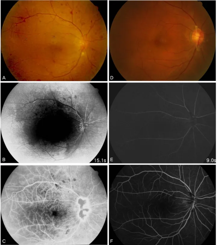
때 5~10분 정도 잘 안 보이는 증상이 우안에 발생하여 내 원하였다. 상기 환자는 당뇨병으로 약 5년간 경구약으로 치 료 중 이었으며, 약 1년 전에 경도의 비증식성당뇨병증으로 진단받았었다. 내원 당시 안통은 호소하지 않았고 우안의 나안시력은 0.8, 좌안은 1.0이었으며, 안압은 압평안압계로 양안 16 mmHg였고, 홍채신생혈관 및 앞방각신생혈관은 관찰되지 않았다. 우안의 안저검사상 유두혈관신생과 망막 동맥 협소, 다수의 점상 망막출혈이 관찰되었으며(Fig. 1A) 좌안에서는 경도의 비증식성당뇨병증 소견 외 특이점은 없 었다. 형광안저촬영 검사상 우안에서 맥락막충만시간 지연 이 관찰되고, 팔망막순환시간이 15.1초 정도로 지연되어 관찰되었으며, 시신경유두 신생혈관과 여러 군데의 망막허 혈부위가 나타났고(Fig. 1B), 후기에서는 망막혈관주위로 누출이 다수 보였다(Fig. 1C). 좌안에서는 이상소견이 발견 되지 않았다. 혈관조영컴퓨터단층촬영검사 및 혈관조영자 기공명영상검사와 경동맥도플러초음파검사상 오른쪽 내경 동맥의 근위부에서 90% 이상의 심한 협착이 관찰되었으며, 왼쪽 내경동맥의 근위부에서도 50% 정도의 협착이 관찰되 었다. 경동맥협착부위에 대한 직접적인 치료가 필요하다고 판단되어 신경과에 의뢰하였으며, 신경과에서 스텐트삽입 혈관성형술을 권유 받았으나 개인적인 사정으로 우선 약물 치료를 시행하였다. 안허혈증후군으로 진단받은 지 약 3개 월 후 시행한 형광안저혈관조영검사에서도 유두혈관신생이
A D
B E
C F
Figure 1.Fundus photograph and fluorescein angiography in the early and late phase before (A, B, C) and after (D, E, F) carotid angioplasty and stenting (CAS). (A) Before CAS, fundus photograph shows multiple dot retinal hemorrhage and arterial narrowing. (B) Early phase in fluorescein angiography shows delayed arm-to-retina time and new vessels on disc. (C) Late phase shows leakage from new vessels on the disc and reveals non-perfusion area. (D) After CAS, fundus photograph shows disappearance of new vessels in the disc which is changed to fibrous tissue. (E) Early phase in fluorescein angiography shows disappearance of new vessels on the disc and no delayed arm-to-retina time. (F) Late phase shows no leakage from the disc and well perfused entire retina.
A B
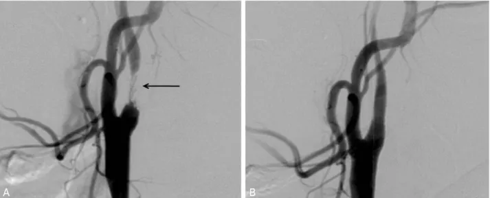
Figure 2. Carotid angiography before (A) and after (B) carotid angioplasty and stenting (CAS). (A) Before CAS, the right internal carotid artery shows stenosis more than 90% but, (B) after CAS, successful stenting and good flow are shown (arrow: stenotic site of the carotid artery).
나타났고, 나안시력은 우안 0.8, 유리체출혈 및 홍채신생혈 관의 발생은 없었으나 일과성 흑암시 증상은 지속되었다.
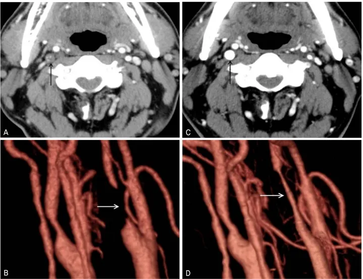
안허혈증후군으로 진단 받은 지 약 6개월이 지난 후 환자의 우측 경동맥의 협착부위에 스텐트삽입혈관성형술을 시행하 였고(Fig. 2), 시행 전 후 환자의 신경학적인 문제나 합병증 은 발생하지 않았다. 수술 후 시행한 경동맥도플러초음파검 사와 혈관조영컴퓨터단층촬영검사상에서 우측 내경동맥의 협착은 없었으며 관류도 정상적이었고(Fig. 3), 좌측은 약 50%정도의 협착이 관찰되었다. 수술을 시행하고 약 24개 월 후 본원 안과 외래에 방문하였다. 나안시력은 양안 1.0, 안압은 양안 20 mmHg, 안저검사상 시신경유두부위 신생 혈관은 퇴화되어 섬유막으로 변화되어 있었다(Fig. 1D).
형광안저촬영상 수술 전 보이던 맥락막충만시간 지연은 사 라지고, 팔망막순환시간은 9초 정도로 정상화되었으며, 시 신경유두주위 누출과 망막허혈부위는 사라지고 주변부까지 혈액순환이 좋게 나타났다(Fig. 1E, F).
고 찰
안허혈증후군은 1963년 Kearns and Hollenhorst1가 내 경동맥의 심한 협착으로 인해 생기는 이차적인 안구의 증 상과 증후를 일컫는다고 처음으로 보고 하였다. 시력상실, 일과성흑암시, 통증 등의 증상이 나타날 수 있고, 홍채혈관 신생, 전포도막염, 각막부종, 망막동맥 협소, 망막정맥 확 장, 망막출혈, 미세혈관류, 유두혈관신생, 망막혈관신생, 면 화반 등이 나타나며, 형광안저촬영상 맥락막충만의 지연과
으로 의심되는 환자는 두경부초음파검사, 경두개색채도플 러영상검사, 뇌자기공명영상 및 혈관조영술로 경동맥주행 로를 검사하여 확진 할 수 있다.10 본 증례에서는 심각한 시 력상실과 통증은 나타나지 않았으나 일과성 흑암시로 의심 되는 증상이 오랫동안 있었다. 일과성 흑암시는 일시적인 망막관류장애나 혈전에 의해 발생하거나 혈관질환으로 나 타날 수 있는 것으로 알려져 있다.11안허혈증후군 진단 시 환자의 약 2/3 정도에서 홍채혈관신생이 관찰되는 것으로 알려져 있는데, 본 증례에서는 관찰되지 않았으며, 각막부 종이나 전포도막염이 뚜렷하지 않았다. 망막동맥 협소, 다 수의 점상 망막출혈, 맥락막충만의 지연과 팔망막순환시간 의 지연이 관찰되었으며, 시신경유두 신생혈관과 여러 군데 의 망막허혈부위, 망막혈관주위로 누출이 다수 보였지만, 심한 황반부종이나 유두혈관신생의 급격한 증가 및 주변부 망막의 혈관신생은 관찰되지 않았다. 이것은 첫째로 본 증 례에서 내경동맥의 협착에 의한 안동맥 관류압의 저하가 다른 경우보다 심하지 않았거나, 둘째로 협착의 진행 및 협 착으로 인한 여러 가지 허혈성 변화들이 발생한지 얼마 안 되었을 가능성도 있을 것으로 생각된다. 하지만 환자가 안 허혈증후군으로 진단된 후 약 3개월이 지난 후 시행한 검사 상에서도 일과성 흑암시의 증상은 있었으나 시력의 저하는 뚜렷하지 않았고 황반부종은 뚜렷이 증가되지 않았으며 유 두혈관신생의 증가나 유리체출혈 등이 발생하지 않은 점과 팔망막순환시간이 15초 정도로 지연이 심하지 않은 것으로 보아 첫 번째 가능성이 조금 더 높을 것으로 생각된다. 이 것의 확인을 위해 본 증례에서는 시행되지는 않았지만 양
A C
B D
Figure 3.CT angiography and reconstructed three dimensional image before (A, B) and after (C, D) carotid angio- plasty and stenting (CAS). Before CAS, the lumen of the right carotid artery is stenotic with artheroma (A) and not visible in reconstructed image (B) but after CAS, the lumen is well enhanced with contrast material (C) and well visible in the reconstructed image (D).
혈증후군의 치료로는 범망막광응고술, 경동맥내막절제술, 신생혈관녹내장에 대한 치료 등이 행해져 왔다. 범망막광응 고술은 홍채혈관신생이 있고 형광안저혈관조영 검사상 유 두혈관신생이 있으면서 모세혈관비관류가 관찰될 때 시도 하기도 하지만 혈관신생이 잘 퇴화되지 않는 경우도 많고, 경동맥내막절제술은 합병증이나 위험성 그리고 성공률 등 에서 여러 의견이 있다.4최근에는 혈관신생과 황반부종 등 의 치료를 위해 항혈관내피세포성장인자인 bevacizumab을 유리체강내로 주입하여 혈관신생의 억제와 황반부종의 감 소 효과를 얻은 보고도 있으나 근본적인 치료라고 보기에 는 힘들다.5이에 근본적인 치료를 위해 내경동맥의 협착부 위에 대한 직접적인 접근들이 이루어져 왔으며, 대표적인 것으로 이전부터 시행된 경동맥내막절제술과 경동맥 스텐 트삽입혈관성형술이 있다.6,7안허혈증후군은 내경동맥의 90%
이상 협착으로 인해 동측 망막중심동맥의 관류압이 50%
이하로 저하되어 증상과 증후를 나타낸다고 하였다.2,3내경 동맥의 협착부위는 경동맥의 분지가 시작되는 곳에서 흔하 게 나타날 수 있으며,12 머리뼈 안쪽으로 주행하는 내경동 맥에서도 협착부위가 발견되는데 특히 동양인에서는 이것 으로 인한 허혈성 변화가 드물지 않은 것으로 보고되고 있 다.12이와 같이 협착부위의 수술적 처치가 힘들 때 경동맥 스텐트삽입혈관성형술을 시행하여 안허혈증후군의 치료와 시력의 호전을 보이는 경우도 보고되고 있다.4,6,7본 증례에 서는 내경동맥협착부위가 근위부에 존재하였으나 당시 환 자의 상태 및 수술의 어려움 등으로 신경과에서 내경동맥 내막절제술보다는 경동맥 스텐트삽입혈관성형술을 시행하 였으며, 이는 스텐트삽입혈관성형술의 효과가 충분히 입증 되었기 때문이라고 여겨진다.6 안허혈증후군에서 스텐트삽 입혈관성형술 후 시력의 호전과 병의 경과가 좋은 경우도 보고되었으나, 본 증례에서와 같이 뒤늦게 시행된 스텐트삽
sive disease of the carotid artery. Proc Staff Meet Mayo Clin 1963;
38:304-12.
2) Brown GC, Magargal LE. The ocular ischemic syndrome. Clini- cal, fluorescein angiographic and carotid angiographic features.
Int Ophthalmol 1988;11:239-51.
3) Mun SJ, Lee KH, Lee DU, Cho NC. Clinical features of ophthal- mic artery hypoperfusion. J Korean Ophthalmol Soc 2007:48:297-302.
4) Sivalingam A, Brown GC, Megargal LE. The ocular ischemic sy- ndrome II. Visual prognosis and the effect of treatment. Int Oph- thalmol 1991;15:15-20.
Neurol 1986;6:194-203.
11) Palmowski AM, Ruprecht KW. Amaurosis fugax. Fortschr Med 1992;110:531-5.
12) Suwanwela NC, Chutinetr A. Risk factors for atherosclerosis of cervicocerebral arteries: intracranial versus extracranial, Neuro- epidemiology 2003;22:37-40.
13) Kawaguchi S, Sakaki T, Iwahashi H et al. Effect of carotid artery stenting on ocular circulation and chronic ocular ischemic syndr- ome. Cerebrovasc Dis 2006;22:402-8.
=ABSTRACT=
Ocular Ischemic Syndrome Successfully Treated With Carotid Angioplasty and Stenting
Yong Seop Han, MD1, Woong Sun Yoo, MD1, In Young Chung, MD1,2, Jong Moon Park, MD, PhD1
Department of Ophthalmology, Gyeongsang National University School of Medicine,1 Jinju, Korea, Gyeongsang Institute of Health Science, Gyeongsang National University2, Jinju, Korea
Purpose: To report a case of ocular ischemic syndrome successfully treated with delayed carotid angioplasty and stenting (CAS).
Case summary: A 52-year-old male was admitted to our hospital because of amaurosis fugax- like symptoms in the right eye for several months. His visual acuity was 0.8 in the right eye and he did not have rubeosis iridis. Neovascularization of the disc, narrowing of the retinal artery and multiple retinal hemorrhages were diagnosed by fundus examination. Fluorescein angiography showed delayed choroidal filling, a delayed arm-to-retina time, prolongation of arteriovenous transit time, neovascularization of the disc, retinal capillary nonperfusion, and staining of the retinal vessels. MR angiography showed severe stenosis in the proximal portion of the right carotid artery. We diagnosed this case as ocular ischemic syndrome. The patient was recommended carotid angioplasty and stenting at the severely narrowed portion of the right carotid artery, but it was postponed about six months after diagnosis because of personal problems. At the final follow-up, 24 months after stenting, the amaurosis fugax symptoms had disappeared, the patient had an improved visual acuity of 1.0, and the new vessels on the disc changed to fibrous tissue. Fluorescein angiography showed resolution of the delayed arm-to-retina time and prolongation of the arteriovenous transit time, disappearances of the leakage around the disc and the retinal capillary nonperfusion.
J Korean Ophthalmol Soc 2010;51(3):447-452
Key Words: Amaurosis fugax, Carotid angioplasty and stenting(CAS), Ocular ischemic syndrome
Address reprint requests to Jong Moon Park, MD, PhD
Department of Ophthalmology, Gyeongsang National University School of Medicine
#90 Chilam-dong, Jinju 660-702, Korea
Tel: 82-55-750-8170, Fax: 82-55-758-4158, E-mail: parkjm@gnu.ac.kr