Korean J Androl. Vol. 29, No. 2, August 2011 http://dx.doi.org/10.5534/kja.2011.29.2.127
127
접수일자: 2011년 8월 1일, 수정일자: 2011년 8월 17일 게재일자: 2011년 8월 19일
Correspondence to: Sae Woong Kim
Department of Urology, The Catholic University of Korea, Seoul St. Mary’s Hospital, 505, Banpo- dong Seocho-gu, Seoul 137-701, Korea Tel: 02-2258-1071, Fax: 02-2258-1080 E-mail: [email protected]
The Effect of Cyanidin-3-O-β-d-glucopyranoside on the Penile Erection and Corpus Cavernosum in a Rat Model of Diabetic Erectile Dysfunction
U Syn Ha1, Joon Sung Koh1, Jang Chun Woo1, Suk Ju Kim1, Su Jin Kim1, Hoon Jang1, Byung Il Yoon1, Seong Yeon Hwang2, Sae Woong Kim1
1Department of Urology, The Catholic University of Korea College of Medicine,
2Korea Bio Medical Science Institute, Seoul, Korea
= Abstract =
Purpose: The aim of this study was to evaluate Cyanidin-3-O-β-d-glucopyranoside on improvement and protection for erectile function.
Materials and Methods: Sprague-Dawley rats (12wks old) were divided into three groups (n=12 in each): normal control, diabetes (DM), and diabetes with Cyanidin-3-O-β-d-glucopyranoside (C3G) concentration materials treatment (DM+C3G). DM and DM+C3G group received a single injection of streptozotocin (50 mg/kg), and 4 wk after induction of diabetes, DM+C3G group were treated with daily C3G (10 mg/kg) dissolved in water for 8 wk. After 12 wk of streptozotocin injections, rats in each group underwent intracavernosal pressure measurement (ICP) and then the corporal tissues were sampled.
Results: DM group showed markedly lower erectile parameters than those in the control group, whereas rats in the DM+C3G group showed improved erectile function by minimizing corporal apoptosis.
Conclusions: The current study is the first to suggest that Cyanidin-3-O-β-d-glucopyranoside may have a potency to improve and protect erectile function in a rat model of diabetic erectile dysfunction.
Key Words: Cyanidin-3-O-β-d-glucopyranoside, Diabetes Mellitus, Erectile dysfunction
Introduction
Numerous studies have estimated the risk of erectile dysfunction (ED) in men with diabetes, and the most common risk factor for ED has been diabetes mellitus (DM).1 An Italian study by Fedele et al.2 in a cohort
of approximately 10,000 men, after correcting for age, reported the prevalence of ED to be 51% among in- dividuals with Type 1 diabetes and 37% among those with Type 2 DM. Further, the Massachusetts Male Aging Study noted the prevalence of ED in diabetic men was 50.7 per 1000 population-years vs. 24.8 in those without diabetes.3
Diabetes harms cavernosal innervation and endothe- lial function, both of which are important for erectile function,4,5 and also decreases nitric oxide production.
Experimental hyperglycaemia has been shown to in- duce many of the pathological consequences observed in DM. Much of neuronal and endothelial damage has been attributed to oxidative stress,6 as hyperglycaemia
induces the overproduction of superoxide (O2
−), serv- ing as an initiating event in the activation of pathways involved in the pathogenesis of the tissue damage from DM. Endothelial damage, resulting from oxidative stress in penile tissues, was found to be a major cause of erectile impairment in diabetic animals. Based on its role in ED, prevention methods aimed at reducing endothelial damage would be a rational strategy against diabetic ED.
Mulberries (Morus alba L., Moraceae) have been used in traditional Oriental medicine to treat and pre- vent diabetes. The root bark of the mulberry tree has long been used in Oriental medicine for anti-inflam- matory, diuretic, antitussive, and antipyretic purposes.7 Berry extracts have been shown to contain high amounts of anthocyanins, a group of naturally occur- ring phenolic compounds responsible for the berry’s color. These extracts are commonly consumed in the diet and are used in some therapeutic applications.8,9 Cyanidin-3-O-β-d-glucopyranoside (C3G) isolated from the berry fruit, which is a aglycon of anthocya- nin, has been demonstrated to exert free radical scav- enging and inflammation suppression activities, and to protect against endothelial dysfunction.10,11
The aim of the present study was to determine the effects of C3G on erectile responses in diabetic rats, as well as to evaluate its protective effects on erectile function and its ability to reverse an established ED.
Materials and Methods
1. Preparation of natural pigment with mulberry fruit
C3G concentration materials, which were extracted from mulberry fruit pigment, used in our experiments was supplied by the Rural Development Administrat- ion, Suwon, Republic of Korea. Natural pigment was extracted and analyzed by the same methods used in previous study.12
2. Animal groups and treatment protocol Thirty-six 12-wk-old male Sprague-Dawley rats were treated under a protocol approved by the Institu-
tional Animal Care and Use Committee (CUMC- 2010-0137-02) and handled according to NIH guidelines. Rats were divided equally into three groups (n=12 in each):: control, diabetes (DM), and diabetes with C3G concentration materials treatment (DM+
C3G). All rats in the DM and DM+C3G groups re- ceived a single intraperitoneal injection of streptozoto- sin (50 mg/kg). Blood glucose and body weight were monitored weekly and significant diabetes (serum glu- cose >250 mg/dl) was confirmed in all rats within 1 wk. The diabetic serum level was maintained through- out the experiment. Four weeks after the induction of diabetes, rats in the DM+C3G group were treated with daily oral C3G (10 mg/kg) dissolved in water for 8 weeks. After the 12 weeks, intracavernosal pressure measurement (ICP) was performed, and after ICP measurement, the skin-denuded middle part of the pen- ile shafts was cut and fixed overnight in 10% formalin, washed, and stored in 70% alcohol at 4oC until proc- essed for paraffin-embedded tissue sectioning (5 μm).
Tissue was stored at −80oC until processing.
3. Intracavernosal pressure measurement Rats were anaesthetized with an intraperitoneal in- jection of 0.2 ml tiletamine (ZoletilⓇ, Virbac, Carros, France). With the rat in the supine position, the penis was dissected and the corpus cavernosum and crus of the penis were exposed. A low, midline abdominal in- cision was made to access the pelvis, and the pelvic ganglion lateral to the right prostate was exposed. For the measurement of ICP, a heparinized 23 G butterfly needle was inserted in the corpus cavernosum of pen- ile proximal portion after the penile skin was degloved and the corpus cavernosum identified. Then, a bipolar electrical stimulator was placed on the ganglion to stimulate the cavernosal nerve for 50 seconds at 10 V and 2.4 mA for 0.5 ms. The cavernosal nerve stim- ulation was conducted at least three times and the in- terval between stimulations was maintained for more than 10 minutes. Both mean arterial pressure (MAP) and ICP were continuously monitored during electrical stimulation. Comparisons were made for ICP/MAP and area under the curve corresponding to the duration of electrical stimulation. After the stimulation test, the
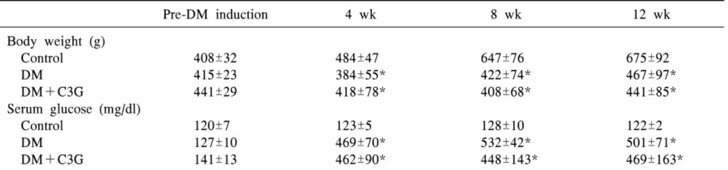
Table 1. Changes in body weight and serum glucose levels in the experimental groups
Pre-DM induction 4 wk 8 wk 12 wk
Body weight (g) Control DM DM+C3G
Serum glucose (mg/dl) Control
DM DM+C3G
408±32 415±23 441±29 120±7 127±10 141±13
484±47 384±55*
418±78*
123±5 469±70*
462±90*
647±76 422±74*
408±68*
128±10 532±42*
448±143*
675±92 467±97*
441±85*
122±2 501±71*
469±163*
DM: diabetes group, DM+C3G: diabetes group treated with Cyanidin-3-O-b-glucopyranoside.
*Significant statistical difference (p<0.05) compared to the control.
corpus cavernosum was removed and divided into two.
The first part was cryopreserved in liquid nitrogen and the other part was fixed in formalin.
4. Terminal deoxynucleotidyl transferase mediated dUTP nick end labeling assay
To assess apoptosis in the corpora tissues, the termi- nal deoxynucleotidyl transferase mediated dUTP nick end labeling (TUNEL) assay was performed using Apop Tag In Situ Apoptosis Detection Kits (Millipore Co., Billerica, MA, US). Tissue preparation for the de- tection of apoptotic bodies was done according to the manufacturer's protocol. After the TUNEL assay, tes- tis tissue sections were examined by light microscopy.
The number of cells positive for the TUNEL assay was counted and the difference among the groups was assessed. For the count of cells positive for the TUNEL assay, 5 sites were selected randomly from a slide of each group, and the number of cells positive for the TUNEL assay was counted as the average of 5 sites under a light microscope at 400× magnification.
5. Masson’s trichrome staining
After ICP, the skin denuded middle part of the pen- ile shafts were fixed overnight in 10% formalin, wash- ed, and stored in 70% alcohol at 4oC until processed for paraffin-embedded tissue sectioning (5 μm). The cavernosal tissue was obtained for the Masson’s tri- chrome staining. After staining, the color distribution of the muscle tissue was approximated by using the Adobe Photoshop CS 8.0. After the entire color dis- tribution of the image was calculated, we selected the
muscle tissue distribution, expressed as the color red.
There were somewhat standard deviations in our cal- culation because of color overlays and ambiguity of the color spectrum of the muscle tissues.
Results
1. General features of diabetes
Table 1 describes changes in body weight and blood glucose levels of the three groups. During the experi- ment, the serum glucose levels of the two DM groups were maintained and showed no significant difference.
With respect to the body weight of DM groups, it was significantly decreased compared with the control group.
2. In vivo assessment of erectile function Peak ICP and ICP/MAP ratios decreased in the DM group compared with the control group and signi- ficantly improved in the C3G treatment group. The control and DM+C3G groups had statistically similar Peak ICP and ICP/MAP ratios, which were signi- ficantly higher than those of the DM group (Table 2).
3. Masson’s trichrome staining
In comparison with the control group, smooth mus- cle content of corpora tissues was decreased and colla- gen deposition was increased in the DM group and smooth muscle content increased and collagen content decreased in the C3G treatment group (Fig. 1). The muscle/collagen ratio was 27.3±3.0 (control), 10.6±4.6 (DM), 21.8±1.7 (DM+C3G). A significant decrease
Table 2. Intracavernosal pressure in response to electrical stimulation of the cavernous nerve in rats from each experimental group
Control DM DM+C3G p-value
Peak ICP MAP
ICP/MAP ratio
83.3±1.9 108.5±3.4 0.77±0.05
35.4±4.5 109.4±2.8 0.32±0.06
58.0±4.6 104.6±4.5 0.55±0.11
0.023*
0.039† 0.004‡ 0.031*
<0.001† 0.023‡ ICP: intracavernosal pressure, MAP: mean arterial pressure.
*One-way ANOVA test, overall comparison, †Comparison between control and DM groups, ‡Omparison between DM and DM+C3G groups.
Fig. 2. Immunohistochemical in situ TUNEL detection of apoptosis in corporal tissue of the control (A), diabetes (B), and diabetes treated with C3G (C) groups. Cells undergoing apoptosis, called apoptotic bodies, show as black or dark brown in the TUNEL assay, while living cells are shown as lighter dots. ×400.
Fig. 1. Masson’s trichrome staining for collagen (blue) and smooth muscle (red) in corporal tissue of the control (A), diabetes (B), and diabetes treated with C3G (C) groups. ×200.
in muscle/collagen ratio was shown in the DM group compared with control group, C3G treatment sig- nificantly increased muscle/collagen ratio.
4. TUNEL assay for apoptosis
The mean apoptotic indices±standard deviation of the three groups as detected by the TUNEL assay were
15.3±3.0 (control), 39.6±4.6 (DM), and 21.3±1.7 (DM
+C3G). The DM group showed a higher mean apop- totic index than that of the control (p<0.05). Com- pared with the DM group, significantly fewer cells stained positively in the TUNEL assay in the C3G ad- ministration groups (p<0.05). Representative pictures are shown in Fig. 2.
Discussion
The main finding of the present study is that C3G improves erectile function by minimizing corporal apoptosis in a rat model of diabetic ED.
This result on apoptosis in the corporal tissue is in accordance with changes in ICP according to the in- duction of diabetic ED and C3G treatment. Nitric oxide has been known as the most important neurotrans- mitter mediating the relaxation of smooth muscle layer present in the corpus cavernosum.13,14 Synthesized within the vascular endothelium, NO is an endothe- lium derived relaxation factor (EDRF), in which nitric oxide synthase (NOS) plays an important role in its metabolism. Nitric oxide exists in three isoforms: neu- ronal NOS (nNOS), endothelial NOS (eNOS), and in- ducible NOS (iNOS). Further, it is currently known to be produced by eNOS in the endothelium of the cor- pus cavernosum and penile artery, and is secreted from nonadrenergic, noncholinergic nerve endings.15 There- fore, increasing the bioactivity of NO in the corpora is the most effective method to prevent and treat ED.
Of the pathophysiology of ED, vasculogenic ED which is caused by the dysfunction of vascular endo- thelium, and neurogenic impairments are currently known to account for the greatest proportion of cases.
Moreover, many recent reports have suggested that apoptosis from oxidative stress damage is the key eti- ology of vascular and neuronal impairments by DM.
Several reports have shown that neuronal and endo- thelial NO-dependent cavernosal smooth muscle relax- ation is diminished in DM.16,17 Experimental hyper- glycemia has been demonstrated to induce many of the pathological consequences observed in both Type 1 and Type 2 DM. Much of the tissue damage has stem- med from the effects of oxidative stress caused by ex- cessive production of free radicals such as O2−
, hydro- gen peroxide (H2O2), and the hydroxyl radical (OH−).
Oxygen free radicals in NO-rich tissues tend to com- bine to form peroxynitrites (O=NOO−), a highly cyto- toxic product of O2−
and NO radical reactions.18 High levels of oxidative stress can initiate a cascade of re- dox reactions, which trigger apoptosis and evoke cyto-
toxic effects on neurons and endothelial cells,19 result- ing in decreased NO production secondary to down- regulation of eNOS and nNOS expression20 and degen- eration of nitrergic nerves.21
Recent reports suggested that C3G, recognized as a potent antioxidant and free radical scavenger, protects endothelial cells against alterations induced by TNF-α, including the activation of NF-κB, increased gene ex- pression of adhesion molecules, leukocyte adhesion to endothelium, and intracellular accumulation of H2O2
and lipid peroxidation byproducts.10,11 In addition, C3G has also been attributed with neuroprotective effects both in vitro and in vivo ischemic oxidative stress.22 Based on the results from the current experiment, it is important to observe that C3G protects endothelial and neuronal damage.
The current study represents the first to suggest that C3G has a potency to protect against diabetes-induced ED in a rat model. As mentioned above, the key path- ophysiology of diabetic ED is the endothelial and neu- ronal damage. Thus, we hypothesize that minimizing corporal apoptosis by C3G may inhibit the progression of ED.
Diabetic ED involves irreversible changes of corporal tissue, and therefore response to medication decreases.
For these reasons, the results of this study are of value given that it is the first to focus on preventing diabetic ED through the use of C3G.
The potential limitations of this study are as follows. First, Streptozotocin-induced diabetes gen- erally reflects a Type 1 diabetes model. Although both Type 1 and Type 2 DM entail hyperglycemia, there are differences in terms of insulin resistance, body mass index, cytokines, and lipid profile between the two types. Of these differences, change in lipid pro- files, in particular, a factor that can affect endothelial damage may result in different pathophysiologic mech- anisms resulting in ED. Second, There were somewhat standard deviations in Masson’s trichrome calculation because of color overlays and ambiguity of the color spectrum of the muscle tissues.
Future work should examine the role of C3G in the treatment of Type 2 diabetes, the predominant form of diabetes. In addition, a composition analysis of natural
fruit pigment, which was used in this experiment, have not been completely done, and therefore the effects of components other than C3G on the results cannot be excluded and remains the subject of future study.
Conclusions
C3G may protect erectile function by minimizing corporal apoptosis.
The current study is the first to suggest that C3G may have a potency to improve and protect erectile function in a rat model of diabetic ED.
Acknowledgements
This research was supported by the Biogreen21 Program (Code#PJ007186), Rural Development Admi- nistration, Suwon, the Republic of Korea.
REFERENCES
1) Burnett AL. Erectile dysfunction. J Urol 2006;175:
S25-31
2) Fedele D, Bortolotti A, Coscelli C, Santeusanio F, Chatenoud L, Colli E, et al. Erectile dysfunction in type 1 and type 2 diabetics in Italy. On behalf of Gruppo Italiano Studio Deficit Erettile nei Diabetici.
Int J Epidemiol 2000;29:524-31
3) Johannes CB, Araujo AB, Feldman HA, Derby CA, Kleinman KP, McKinlay JB. Incidence of erectile dysfunction in men 40 to 69 years old: longitudinal results from the Massachusetts male aging study. J Urol 2000;163:460-3
4) Blanco R, Saenz de Tejada I, Goldstein I, Krane RJ, Wotiz HH, Cohen RA. Dysfunctional penile chol- inergic nerves in diabetic impotent men. J Urol 1990;144:278-80
5) Pegge NC, Twomey AM, Vaughton K, Gravenor MB, Ramsey MW, Price DE. The role of endothelial dysfunction in the pathophysiology of erectile dys- function in diabetes and in determining response to treatment. Diabet Med 2006;23:873-8
6) Jeremy JY, Jones RA, Koupparis AJ, Hotston M, Persad R, Angelini GD, et al. Reactive oxygen spe- cies and erectile dysfunction: possible role of
NADPH oxidase. Int J Impot Res 2007;19:265-80 7) Asano N, Yamashita T, Yasuda K, Ikeda K, Kizu
H, Kameda Y, et al. Polyhydroxylated alkaloids iso- lated from mulberry trees (Morusalba L.) and silk- worms (Bombyx mori L.). J Agric Food Chem 2001;49:4208-13
8) Dugo P, Mondello L, Errante G, Zappia G, Dugo G.
Identification of anthocyanins in berries by nar- row-bore high-performance liquid chromatography with electrospray ionization detection. J Agric Food Chem 2001;49:3987-92
9) Mitcheva M, Astroug H, Drenska D, Popov A, Kassarova M. Biochemical and morphological stud- ies on the effects of anthocyans and vitamin E on carbon tetrachloride induced liver injury. Cell Mol Biol (Noisy-le-grand) 1993;39:443-8
10) Seeram NP, Momin RA, Nair MG, Bourquin LD.
Cyclooxygenase inhibitory and antioxidant cyanidin glycosides in cherries and berries. Phytomedicine 2001;8:362-9
11) Serraino I, Dugo L, Dugo P, Mondello L, Mazzon E, Dugo G, et al. Protective effects of cyanidin- 3-O-glucoside from blackberry extract against perox- ynitrite-induced endothelial dysfunction and vascular failure. Life Sci 2003;73:1097-114
12) Kim HB, Kim SL, Koh SH, Seok YS, Kim YS, Sung GB, et al. The deelopmentv of natural pigment with mulberry fruit as a food additive. Korean J Crop Sci 2011;56:18-22
13) Becker AJ, Uckert S, Stief CG, Scheller F, Knapp WH, Machtens SA, et al. Systemic and cavernous plasma levels of vasoactive intestinal polypeptide during sexual arousal in healthy males. World J Urol 2002;20:59-63
14) Palmer RM, Ferrige AG, Moncada S. Nitric oxide release accounts for the biological activity of endo- thelium-derived relaxing factor. Nature 1987;327:
524-6
15) Rajfer J, Aronson WJ, Bush PA, Dorey FJ, Ignarro LJ. Nitric oxide as a mediator of relaxation of the corpus cavernosum in response to nonadrenergic, noncholinergic neurotransmission. N Engl J Med 1992;326:90-4
16) Saenz de Tejada I, Goldstein I, Azadzoi K, Krane RJ, Cohen RA. Impaired neurogenic and endothe- lium-mediated relaxation of penile smooth muscle from diabetic men with impotence. N Engl J Med
1989;320:1025-30
17) Keegan A, Cotter MA, Cameron NE. Effects of che- lator treatment on aorta and corpus cavernosum from diabetic rats. Free Radic Biol Med 1999;27:536-43 18) Qutub AA, Popel AS. Reactive oxygen species regu- late hypoxia-inducible factor 1alpha differentially in cancer and ischemia. Mol Cell Biol 2008;28:5106-19 19) Evgenov OV, Liaudet L. Role of nitrosative stress
and activation of poly (ADP-ribose) polymerase-1 in cardiovascular failure associated with septic and hemorrhagic shock. Curr Vasc Pharmacol 2005;3:
293-9
20) Akingba AG, Burnett AL. Endothelial nitric oxide synthase protein expression, localization, and activity in the penis of the alloxan-induced diabetic rat. Mol Urol 2001;5:189-97
21) Cellek S, Foxwell NA, Moncada S. Two phases of nitrergic neuropathy in streptozotocin-induced dia- betic rats. Diabetes 2003;52:2353-62
22) Kang TH, Hur JY, Kim HB, Ryu JH, Kim SY.
Neuroprotective effects of the cyanidin-3-O-beta-d- glucopyranoside isolated from mulberry fruit against cerebral ischemia. Neurosci Lett 2006;391:122-6