논문접수일 :2017년 9월 25일 논문수정일 :2017년 10월 25일 심사완료일 :2017년 11월 16일
교신저자 :최정환, 01757 서울 노원구 동일로 1342 인제대학교 의과대학 상계백병원 이비인후과학교실 전화 :(02) 950-1104・전송 :(02) 935-6220 E-mail:choijh92@paik.ac.kr
Introduction
Kawasaki disease (KD) which was first described in 1967,
1)also known as an acute febrile mucocuta- neous lymph node syndrome, is a systemic vasculitis of unknown etiology. KD is notorious for serious com- plications of the coronary artery aneurysm (CAA), acute myocardiac infarction, and even sudden cardiac failure. Because treatment with high-dose intravenous immunoglobulin (IVIG) with aspirin in the acute stage can effectively reduce the incidence of coronary
artery involvement,
2)it is very important to get an early diagnosis of KD. However, due to the lack of a specific diagnostic test, diagnosis is only based on a symptom pattern,
1,3,4)that includes high fever, poly- morphous skin rash, conjunctival injection, erythema of the lips and oral mucosa, cervical lymphadenopa- thy, and changes in the extremities like desquamation of hands and feet. Furthermore, due to variable presen- tation of the disease, the diagnosis is often incorrect initially.
5)Therefore, only 40% of KD patients met the diagnostic criteria.
6)Approximately 20% of KD cases with atypical presentations are diagnosed delayed or missed.
7)Of these symptoms, the most infrequent symptom which appears in less than half of case, is cervical lymphadenopathy,
8)while the others occur in 90% of the case.
8)KD with only lymphadenopathy is consid- ered to have an atypical presentation with unfavor- able outcome.
9)Reports of KD presenting as a retro-
The Neglected Diagnosis in Retropharyngeal Abnormalities : Kawasaki Disease
Changyun Kwon, MD and Jeong Hwan Choi, MD
Department of Otorhinolaryngology-Head and Neck Surgery, Sanggye Paik Hospital, College of Medicine, Inje University, Seoul, Korea
- ABSTRACT -
Background and Objectives:In patients with Kawasaki disease, retropharyngeal involvements (KDWRPI) is a rare complication. Most reported cases were diagnosed lately because those were often misdiagnosed as bac- terial retropharyngeal abscess (BRA). The purpose of this study was to differentiate KDWRPI from BRA in ad- vance. Materials and Methods:We performed a retrospective study comparing children with KDWRPI to those with BRA hospitalized at the university teaching hospital between January 2008 and September 2013.
From our retrospectively collected database, we compared clinical, laboratory, and imaging characteristics of KDWRPI and BRA. Results: The study include 11 patients with retropharyngeal involvement on neck comput- erized tomography (CT) which were divided into two groups. Group A was classified as KDWRPI (n=6) and group B was classified as BRA (n=5). Compared with group B, patients with KDWRPI had lower sodium and albumin (p=0.0176 and 0.0828, respectively). Conclusions:Careful attention to manifestations and close anal- yses of laboratory findings and CT images may allow otorhinolaryngologists to differentiate KDWRPI from BRA. In the case of retropharyngeal edema on CT, the diagnosis of KDWRPI should not be neglected. (J Clini- cal Otolaryngol 2017;28:21-218)
KEY WORDS : Kawasaki diseaseㆍRetropharyngeal edemaㆍLymphadenitisㆍRetropharyngeal abscess.
J Clinical Otolaryngol 2017;28:211-218
원 저
pharyngeal involvement such as cellulitis or abscess are rare.
4,6,10-13)Neck computerized tomography (CT) of the neck is not a routine diagnostic modality for KD patients, just in case of severe cervical lymphade- nopathy implying deep-neck infections. Thus, we could not know the true incidence of retropharyngeal space involvement in the overall KD cohort. One study re- ported the retropharyngeal low-density lesions were identified by Neck CT in 3.6% of the KD patients.
14)Moreover, radiological reports suggested that the ret- ropharyngeal low-density lesions were not abscesses, but rather were due to soft-tissue reactions that im- proved, as do other KD symptoms, after treatment.
Those diagnostic delay and inappropriate treatment may cause the development of CAA finally.
15)Thus, early diagnosis and differentiation of KDWRPI from bacterial retropharyngeal abscess (BRA) is essential to avoid disastrous consequence.
To the best of my knowledge, the clinical and radio- logical differences between KDWRPI and BRA have not been thoroughly studied.
13)The object of this study was to compare the clinical characteristics, laboratory results of patients with KDWRPI to those with BRA.
Materials and Methods
Study design
We performed a retrospective study comparing children with KDWRPI to those with BRP hospital- ized at the university teaching hospital between Janu- ary 2008 and September 2013. We reviewed the radi- ology reports using the search term “retropharyngeal abscess”, “retropharyngeal edema”, and “retropha- ryngeal low density area” in those groups. Cases with neck CT showed specific retropharyngeal le- sions suggesting other diagnoses were excluded. All of 11 patients had complete medical records insuring the correct diagnosis. The 11 patients with retropha- ryngeal lesion on neck CT were categorized into two groups: group A was classified as KDWRPI and group B as BRA. KDWRPI (group A) was diagnosed when patients fulfilled the diagnostic criteria of KD.
16)We compared the group A and group B with regard to clinical and demographic data (age, sex), physical findings, the earliest available laboratory data pro- spectively collected during the hospital admission and neck CT findings. Laboratory test results includ- ed white blood cell (WBC) counts, hemoglobin con- centrations (Hgb), platelet count, erythrocyte sedimen- tation rate (ESR), C-reactive protein (CRP), serum levels of alanine aminotransferase (ALT) level, albu- min, and sodium (Na).
The Institutional Review Board of our university hospital approved this study and granted a waiver of informed consent. The approval included viewing im- ages and medical records of the patients.
Statistical analysis
Data are expressed as median and interquartile range (IQR, 25%ile-75%ile). Because of nonnor- mality of some variables, the Mann-Whitney U test was used for comparison between groups. p values
<0.05 were considered statistically significant. All statistical analyses were performed using MedCalc for Windows, version 15.0 (MedCalc Software, Os- tend, Belgium).
Results
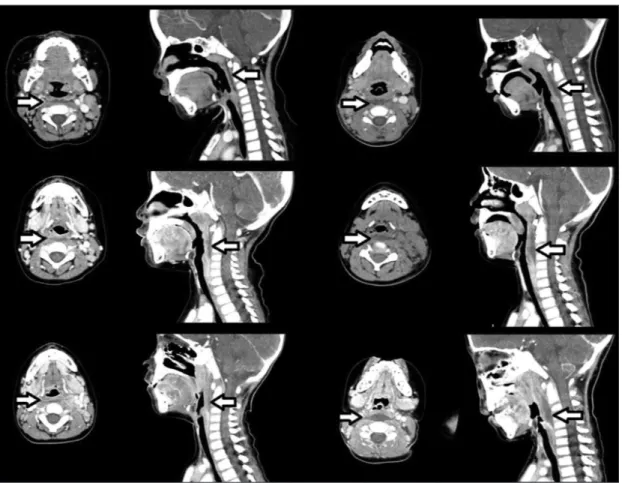
We identified 6 patients in group A (KDWRPI) and 5 patients in group B (BRA). Group A patients were all in the febrile conditions with retropharyngeal swell- ing or edema (Fig. 1). All the patients were initially treated with antibiotics but were not responsive and one of them even underwent unproductive, culture- negative surgical drainage of the retropharyngeal space.
Following the diagnosis of KD, all patients received high dose IVIG with aspirin. Group B patients who did not fulfill the criteria for KD following neck CT (Fig. 2), were diagnosed as having BRA and treated with antibiotic therapy and/or surgical intervention.
Neck CT of both groups demonstrated low-attenua-
tion area without contrast rim enhancement at the ret-
ropharyngeal space (Fig. 1, 2).
The clinical characteristics and laboratory date of both groups are shown in Table 1. There were no sig- nificant differences in sex ratio between the two groups. In a comparison of two groups, group A pa- tients had low serum sodium level (135.0 vs. 139.0 mEq/L) with statistical significance (p=0.0176). Al- bumin level was also lower in group A (3.75 vs. 4.00 g/dL), but statistically less significance (p=0.0828).
However, WBC, ANC, platelet counts, ESR, and ALT level did not differ between the two groups statistical- ly (Fig. 3).
Discussion
KD is often difficult to diagnose because of lack of
specific laboratory test for this disease, and further- more, symptoms that match the diagnostic criteria for KD might not be present at the same time, but rather can appear in random pattern. As both KD and retro- pharyngeal abscess can be fatal if an appropriate treatment is not applied. When otolaryngologists are consulted for the Image of retropharyngeal low-den- sity area, we pay special attention to this condition.
Because these findings are often observed in the cas- es of retropharyngeal abscess and edema usually as- sociated to incoming lethal condition such as airway obstruction, mediastinitis, and sepsis, if not appropri- ately treated. So we prefer treating it with prompt sur- gical intervention.
17)But, in the case of KD, some re- ports of KD patients with retropharyngeal lesions
Fig. 1. Neck CT with contrast enhancement of group A patients (retropharyngeal involvements in Kawasaki disease)
shows low-attenuation area without contrast rim enhancement at the retropharyngeal space.
revealed drainage or aspiration findings failing the present of fluid or bacterial infection.
18-20)In the absence of a definite diagnostic test for KD, the differentiation of KDWRPI from BRA remains challenging. In such cases, clinicians should pay at- tention to other clinical or laboratory findings not in- cluded in the diagnostic criteria that are useful in cor- roborating the diagnosis. Because KD sometimes manifests with only fever and cervical adenopathy be- fore other clinical signs and symptoms appear.
6)As KD is mainly diagnosed and treated by pediatri- cians, these children are extremely rarely seen by an otorhinolaryngologist. However, as many of the man- ifestations occur in the head and neck, an otorhino- laryngologist may be the first medical professional to see a patient with KD. KD may begin with fever and
cervical adenopathy with other clinical signs appear- ing later.
21)Otolaryngologists should be considered a rare symp- tom of KD in dealing with the patients, especially children with cervical lymphadenopathy, retropha- ryngeal edema, and the presence of low-density lesions in the retropharyngeal space observed by neck CT.
Furthermore, when the febrile patient with leukocy- tosis, lymphadenopathy, and edema of the retropha- ryngeal space visit clinic, retropharyngeal infection might be initially suspected, but retropharyngeal ab- normalities as atypical manifestations of KD should also be considered.
The posterior pharyngeal wall adenitis symptoms such as fever, neck pain, torticollis, and cervical lymph- adenopathy are all shared with Kawasaki disease.
Fig. 2. Neck CT with contrast enhancement of group B patients [bacterial retropharyngeal abscess (BRA)] shows cer-
vical lymph node enlargement with retropharyngeal swelling.
Thus it is not surprising that Kawasaki disease can be misdiagnosed as retropharyngeal abscess.
More than half of KD patients have atypical pre- sentations that often result in erroneous or delayed diagnosis and appropriate treatment.
6,7)Furthermore, due to the slow and variable evolution of the disease, the initial diagnosis is often incorrect.
5)Such errone- ous or delayed diagnosis can lead to increased devel- opment of CAA that have been reportedly to occur in 20% of KD patients with up to 2% of overall mortal-
ity.
18,32)With regards to the diagnosis of a retropharyngeal abscess, CT may be a sensitive tool for the detection of deep neck lesions.
4)CT scan provides the most ac- curate information on the extent and exact anatomi- cal location of the lesion.
22)In the presence of acute febrile illness, lymph nodes in the retropharyngeal space can become enlarged and have enhancing sur- rounding with a hypodense core on CT scan which could be interpreted as an abscess.
23)Thus, a hypoat-
Fig. 3. Comparison of demographic characteristics and laboratory data of group A [patients with retropharyngeal involvements in Kawasaki disease (KDWRPI)] and group B [patients with bacterial retropharyngeal abscess (BRA)] In a comparison of two groups, group A patients had low serum sodium level with statistical significance (p=0.0176). Al- bumin level was also lower in group A, but statistically less significance (p=0.0828). However, WBC, ANC, platelet counts, ESR, and ALT level did not differ between the two groups statistically. Data are expressed as median and in- terquartile range (IQR, 25%ile-75%ile). The statistical analysis was performed by the Mann-Whitney U test for compari- son between groups. p values <0.05 were considered statistically significant. cal_age : calculated age, WBC : white blood cell counts, Hb : hemoglobin concentrations, PLT : platelet count, ESR : erythrocyte sedimentation rate, CRP : C-reactive protein, ALT : serum levels of alanine aminotransferase level, Alb : albumin, Na : sodium.
8 7 6 5 4 3 2 1
cal_age_BRA cal_age_KDWRPI
14.0 13.5 13.0 12.5 12.0 11.5 11.0 10.5 10.0
Hb_BRA Hb_KDWRPI 25,000
20,000 15,000 10,000 5,000 0
WBC_BRA WBC_KDWRPI
120 100 80 60 40 20
ESR_BRA ESR_KDWRPI
35 30 25 20 15 10 5 0
CRP_BRA CRP_KDWRPI 600,000
500,000 500,000 450,000 400,000 350,000 300,000 250,000 200,000 150,000
PLT_BRA PLT_KDWRPI
18 16 14 12 10 8 6 4
ALP_BRA ALP_KDWRPI
4.5 4.4 4.3 4.2 4.1 4.0 3.9 3.8 3.7 3.6
Alo_BRA Alo_KDWRPI
140 139 138 137 136 135 134 133
Na_BRA Na_KDWRPI
tenuating rim-enhancing retropharyngeal collection on CT scan is predictive, but not diagnostic of ab- scess.
24)The lack of enhancement around the fluid collection in the KDWRPI may be helpful to distinguish the BRA.
4,13)However, if the fluid collection is accompa- nied by necrotic lymphadenopathy, it is difficult to distinguish KDWRPI and BRA.
25)Both the pathogenesis of KD and the etiology of retropharyngeal low-density lesions remain unclear.
However, considering the operative findings of un- productive surgical drainage, negative culture results for drainage specimens and the responses to immu- noglobulin, inflammation and edema are considered as one of the pathogenesis.
26)KD starts with vasculi- tis increasing microvascular permeability, causing extravascular albumin leakage and edema.
27)Vasculi- tis of microvessels with tissue edema and inflamma- tion is hypothesized to cause extensive KDWRPI.
28)During the acute phase of KD, mucosal immune sys- tem produce inflammatory cytokines.
29)Retropha- ryngeal lesions in KD might be explained by this mechanism. RPE formation could be a risk factor for developing CAA.
10)The retropharyngeal space consists of lymphatic vessels and adipose tissue. This space was surrounded by alar and prevertebral fas- cia.
30)Besides retropharyngeal abscess, retropharyn- geal hypodense lesion in the CT can be observed in
various pathogenesis related with cervical lymph node swelling.
23)Thus, this lesion on CT might be a type of nonsuppurative inflammation rather than an infec- tion
4)as proved by my surgical cases.
Laboratory tests can be performed to help with the diagnosis. Blood test may show leukocytosis and el- evated acute-phase reactants, such as CPR, ESR, nor- mocytic normochromic anemia, thrombocytosis. Oth- er findings include elevated liver enzymes (ALT), decreased cholesterol and high-density lipoprotein levels, increased triglycerides level,
31)hypoalbumin- emia, hyponatremia, and more rarely hyperbilirubi- nemia. Urinalysis may reveal sterile pyuria, whereas the analysis of cerebrospinal fluid shows evidence of aseptic meningitis. Our study identified lower sodium level in serum was the most reliable variables distin- guishing KDWRPI from BRA. Lower serum albumin level also was taken into consideration. Unnecessary pharyngeal exploration or unproductive surgical drain- age for patients with KDWRPI can be avoided.
This is the first study that has compared the clinical characteristics of patients of KDWRPI with patients of BRA with significant different laboratory findings unlike previous study.
13)Distinguishing patients with KDWRPI from patients with BRA in the early phase is difficult. However, our study showed that KDWR- PI and BRA could be distinguished by careful evalu- ation of clinical manifestations serially.
Table 1. Comparison of demographic characteristics and laboratory data of group A [patients with retropharyngeal involvements (KDWRPI) in Kawasaki disease] and group B [patients with bacterial retropharyngeal abscess (BRA)]
Clinical and laboratory characteristics KDWRPI (n=6) BRA (n=5) p value
Age, y, median (IQR) 4.2 (3.6-4.9) 6.4 (2.95-6.95) 0.5839
Males, No. (%) 3 (50) 3 (60)
WBC, ×10
6cells/mL, median (IQR) 14,745 (13430-17,900) 18,440 (12287.5-20,865) 0.7150 Hemoglobin, g/dL, median (IQR) 10.800 (10.400-12.000) 12.200 (11.325-12.600) 0.1709 Platelet count, cells/mL, median (IQR) 311,500 (220,000-334,000) 365,000 (316000-415,000) 0.2733
ESR, mm/h, median (IQR) 106 (78-120) 63 (51-94) 0.3314
CRP, mm/h, median (IQR) 12.45 (6.3-30.6) 13.8 (2.75-14.3) 0.2733
ALT level, U/L, median (IQR) 9.5 (6-12) 11 (8.75-16.5) 0.2733
Albumin, g/dL, median (IQR) 3.75 (3.7-3.9) 4.0 (3.95-4.2) 0.0828
Na, mEq/L, median (IQR) 135 (134-136) 139 (136.75-139.25) 0.0176
IQR : interquartile range, WBC : white blood cell counts, ESR : erythrocyte sedimentation rate, CRP : C-reactive
protein, ALT : serum levels of alanine aminotransferase level, Na : sodium
Our study had some limitations. A sample size was too small for conclusions and generalizations to be representative of all patients. However, this may be a starting point for further research seeking variables significantly leading to proper patient management and preventing unnecessary surgical intervention.
Furthermore, we diagnosed KD and BRA clinically.
Thus, there was no definitive diagnostic standard for BRA. Further research is needed to eliminate these limitations.
Conclusion
KD sometimes presents with symptoms first in the head and neck region and the patient may initially be admitted to ORL ward. The other symptoms leading to diagnosis of KD may appear only after several days.
The diagnosis requires a high level of awareness. The possibility of KD should be kept in mind, at least pri- or to surgery, in pediatric patients with suspicion of retropharyngeal infections. Clinicians who treat febrile children with cervical adenopathy should keep KD- WRPI in the differential diagnosis. Clinicians should be alert to the occurrence of retropharyngeal edema and multiple solid enlarged nodes KDWRPI when in- terpreting CT of nodes to ensure the timely diagnosis of KD. Prompt diagnosis of KD and appropriate treat- ment with IVIG can lead to proper patient manage- ment and prevent unnecessary surgical intervention as well as serious cardiac complications.
This work was supported by Grant from Inje University, 2008.
REFERENCES
1) Kawasaki T. Acute febrile mucocutaneous syndrome with
lymphoid involvement with specific desquamation of the fingers and toes in children. Arerugi 1967;16:178-222.
2) Newburger JW, Takahashi M, Burns JC, Beiser AS, Chung KJ, Duffy CE, et al. The treatment of Kawasaki syndrome
with intravenous gamma globulin. N Engl J Med 1986;315 (6):341-7.
3) Dajani AS, Taubert KA, Gerber MA, Shulman ST, Ferri- eri P, Freed M, et al. Diagnosis and therapy of Kawasaki
disease in children. Circulation 1993;87(5):1776-80.
4) Hung M, Wu K, Hwang B, Lee P, Meng CL. Kawasaki
disease resembling a retropharyngeal abscess-case report and literature review. Int J Cardiol 2007;115(2):e94-6.
5) Murrant N, Cook J, Murch S. Acute ENT admission in Ka-
wasaki disease. J Laryngol Otol 1990;104(7):581-4.
6) Park AH, Batchra N, Rowley A, Hotaling A. Patterns of
Kawasaki syndrome presentation. Int J Pediatr Otorhino- laryngol 1997;40(1):41-50.
7) Manlhiot C, Christie E, McCrindle BW, Rosenberg H, Chahal N, Yeung RS. Complete and incomplete Kawasa-
ki disease: two sides of the same coin. Eur J Pediatr 2012;
171(4):657-62.
8) Burgner D, Festa M, Isaacs D. Delayed diagnosis of Ka-
wasaki disease presenting with massive lymphadenopa- thy and airway obstruction. BMJ 1996;312(7044):1471-2.
9) Nomura Y, Arata M, Koriyama C, Masuda K, Morita Y, Hazeki D, et al. A severe form of Kawasaki disease pre-
senting with only fever and cervical lymphadenopathy at admission. J Pediatr 2010;156(5):786-91.
10) Kritsaneepaiboon S, Tanaanantarak P, Roymanee S, Lee EY. Atypical presentation of Kawasaki disease in young
infants mimicking a retropharyngeal abscess. Emerg Ra- diol 2012;19(2):159-63.
11) Rooks VJ, Burton BS, Catalan JN, Syms MJ. Kawasaki
disease presenting as a retropharyngeal phlegmon. Pedi- atr Radiol 1999;29(11):875-6.
12) Kim JS, Kwon SH. Atypical Kawasaki disease presenting
as a retropharyngeal abscess. Braz J Otorhinolaryngol 2016;82(4):484-6.
13) Nomura O, Hashimoto N, Ishiguro A, Miyasaka M, No- saka S, Oana S, et al. Comparison of patients with Kawa-
saki disease with retropharyngeal edema and patients with retropharyngeal abscess. Eur J Pediatr 2014;173(3):
381-6.
14) Tona R, Shinohara S, Fujiwara K, Kikuchi M, Kanazawa Y, Kishimoto I, et al. Risk factors for retropharyngeal cel-
lulitis in Kawasaki disease. Auris Nasus Larynx 2014;41 (5):455-8.
15) Muta H, Ishii M, Yashiro M, Uehara R, Nakamura Y. Late
intravenous immunoglobulin treatment in patients with Kawasaki disease. Pediatrics 2012;129(2):e291-7.
16) Newburger JW, Takahashi M, Gerber MA, Gewitz MH, Tani LY, Burns JC, et al. Diagnosis, treatment, and long-
term management of Kawasaki disease: a statement for health professionals from the Committee on Rheumatic Fever, Endocarditis and Kawasaki Disease, Council on Cardiovascular Disease in the Young, American Heart Association. Circulation 2004;110(17):2747-71.
17) Craig FW, Schunk JE. Retropharyngeal abscess in chil-
dren: clinical presentation, utility of imaging, and current management. Pediatrics 2003;111(6 Pt 1):1394-8.
18) Homicz MR, Carvalho D, Kearns DB, Edmonds J. An atyp-
ical presentation of Kawasaki disease resembling a retro- pharyngeal abscess. Int J Pediatr Otorhinolaryngol 2000;
54(1):45-9.
19) Pontell J, Rosenfeld RM, Kohn B. Kawasaki disease mim-
icking retropharyngeal abscess. Otolaryngol Head Neck Surg 1994;110(4):428-30.
20) Gross M, Eliashar R, Attal P, Sichel JY. Radiology quiz
case 2: Kawasaki disease (KD) mimicking a retropharyn- geal abscess. Arch Otolaryngol Head Neck Surg 2001;127 (12):1507, 1508-9.
21) Kanegaye JT, Van Cott E, Tremoulet AH, Salgado A, Shi- mizu C, Kruk P, et al. Lymph-node-first presentation of
Kawasaki disease compared with bacterial cervical ade- nitis and typical Kawasaki disease. J Pediatr 2013;162(6):
1259-63. e1-2.
22) Vural C, Gungor A, Comerci S. Accuracy of computer-
ized tomography in deep neck infections in the pediatric population. Am J Otolaryngol 2003;24(3):143-8.
23) Boucher C, Dorion D, Fisch C. Retropharyngeal abscess-
es: a clinical and radiologic correlation. J Otolaryngol 1999;28(3):134-7.
24) Stone ME, Walner DL, Koch BL, Egelhoff JC, Myer CM.
Correlation between computed tomography and surgical findings in retropharyngeal inflammatory processes in children. Int J Pediatr Otorhinolaryngol 1999;49(2):121-5.
25) Tomita H, Yamashiro T, Ikeda H, Fujikawa A, Kurihara Y, Nakajima Y. Fluid collection in the retropharyngeal
space: a wide spectrum of various emergency diseases.
Eur J Radiol 2016;85(7):1247-56.
26) Langley EW, Kirse DK, Barnes CE, Covitz W, Shetty AK.
Retropharyngeal edema: an unusual manifestation of Ka- wasaki disease. J Emerg Med 2010;39(2):181-5.
27) Ueda Y, Saita Y, Matsuzawa T, Wada T, Kanai N, Kobayas- hi I. Six patients with Kawasaki disease showing retropha-
ryngeal low-density areas on computed tomography. Pe- diatr Int 2010;52(4):e187-9.
28) Roh K, Lee SW, Yoo J. CT analysis of retropharyngeal ab-
normality in Kawasaki disease. Korean J Radiol 2011;12 (6):700-7.
29) Abe J. Immunological aspects of Kawasaki disease. Ni-
hon Rinsho 2008;66(2):267-71.
30) Philpott C, Selvadurai D, Banerjee A. Paediatric retropha-
ryngeal abscess. J Laryngol Otol 2004;118(12):919-26.
31) Chiang A, Hwang B, Shaw G, Lee B, Lu J, Meng C, et al.
Changes in plasma levels of lipids and lipoprotein compo- sition in patients with Kawasaki disease. Clin Chim Act 1997;260(1):15-26.
32) Moon JH, Ahn HY, Chang MK, Cha CI. Cervical lymph-
![Fig. 2. Neck CT with contrast enhancement of group B patients [bacterial retropharyngeal abscess (BRA)] shows cer- cer-vical lymph node enlargement with retropharyngeal swelling.](https://thumb-ap.123doks.com/thumbv2/123dokinfo/5427395.229902/4.799.90.714.129.598/contrast-enhancement-patients-bacterial-retropharyngeal-enlargement-retropharyngeal-swelling.webp)
![Fig. 3. Comparison of demographic characteristics and laboratory data of group A [patients with retropharyngeal involvements in Kawasaki disease (KDWRPI)] and group B [patients with bacterial retropharyngeal abscess (BRA)] In a comparison of two groups,](https://thumb-ap.123doks.com/thumbv2/123dokinfo/5427395.229902/5.799.91.712.128.614/comparison-demographic-characteristics-laboratory-retropharyngeal-involvements-retropharyngeal-comparison.webp)
![Table 1. Comparison of demographic characteristics and laboratory data of group A [patients with retropharyngeal involvements (KDWRPI) in Kawasaki disease] and group B [patients with bacterial retropharyngeal abscess (BRA)]](https://thumb-ap.123doks.com/thumbv2/123dokinfo/5427395.229902/6.799.92.708.162.372/comparison-demographic-characteristics-laboratory-retropharyngeal-involvements-kawasaki-retropharyngeal.webp)
