© 2014 Korean Breast Cancer Society. All rights reserved. http://ejbc.kr | pISSN 1738-6756
INTRODUCTION
Surgery for breast cancer has changed from the radical ap- proach of removing the skin, nipple, and underlying glandular tissue of the breast to a more conservative approach. Studies have shown that more than 60% of women diagnosed with early breast cancer have had breast conserving surgery [1].
However, patients with large tumors (>5 cm), multifocal/
multicentric tumors, or those with a contraindication to radi- ation therapy, need to have most of their breast tissue removed for proper treatment.
While oncological safety can be achieved for patients un-
dergoing radical mastectomy, the psychological effects and cosmetic outcomes are generally poor [2,3]. To address this issue, breast reconstruction techniques have been developed that can be carried out immediately following mastectomy, and have shown good results for both oncological safety and patient satisfaction [4]. In particular, the skin sparing mastec- tomy (SSM) procedure has become widely accepted because of its favorable outcomes.
A modification of the SSM, the nipple sparing mastectomy (NSM), where the nipple-areolar complex (NAC) is left intact, provides even better cosmetic results [5]. Originally developed as a treatment for patients with benign breast disease [6], NSM has been shown to have a low rate of local recurrence for breast cancer patients in several studies [4,5].
However, there is a potential risk of leaving an occult tumor within the nipple with NSM, which could lead to cancer re- lapse and a poor prognosis for the patient. Therefore, to ensure the safety of this procedure, careful selection of surgical candi- dates is needed. Thus far, there is no definite method of identi-
Magnetic Resonance Imaging and Clinicopathological Factors for the Detection of Occult Nipple Involvement in Breast Cancer Patients
Wooseok Byon, Eunyoung Kim, Junseong Kwon, Yong Lai Park, Chanheun Park
Department of Surgery, Kangbuk Samsung Hospital, Sungkyunkwan University School of Medicine, Seoul, Korea ORIGINAL ARTICLE
Purpose: Nipple sparing mastectomy provides good cosmetic results and low local recurrence rates for breast cancer patients.
However, there is a potential risk of leaving an occult tumor within the nipple, which could lead to cancer relapse and poor prognosis for the patient. The objective of this study was to in- vestigate the occult nipple involvement rate in mastectomy specimens, and to identify preoperative magnetic resonance im- aging (MRI) findings and the clinicopathological characteristics of the primary tumor that may correlate with nipple invasion.
Methods: Four hundred sixty-six consecutive mastectomy sam- ples with grossly unremarkable nipples were evaluated. Demo- graphic and clinicopathological data were collected. Nipple in- volvement was evaluated using serial histological sections. The tumor size and tumor-nipple distance were measured using pre- operative MRI images. Results: Thirty-six of the 466 therapeutic mastectomy specimens (7.7%) were found to have occult nipple
involvement. In univariate analysis, tumor size, tumor-nipple dis- tance, lymph node status, p53 mutation, and lymphovascular in- vasion (LVI) were found to influence the likelihood of nipple in- volvement. Multivariate logistic regression analysis, adjusted by lymph node status, p53 mutation, and LVI, showed that tumor size and tumor-nipple distance were predictive factors indicating nipple involvement. With regard to tumor location, only tumors in the central area of the breast showed a significant association with nipple involvement. Conclusion: In this study, a statistically significant association was found between occult nipple involve- ment and tumor size, tumor-nipple distance, axillary lymph node status, LVI, and p53 mutation. A cutoff point of 2.2 cm for tumor size and 2 cm for tumor-nipple distance could be used as pa- rameters to predict occult nipple involvement.
Key Words: Breast, Carcinoma, Magnetic resonance imaging, Mastectomy
Correspondence to: Chanheun Park
Department of Surgery, Kangbuk Samsung Hospital, Sungkyunkwan University School of Medicine, 29 Saemunan-ro, Jongno-gu, Seoul 110-746, Korea
Tel: +82-2-2001-1730, Fax: +82-2-2001-1883 E-mail: [email protected]
Received: May 7, 2014 Accepted: December 8, 2014
Cancer
fying tumor invasion arising from clinically unremarkable nipples except by direct tissue biopsy and sequential patholog- ical evaluation.
Magnetic resonance imaging (MRI) is a useful tool for the assessment of disease extent in patients with a recent diagno- sis of breast cancer [7,8]. In particular, occult invasive tumors that were not detected by clinical examination or conventional imaging methods, have been identified with MRI imaging [9].
The objective of this study was to investigate the rate of oc- cult nipple involvement in mastectomy specimens, and to identify preoperative MRI findings and the clinicopathologi- cal characteristics of the primary tumor that may correlate with nipple invasion.
METHODS
Patients who received radical mastectomy for breast cancer between January 2005 and December 2010 were considered eligible for the present study. Data from a total of 511 mastec- tomy samples were retrieved. Patients with clinically evident nipple involvement, Paget’s disease, or inflammatory breast cancer were excluded. In addition, patients who underwent neoadjuvant chemotherapy and patients without preoperative MRI imaging were not included. Overall, 466 consecutive mastectomy samples with grossly unremarkable nipples were selected and retrospectively analyzed.
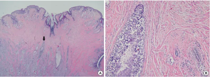
The NAC was routinely inspected in all mastectomy sam- ples. Nipple specimens were fixed in formalin, and subse- quently, a single sagittal section through the nipple was made.
A hematoxylin and eosin-stained section was prepared from each block and reviewed by a breast pathologist (Figure 1). If
necessary, additional sections or immunohistochemical stains were performed for diagnosis. Occult nipple involvement was defined as the detection of invasive ductal or lobular carcino- ma and/or ductal or lobular carcinoma in situ in the nipple section, including the subareolar margin.
We reviewed patient clinical and pathological data includ- ing age, histologic type, histologic grade, lymph node status, hormone receptor status, human epidermal growth factor re- ceptor 2 (HER2) status, p53 mutation, and lymphovascular invasion (LVI).
Dynamic contrast-enhanced breast MRI examinations were performed using 3.0 T equipment (Achieva; Philips Medical System, Best, The Netherlands) and a dedicated 7-channel SENSE breast coil. Dynamic contrast-enhanced images were acquired following intravenous injection of 1.0 M gadobutrol (7.5 mL, Gadavist; Bayer Schering Pharma, Berlin, Germany).
Dynamic subtraction (i.e., postcontrast images minus precon- trast images) and three-dimensional maximum intensity pro- jection images were generated for all studies. The diameter of the lesion was defined as the maximum extent of suspicious contrast enhancement. In the case of multifocal or multicen- tric lesions, the diameter of the largest lesion was measured.
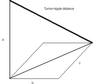
Tumor-nipple distance was determined by measuring four distinctive distances in each case (Figure 2). All measure- ments were made using digital images on flat-screen liquid- crystal display monitors. First, two images were selected, one which included the nipple in its most convex state and anoth- er which included the lesion shown with the maximum extent of contrast enhancement. Using the first image, a vertical line was drawn from the center of the nipple to the chest wall and the distance from the base of the NAC to the chest wall was
A B
Figure 1. Microscopic findings of nipple involvement. (A) The sagittal section of nipple shows involvement of tumor (black arrow) without epidermal in- volvement (H&E stain, ×2.5). (B) Magnified view shows underlying lactiferous duct with ductal carcinoma in situ and invasive cancer in stroma (H&E stain, ×100).
measured. A ruler was placed on the flat-screen monitor per- pendicular to this line and using this as a reference point, the vertical distance from the uppermost point of the lesion to the chest wall and the horizontal distance from this vertical line to the lesion was measured. By subtracting the lesion-chest wall
distance from the NAC base-chest-wall distance, measure- ment “a” was acquired (Figure 3). Measurement “b” was the horizontal distance mentioned above. Measurement “c” was determined by calculating the number of images between the selected images multiplied by the cut width of each MRI im- age. The tumor-nipple distance was calculated by substituting the measurements above into the following equation:
Tumor-nipple distance=√(a2+b2+c2)
The location of the tumor was categorized into nine areas.
For large tumors, the location of the center of the lesion was used for assessment. In the case of multifocal and/or multi- centric lesions, the location of the largest lesion was used for evaluation.
Categorical data are presented as frequencies and percent- ages, and numerical data as the mean±standard deviation.
For comparison of categorical variables, the chi-square test or Fisher exact test was used. The Student t-test was used for nu- merical variables. Multivariate logistic regression analysis with stepwise selection was performed for nipple involvement on variables that were significant in the univariate setting. Statis- tical calculations were carried out using Microsoft Excel (Mi- crosoft Corp., Redwood City, USA) and SAS statistical soft- ware (SAS Institute, Cary, USA), and p-values <0.05 were considered statistically significant. The study was approved by the institutional review board of Kangbuk Samsung Hospital (KBSMC IRB 2014-01-250).
A B
Figure 2. Measurement of tumor-nipple distance in MRI imaging. (A) A vertical line is drawn from the center of the nipple to the chest wall and the dis- tance from the base of the nipple-areolar complex to the chest wall is measured (red line). (B) The vertical distance from the upper most point of the lesion to the chest wall (orange line) and the horizontal distance from this vertical line to the lesion is measured (blue line).
MRI=magnetic resonance imaging.
Figure 3. Diagram of calculation method of tumor-nipple distance.
Measurement “a” is calculated by subtracting the lesion-chest wall dis- tance from the nipple-areolar complex base-chest wall distance. Mea- surement “b” is the horizontal distance from the lesion to the vertical line drawn from the center of the nipple to the chest wall. Measurement “c”
is acquired by calculating the number of images between the selected images multiplied by the cut width of each MRI image.
MRI=magnetic resonance imaging.
Tumor-nipple distance
c
b a
RESULTS
Four hundred sixty-six mastectomy specimens were evalu- ated in this study. All radical mastectomies were performed for therapeutic intent for previously diagnosed breast carcino- ma. No prophylactic surgical specimens from highrisk pa- tients were included.
Table 1 lists the clinical and pathological characteristics of the patients and the results of the histologic evaluation of the specimens.
Thirty-six of the 466 therapeutic mastectomy specimens (7.7%) were found to have occult nipple involvement. The pa- tients’ ages ranged from 30 to 82 years with an overall mean age of 53.1±10.5 years. The mean age of the patients with nipple involvement was 55.3±12.9 years, and that of the unaf- fected group was 52.9±10.3 years (p=0.274).
When comparing the occult nipple involvement and unaf- fected groups, age distribution, histologic type of cancer, his- tologic grade, estrogen or progesterone receptor status, HER2 status, and multifocality or multicentricity on MRI imaging, did not show significant differences between the two groups.
However, tumor size, tumor-nipple distance, lymph node status, p53 mutation, and LVI all influenced the likelihood of nipple involvement. A median tumor size of 2.2 cm was cho- sen as a cutoff point to dichotomize tumor size and this re- sulted in a significant difference between the two groups (p=
0.006). In addition, a median tumor-nipple distance of 2 cm was used as a cutoff point. Both tumor size and tumor-nipple distance were statistically significant when evaluated as con- tinuous variables (p=0.001 and p<0.001, respectively).
Multivariate logistic regression analysis adjusted for lymph node status, p53 mutation, and LVI, showed that both ‘increas- ing’ tumor size and ‘closer’ tumor-nipple distance were predic- tive factors indicating nipple involvement (odds ratio [OR], 2.43; 95% confidence interval [CI], 1.12–5.27; p=0.025; and OR, 3.18; 95% CI, 1.38–7.34; p=0.007, respectively) (Table 2).
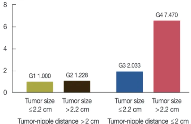
We decided to categorize the population into four groups based on tumor size and tumor-nipple distance, because of the strong statistical association of both of these variables with nipple involvement (Table 3). Group 1 was categorized as tu- mor size ≤2.2 cm/tumor-nipple distance >2 cm; group 2 as tumor size >2.2 cm/tumor-nipple distance >2 cm; group 3 as tumor size ≤2.2 cm/tumor-nipple distance ≤2 cm; and group 4 as tumor size >2.2 cm/tumor-nipple distance ≤2 cm.
Logistic regression analysis was carried out on the four groups and showed that group 4 was statistically associated with a much higher likelihood of nipple involvement than group 1 (OR, 7.47; 95% CI, 2.46–22.64; p<0.001). Although group 2 and group 3 did not show statistically significant re-
Table 1. Correlation between clinicopathologic characteristics and nip- ple involvement
Characteristic
Nipple involvement
p-value Yes (n=36)
No. (%) No (n=430) No. (%)
Age (yr) 0.853
<50 15 (41.7) 186 (43.3)
≥50 21 (58.3) 244 (56.7)
Histologic type 0.479
IDC 27 (75.0) 331 (77.0)
mIDC and DCIS 6 (16.7) 12 (2.8)
DCIS 2 (5.6) 50 (11.6)
ILC 0 13 (3.0)
Other 1 (2.8) 24 (5.6)
Histologic grade 0.954
1 8 (22.2) 98 (24.3)
2 15 (41.7) 167 (41.4)
3 13 (36.10) 138 (34.2)
T stage <0.001
T1 11 (30.6) 210 (48.8)
T2 14 (38.9) 203 (47.2)
T3 11 (30.6) 17 (4.0)
Tumor size (cm) 0.006
≤2.2 12 (33.3) 246 (57.2)
>2.2 24 (66.7) 184 (42.8)
Tumor-nipple distance (cm) 0.001
≤2 28 (77.8) 205 (47.7)
>2 8 (22.2) 225 (52.3)
Lymph node status 0.008
N0 18 (50.0) 246 (57.5)
N1 5 (13.9) 111 (25.9)
N2 10 (27.8) 38 (8.9)
N3 3 (8.3) 33 (7.7)
Estrogen receptor 0.335
Positive 23 (63.9) 306 (71.5)
Negative 13 (36.1) 122 (28.5)
Progesterone receptor 0.598
Positive 20 (55.6) 257 (60.0)
Negative 16 (44.4) 171 (40.0)
HER2 0.432
Positive 12 (33.3) 116 (27.2)
Negative 24 (66.7) 310 (72.8)
p53 mutation 0.010
Positive 28 (77.8) 237 (55.6)
Negative 8 (22.2) 189 (44.4)
Lymphovascular invasion 0.029
Positive 19 (52.8) 133 (34.5)
Negative 17 (47.2) 253 (65.5)
Multifocality 0.425
Positive 8 (22.2) 73 (17.0)
Negative 28 (77.8) 357 (83.0)
Multicentricity 0.135
Positive 6 (16.7) 38 (8.8)
Negative 30 (83.3) 392 (91.2)
IDC =invasive ductal carcinoma; mIDC =microinvasive ductal carcinoma;
DCIS=ductal carcinoma in situ; ILC=invasive lobular carcinoma; HER2=
human epidermal growth factor receptor 2.
sults (OR, 1.23; 95% CI, 0.3–5.032; p=0.776; and OR, 2.03;
95% CI, 0.6–6.93; p=0.257, respectively), the OR showed a sequential increase in the likelihood of nipple involvement from group 1 to group 4, which was statistically significant (p<0.001) (Figure 4).
With regard to the tumor location, only tumors located in the center of the breast showed a significant association with nipple involvement (p<0.001) (Table 4).
DISCUSSION
MRI imaging has been found to have a vital role in the pre- operative evaluation of breast cancer patients. In a study by Bedrosian et al. [7], preoperative MRI studies had an overall sensitivity of 95% for detecting primary malignant breast tu- mors. Schnall et al. [8] reported that MRI identified additional disease at least 2 cm from the index malignant lesion in 18%
of 426 breast cancer patients included in the study. Further- more, compared to mammography or ultrasonography, which are both two-dimensional imaging modalities, breast MRI provides a three-dimensional outline of the whole breast and tumor, which is needed to accurately measure tumor size and other values. With the advantages of preoperative MRI, it is possible to make a valid prediction of the extent of disease, and in particular, occult nipple invasion.
In our study, we found a 7.7% (36/466) rate of occult nipple invasion. The rate of tumor involvement of the nipple in previ- ous series ranged from 0% to 58% [10-18]. Differences in pa-
tient selection, sample size, tissue processing, and histopatho- logic analysis may be the reason for this wide variation in re- sults. In addition, indications for mastectomy have changed over the years [16], which could have influenced the patient population. Furthermore, after the National Institutes of Health (NIH)’s announcement in 1991, more than half of eligible breast cancer patients needing surgery have received conserva- tive treatment instead of a mastectomy [19]. Therefore, studies predating this landmark shift in treatment might not reflect the prevalence of occult nipple involvement. Studies after 1991 show occult nipple invasion to a lesser extent, with the preva- lence ranging from 0% to 16%, which is consistent with our study [16-18].
The present study shows that occult nipple involvement is significantly associated with tumor size, tumor-nipple dis- tance, axillary lymph node status, LVI, and p53 mutation.
With regard to tumor size, several studies have reported a cor- relation between nipple involvement and the size of the tumor [10,12,14,18]. However, these studies determined tumor size by measuring postoperative pathology specimens, which are Table 2. Results of the multivariate analysis (nipple involvement as de-
pendent variable)
Variable p-value OR (95% CI)
Tumor size 0.025 2.43 (1.12–5.27)
Tumor-nipple distance 0.007 3.18 (1.38–7.34) Lymph node status 0.827 1.085 (0.53–2.24)
p53 mutation 0.075 2.154 (0.93–5.01)
Lymphovascular invasion 0.356 1.50 (0.64–3.52) OR=odds ratio; CI=confidence interval.
Table 3. Categorization of groups regarding tumor size and tumor-nip- ple distance
Tumor-nipple distance (cm)
Total no.
≤2 (No.) >2 (No.) Tumor size (cm)
≤2.2 G3 (130) G1 (128) 258
>2.2 G4 (103) G2 (105) 208
Total No. 233 233 466
G1 =tumor size ≤2.2 cm/tumor-nipple distance >2 cm; G2 =tumor size
>2.2 cm/tumor-nipple distance >2 cm; G3=tumor size ≤2.2 cm/tumor-nip- ple distance ≤2 cm; G4=tumor size >2.2 cm/tumor-nipple distance ≤2 cm.
Table 4. Correlation between tumor location and nipple involvement Tumor location
Nipple involvement
p-value Yes (n=36)
No. (%) No (n=430) No. (%)
UOQ 11 (30.6) 186 (43.3) 0.138
UIQ 3 (8.3) 70 (16.3) 0.208
LOQ 3 (8.3) 51 (11.9) 0.786
LIQ 3 (8.3) 32 (7.4) 0.745
Upper central 2 (5.6) 26 (6.0) 1.000
Lower central 0 4 (0.9) 1.000
Medial central 0 6 (1.4) 1.000
Lateral central 1 (2.8) 22 (5.1) 1.000
Center 13 (36.1) 33 (7.7) <0.001
UOQ=upper outer quadrant; UIQ=upper inner quadrant; LOQ=lower outer quadrant; LIQ=lower inner quadrant.
G4 7.470
G3 2.033
Tumor size
≤2.2 cm
Tumor size
>2.2 cm Tumor-nipple distance ≤2 cm G1 1.000 G2 1.228
Tumor size
≤2.2 cm
Tumor-nipple distance >2 cm Tumor size
>2.2 cm 8
6
4
2
0
Figure 4. Odds ratio of nipple involvement according to tumor size and tumor-nipple distance.
not accessible before surgery. The tumor size in this study was defined as the maximum extent of suspicious contrast en- hancement on MRI imaging.
Although, MRI, especially contrast-enhanced MRI, has gained increased acceptance for accurate preoperative assess- ment of disease extent [20], there have been reports showing overestimation of the lesion size. Onesti et al. [21] demon- strated that, although MRI tumor size correlates with pathol- ogy size, a significant overestimation exists, particularly for tumors >2 cm.
However, the disease extent shown on MRI imaging could be a more accurate predictor of occult disease of the nipple because ductal carcinoma in situ (DCIS) is the most common form of nipple involvement [11-13,15]. Overestimation of tu- mor size on MRI is partially due to the DCIS component of the tumor [22]. Enhancement of malignant tumors on MRI is caused by the presence of tumor-induced angiogenesis. DCIS has been shown to have an increased amount of stromal mi- cro vessels [23], which increase blood flow, resulting in greater contrast enhancement. Thus, MRI tumor size measurements have the potential to represent the extent of DCIS, in addition to the invasive components.
The tumor-nipple distance was the most significant charac- teristic related to nipple involvement in both univariate and multivariate analysis in this study. Since our data suggest that tumor-nipple distance is the most important predictive factor, we tried to define the best cutoff value that would predict the risk of NAC involvement. Previous studies have had variable results; some supported a distance of 1 cm [5], and others, 2 cm [4,24]. In a review article on NSM [24], a tumor-nipple distance of ≥2 cm and tumor size of ≥5 cm with no cancer involvement of the retroareolar tissue were proposed as inclu- sion criteria for this surgical method, which is consistent with our findings.
An interesting result in our study was the correlation of p53 mutation with nipple involvement: 77.8% of patients with oc- cult nipple involvement had p53 mutations compared with 55.6% of those without involvement. The presence of p53 mu- tations is a prognostic marker in breast cancer, and is associat- ed with shorter disease-free and overall survival [25].
Although the correlation of p53 mutations with response to therapy has been controversial, a study by Jansson et al. [26]
showed that node-negative breast cancer patients with p53 mutations had significantly improved relapse-free survival, breast cancer-corrected survival, and overall survival rates when treated with locoregional radiotherapy. NSM and SSM usually involve insertion of breast implants for reconstruction, and postoperative radiation therapy, although not an absolute contraindication, is usually avoided due to complications such
as alteration in cosmetic results and increased risk of infection [27,28]. Therefore, according to our results, a subset of patients with p53 mutations may require more radical surgical treat- ment due to the risk of occult nipple involvement and may need radiation treatment in order to ensure a better outcome.
LVI is a good predictor of locoregional recurrence and axil- lary lymph node involvement. Vlajcic et al. [29] and Vyas et al. [17] have shown that LVI status is also predictive of NAC invasion, which supports the findings of this study.
Although the results of our study are consistent with prior research reporting a correlation between nipple involvement and the clinicopathological characteristics of the tumor, fac- tors such as p53 and LVI need gross tissue, and a thorough histological examination to yield results. However, it is very difficult to use these parameters in the preoperative setting, as prediction of nipple involvement must be made prior to sur- gery in order to select the appropriate surgical approach for the patient.
There are several limitations to our study. First, its retro- spective design made it vulnerable to selection bias. Second, nipple involvement of the tumor was evaluated based only on a single sagittal section of the nipple. Although standard guidelines recommend this technique for histopathology [30], there is a chance that occult nipple involvement may have been underestimated in this study.
In conclusion, a statistically significant association was found between occult nipple involvement and tumor size, tu- mor-nipple distance, axillary lymph node status, LVI, and p53 mutation. Tumor size and tumor-nipple distance are the only two factors that were associated with nipple involvement after multivariate analysis.
At present, there is no definite method of assessing nipple involvement prior to NSM. This study showed that cutoff points of 2.2 cm for tumor size and 2 cm for tumor-nipple distance could be used as parameters to predict occult nipple involvement. Although the feasibility and oncological safety of NSM have been demonstrated, further randomized pro- spective studies and long term follow-up data are required to validate this recommendation.
CONFLICT OF INTEREST
The authors declare that they have no competing interests.
REFERENCES
1. Foster RS Jr, Farwell ME, Costanza MC. Breast-conserving surgery for breast cancer: patterns of care in a geographic region and estimation of potential applicability. Ann Surg Oncol 1995;2:275-80.
2. Curran D, van Dongen JP, Aaronson NK, Kiebert G, Fentiman IS, Mi- gnolet F, et al. Quality of life of early-stage breast cancer patients treated with radical mastectomy or breast-conserving procedures: results of EORTC Trial 10801. The European Organization for Research and Treatment of Cancer (EORTC), Breast Cancer Co-operative Group (BCCG). Eur J Cancer 1998;34:307-14.
3. Bartelink H, van Dam F, van Dongen J. Psychological effects of breast conserving therapy in comparison with radical mastectomy. Int J Radiat Oncol Biol Phys 1985;11:381-5.
4. Gerber B, Krause A, Reimer T, Müller H, Küchenmeister I, Makovitzky J, et al. Skin-sparing mastectomy with conservation of the nipple-areola complex and autologous reconstruction is an oncologically safe proce- dure. Ann Surg 2003;238:120-7.
5. Sacchini V, Pinotti JA, Barros AC, Luini A, Pluchinotta A, Pinotti M, et al. Nipple-sparing mastectomy for breast cancer and risk reduction: on- cologic or technical problem? J Am Coll Surg 2006;203:704-14.
6. Freeman BS. Subcutaneous mastectomy for benign breast lesions with immediate or delayed prosthetic replacement. Plast Reconstr Surg Transplant Bull 1962;30:676-82.
7. Bedrosian I, Mick R, Orel SG, Schnall M, Reynolds C, Spitz FR, et al.
Changes in the surgical management of patients with breast carcinoma based on preoperative magnetic resonance imaging. Cancer 2003;98:
468-73.
8. Schnall MD, Blume J, Bluemke D, Smazal S, Deangelis G, Harms S, et al.
MRI detection of multi focal breast carcinoma: report from the Interna- tional Breast MRI Consortium. J Clin Oncol 2004;22(14 Suppl):504.
9. Orel SG, Weinstein SP, Schnall MD, Reynolds CA, Schuchter LM, Fraker DL, et al. Breast MR imaging in patients with axillary node me- tastases and unknown primary malignancy. Radiology 1999;212:543-9.
10. Smith J, Payne WS, Carney JA. Involvement of the nipple and areola in carcinoma of the breast. Surg Gynecol Obstet 1976;143:546-8.
11. Andersen JA, Pallesen RM. Spread to the nipple and areola in carcino- ma of the breast. Ann Surg 1979;189:367-72.
12. Wertheim U, Ozzello L. Neoplastic involvement of nipple and skin flap in carcinoma of the breast. Am J Surg Pathol 1980;4:543-9.
13. Morimoto T, Komaki K, Inui K, Umemoto A, Yamamoto H, Harada K, et al. Involvement of nipple and areola in early breast cancer. Cancer 1985;55:2459-63.
14. Santini D, Taffurelli M, Gelli MC, Grassigli A, Giosa F, Marrano D, et al.
Neoplastic involvement of nipple-areolar complex in invasive breast cancer. Am J Surg 1989;158:399-403.
15. Menon RS, van Geel AN. Cancer of the breast with nipple involvement.
Br J Cancer 1989;59:81-4.
16. Verma GR, Kumar A, Joshi K. Nipple involvement in peripheral breast carcinoma: a prospective study. Indian J Cancer 1997;34:1-5.
17. Vyas JJ, Chinoy RF, Vaidya JS. Prediction of nipple and areola involve- ment in breast cancer. Eur J Surg Oncol 1998;24:15-6.
18. Simmons RM, Brennan M, Christos P, King V, Osborne M. Analysis of nipple/areolar involvement with mastectomy: can the areola be pre- served? Ann Surg Oncol 2002;9:165-8.
19. Lazovich D, White E, Thomas DB, Moe RE, Taplin S. Change in the use of breast-conserving surgery in western Washington after the 1990 NIH Consensus Development Conference. Arch Surg 1997;132:418-23.
20. Lehman CD, DeMartini W, Anderson BO, Edge SB. Indications for breast MRI in the patient with newly diagnosed breast cancer. J Natl Compr Canc Netw 2009;7:193-201.
21. Onesti JK, Mangus BE, Helmer SD, Osland JS. Breast cancer tumor size:
correlation between magnetic resonance imaging and pathology mea- surements. Am J Surg 2008;196:844-8.
22. Schouten van der Velden AP, Boetes C, Bult P, Wobbes T. Magnetic res- onance imaging in size assessment of invasive breast carcinoma with an extensive intraductal component. BMC Med Imaging 2009;9:5.
23. Guidi AJ, Fischer L, Harris JR, Schnitt SJ. Microvessel density and distri- bution in ductal carcinoma in situ of the breast. J Natl Cancer Inst 1994;
86:614-9.
24. Garcia-Etienne CA, Cody Iii HS 3rd, Disa JJ, Cordeiro P, Sacchini V.
Nipple-sparing mastectomy: initial experience at the Memorial Sloan- Kettering Cancer Center and a comprehensive review of literature.
Breast J 2009;15:440-9.
25. Fitzgibbons PL, Page DL, Weaver D, Thor AD, Allred DC, Clark GM, et al. Prognostic factors in breast cancer. College of American Patholo- gists Consensus Statement 1999. Arch Pathol Lab Med 2000;124:966- 78.
26. Jansson T, Inganäs M, Sjögren S, Norberg T, Lindgren A, Holmberg L, et al. p53 Status predicts survival in breast cancer patients treated with or without postoperative radiotherapy: a novel hypothesis based on clinical findings. J Clin Oncol 1995;13:2745-51.
27. Kuske RR, Schuster R, Klein E, Young L, Perez CA, Fineberg B. Radio- therapy and breast reconstruction: clinical results and dosimetry. Int J Radiat Oncol Biol Phys 1991;21:339-46.
28. Contant CM, van Geel AN, van der Holt B, Griep C, Tjong Joe Wai R, Wiggers T. Morbidity of immediate breast reconstruction (IBR) after mastectomy by a subpectorally placed silicone prosthesis: the adverse effect of radiotherapy. Eur J Surg Oncol 2000;26:344-50.
29. Vlajcic Z, Zic R, Stanec S, Lambasa S, Petrovecki M, Stanec Z. Nipple- areola complex preservation: predictive factors of neoplastic nipple- areola complex invasion. Ann Plast Surg 2005;55:240-4.
30. Lester SC. Manual of Surgical Pathology. 2nd ed. Philadelphia: Elsevier Churchill Livingstone; 2006.