Propofol Post-conditioning Protects against COS-7 Cells in Hypoxia/reoxygenation Injury by Induction of Intracellular Autophagy
Jin-Won Kwak, Eok-Nyun Kim, Bong-Soo Park*, Yong-Ho Kim*, Yong-Deok Kim†, Ji-Uk Yoon‡, Cheul-Hong Kim, Ji-Young Yoon
Department of Dental Anesthesia and Pain Medicine, School of Dentistry, Pusan National University, Dental Research Institude,
*Department of Oral Anatomy School of Dentistry, †Department of Oral and Maxillofacial Surgery School of Dentistry,
‡Department of Anesthesia and Pain Medicine School of Medicine, Pusan National University, Busan, Korea
Background:Propofol (2.6-diisopropylphenol) is a widely used intravenous anesthetic agent for the induction and maintenance of anesthesia during surgeries and sedation for ICU patients. Propofol has a structural similarity to the endogenous antioxidant vitamin E and exhibits antioxidant activities.13) However, the mechanism of propofol on hypoxia/reoxygenation (H/R) injury has yet to be fully elucidated. We investigated how P-PostC influences the autophagy and cell death, a cellular damage occurring during the H/R injury.
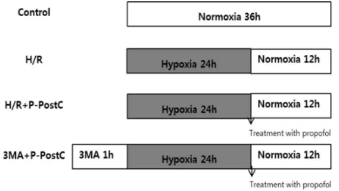
Methods:The groups were randomly divided into the following groups: Control: cells were incubated in normoxia (5% CO2, 21% O2, and 74% N2) without propofol treatment. H/R: cells were exposed to 24 h of hypoxia (5% CO2, 1% O2, and 94%
N2) followed by 12 h of reoxygenation (5% CO2, 21% O2, and 74% N2). H/R + P-PostC: cells post-treated with propofol were exposed to 24 h of hypoxia followed by 12 h of reoxygenation. 3-MA + P-PostC: cells pretreated with 3-MA and post-treated propofol were exposed to 24 h of hypoxia followed by 12 h of reoxygenation
Results:The results of our present study provides a new direction of research on mechanisms of propofol-mediated cytoprotection.
There are three principal findings of these studies. First, the application of P-PostC at the onset of reoxygenation after hypoxia significantly increased COS-7 cell viability. Second, the cellular protective effect of P-PostC in H/R induced COS-7 cells was probably related to activation of intra-cellular autophagy. And third, the autophagy pathway inhibitor 3-MA blocked the protective effect of P-PostC on cell viability, suggesting a key role of autophagy in cellular protective effect of P-PostC.
Conclusions: These data provided evidence that P-PostC reduced cell death in H/R model of COS-7 cells, which was in agreement with the protection by P-PostC demonstrated in isolated COS-7 cells exposed to H/R injury. Although the this study could not represent the protection by P-PostC in vivo, the data demonstrate another model in which endogenous mechanisms evoked by P-PostC protected the COS-7 cells exposed to H/R injury from cell death.
Key Words: Cos-7 cells; Hypoxia reoxygenation; Post-Treated; Propofol
Received: 2014. 3. 27•Revised: 2014. 4. 1•Accepted: 2014. 4. 2 Corresponding Author: Ji Young Yoon, Department of Dental Anesthesia and Pain Medicine, School of Dentistry, Pusan National University, Beomeo-ri, Mulgeum-eup, Yangsan-si, Gyeongsangnam-do 626-787, Korea
Tel: +82.55.360-5379 Fax: +82.55.360.5369 email: [email protected]
* Thesis for the degree of Master of Science in Dentistry
INTRODUCTION
Tissue Autophagy is an evolutionary conserved process involved in degradation of long-lived or damaged proteins and organelles [1]. Autophagy has been impli- cated in tissue ischemia/reperfusion (I/R). Nevertheless, the exact functional role of autophagy in the cell survival and death pathways associated with tissue damage is still not clear. Some observations suggest that stimulation of autophagy can promote cell survival in response to stress
conditions in cardiomyocytes [2,3]. On the other hand, the excessive and long-term upregulation of autophagy under certain conditions inhibit cell survival, rather greatly accelerating the cell death in the heart [4]. The term ‘‘ischemic pre-conditioning’’ (IPC) was first coined
by Murry [5] to describe a phenomenon whereby brief episodes of I/R could attenuate tissue injury, but its clinical application has been disappointing due to the unpredictability of ischemia. Ischemic post-conditioning (I-PostC) [6] performed after ischemia has solved the problem of unpredictability. Furthermore, pharmacolo- gical post-conditioning, which only requires a drug given as an adjunctive intervention during early reperfusion, can also achieve tissue protection and is easier to implement [7-9]. The mechanisms responsible for post- conditioning are largely unknown, but some previous studies have suggested that signal transduction pathways for survival such as activation of the extracellular signal- regulated kinase (ERK) [10] and of the phosphoino- sitide-3-kinase/Akt (PI3K/Akt) pathways [11] may play an important role in the protective effects of post-condi- tioning, by stimulating the endogenous anti-injury capacity of cells to protect themselves [12].
Propofol (2.6-diisopropylphenol) is a widely used intravenous anesthetic agent for the induction and maintenance of anesthesia during surgeries and sedation for ICU patients. Propofol has a structural similarity to the endogenous antioxidant vitamin E and exhibits antioxidant activities [13]. Wang et al. [14] found that infusing propofol at the onset of reperfusion for 30 min could provide neuroprotection to transient middle cerebral artery occlusion (MCAO) in rats partly by activation of the PI3K/Akt pathway, and He reported that post-conditioning with propofol (P-PostC) has cardiopro- tective effects against I/R injury of the heart, which is associated with inhibition of mitochondrial permeability transition pore (MPTP) opening [15]. However, the mechanism of propofol on hypoxia/reoxygenation (H/R) injury has yet to be fully elucidated. COS-7 cell is the African green monkey kidney fibroblast-like cell line has been established from CV-1 cells which have been transformed by an origin-defective mutant of SV40 coding
for wild-type T antigen. This line contains T antigen, retains complete permissiveness for lytic growth of SV40.
Here we investigated how P-PostC influences the autophagy and cell death, a cellular damage occurring during the H/R injury.
MATERIALS AND METHODS
1. Reagents
Propofol (2, 6-diisopropylphenol) diluted with dime- thyl sulfoxide (DMSO). The following reagents were obtained commercially: 3-[4,5-dimethylthiazol -2-yl]2,5- diphenyl tetrazolium bromide (MTT), acridine orange, monodansylcadaverine (MDC), 3-methyladenine (3-MA, class III PI3K inhibitor) was obtained from Calbiochem (La Jolla, CA, USA). Antibodies used in the study were as follows: LC3-II (1:3,000), Beclin-1 (1:1,000) from Abcam, and p62 (1:1,000), Atg5 (1:500) from Santa Cruz.
Secondary antibodies against rabbit (1:3,000) and mouse (1:3,000) immunoglobulins were purchased from Bio- Rad.
2. Cell culture
COS-7 cells (African green monkey kidney fibroblast- like cell line) were obtained from the American Type Culture Collection (ATCC, Manassas, USA). Dulbecco's modified Eagle’s medium (DMEM, GIBCO) supplemented with 10% inactivated fetal bovine serum (FBS, GIBCO) containing 500 μg/ml penicillin and 500 μg/ml strepto- mycin (GIBCO), and cells were incubated at 37°C in a humidified atmosphere with 5% CO2. Media were changed every 3 days.
3. Treatment of propofol postconditioning
Propofol which were made by dissolving them in DMSO were kept frozen at -4°C until use. The stock was diluted to their concentration with DMEM when needed.
Fig. 1. Experimental protocol for propofol post-conditioning.
H/R, Hypoxia/Reoxygenation; P-PostC, post-condi- tioning with propofol; 3-MA, 3-Methyladenine.
Prior to propofol treatment cells were grown to about 80% confluence and then exposed to propofol at different concentrations (0, 25, 50, 100 μM) for 2 h. Cells grown in medium containing an equivalent amount of DMSO without propofol served as control (Fig. 1).
4. MTT assay
Cell viability was measured using a quantitative colo- rimetric assay with thiazolyl blue tetrazolium bromide (MTT, AMResco), showing the mitochondrial activity of living cells. COS-7 cells (3 × 10⁴) were seed in 96-well plates. After drug treatment as indicated, cells were incubated with 300 μl MTT (final concentration 0.5 mg/mL) for 1.5 h at 37°C. The reaction was terminated by addition of 200 μl DMSO. Cell viability was measured by an ELISA reader (Tecan, Männedorf, Switzerland) at 570 nm excitatory emission wavelength.
5. Fluorescence microscopy
Cells were grown on coverslips and treated with COS-7 cells. After 24 h, cells were stained with 0.05 mM MDC, a selective fluorescent marker for autophagic vacuoles, at 37°C for 1 h. The cellular fluorescence changes were observed using a fluorescence microscope (Axioskop, Carl Zeiss, Germany). For further detection of the acidic cellular compartment, we used acridine orange, which
emits bright red fluorescence in acidic vesicles but fluoresces green in the cytoplasm and nucleus. Cells were stained with 1 μg/mL acridine orange for 15 min and washed with PBS. AVOs (autophagic vesicles organelles) formation was obtained under a confocal microscope LSM 700 (Carl Zeiss, Germany).
6. Western blot analysis
Cells (2 × 10⁶) were washed twice in ice-cold PBS, resuspended in 200 μl ice-cold solubilizing buffer [300 mM NaCl, 50 mM Tris-Cl (pH 7.6), 0.5% Triton X-100, 2 mM PMSF, 2 μl/ml aprotinin and 2 μl/ml leupeptin]
and incubated at 4°C for 30 min. The lysates were centrifuged at 14,000 revolutions per min for 15 min at 4°C. Protein concentrations of cell lysates were determined with Bradford protein assay (Bio-Rad, Richmond, CA, USA) and 20 μg of proteins were resolved by 10% SDS/PAGE.
The gels were transferred to polyvinylidene fluoride (PVDF) membranes (Millipore, Billerica, MA, USA) and reacted with appropriate primary antibodies. Immuno- staining with secondary antibodies was detected using SuperSignal West Femto (Pierce, Rockford, IL, USA) enhanced chemiluminescence substrate and detected with Alpha Imager HP (Alpha Innotech, Santa Clara, USA).
7. Statistical analysis
Data were expressed as means ± SEM. The statistical significance was assessed by one-way ANOVA followed by Tukey’s post hoc t-test. (P< 0.05 was considered statistically significant)
RESULT
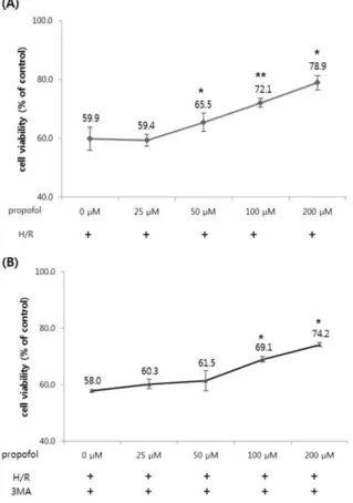
1. P-PostC improved the cell viability of H/R-induced COS-7 cells
The effect of P-PostC and H/R injury on COS-7 cells was investigated. Propofol did not show any significant
Fig. 3. Effect of propofol post-conditioning on cell viability.
The cells were post-conditioned with increasing concentrations of propofol (0-200 μM) after 24 h of hypoxia and before 12 h of reoxygenation. Cell viability was determined by 3- (4,5-dimethylthiazol-2-yl) -2,5-diphenylterazolium bromide (MTT) assay, as previously described. Values are represented as the percentage of viable cells ± SD; vehicle-treated cells were considered as 100% viable. *P < 0.05, **P < 0.01 compared with the hypoxia/reoxygenation (H/R) group.
Fig. 2. Effect of propofol post-conditioning on cell viability.
The normal COS-7 cells were treated with different concentrations (0-200 μM) of propofol. Cell viability was determined by 3-(4,5-dimethylthiazol-2-yl)- 2,5- diphenylterazolium bromide (MTT) assay, as previously described. Values are represented as the percentage of viable cells; vehicle-treated cells were considered as 100% viable. The data represented are mean percentages of viable cells ± SD of three independent experiments.
* P < 0.05, ** P < 0.01 compared with the hypoxia/
reoxygenation (H/R) group.
toxic effect on the COS-7 cells (Fig. 2). After COS-7 cells were exposed to hypoxia and post-treated with various doses of propofol and exposed to reoxygenation, we measured cell viability by MTT assay again to investigate the effect of various concentrations of P-PostC (0, 25, 50, 100 and 200 μM) on the H/R-induced COS-7 cells and discovered that P-PostC significantly protected the COS-7 cells from H/R-induced cytotoxicity and our results showed that the viability of P-PostC COS-7 cells was increased in a dose-dependent manner (Fig. 3A). As showed in Fig. 3A, compared with control, the cell proliferation was markedly inhibited after H/R treatment (P < 0.05), which was significantly improved by P-PostC.
Among all of the concentrations, 100 μM Propofol represented the optimal effect, improving cell viability by approximately 20% (P < 0.05). Based on this result, all subsequent experiments were performed with 100 μM propofol. The role of autophagy in the H/R damage of COS-7 cells was further confirmed by the autophagy inhibitor 3-MA, an inhibitor of class III phosphoinositide
3-kinase (PI3K). As shown in Fig. 3B, 3-MA (5 mM) 1 h before H/R inhibited protective effect of P-PostC and significantly increased H/R induced COS-7 cell death. This decrease in the cell viability was accompanied by the autophagy inhibitor 3-MA, indicating that the inhibition of autophagy by 3-MA sensitized COS-7 cells to H/R injury.
2. P-PostC leads to induction of autophagy in COS-7 cells
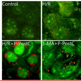
Prominent accumulation of autophagic specific stain- ing MDC was observed around the nuclei in P-PostC group COS-7 cells (Fig. 4). Similarly, AO staining, red
Fig. 4. Fluorescence microscopic (× 400) analysis of auto- phagosome in the H/R injured COS-7 cells. H/R caused accumulation of autophagosomes containing partially digested cytoplasmic contents compared to the control group. The P-PostC during I/R dramatically increased formation of autophagosomes and the autophagy pathway inhibitor 3-MA blocked formation of auto- phagosomes by P-PostC. H/R, Hypoxia/Reoxygenation;
P-PostC, post-conditioning with propofol; 3-MA, 3- Methyladenine.
Fig. 5. Fluorescence microscopic (× 400) analysis of auto- phagosome in the H/R injured COS-7 cells. Stained with acridine orange the green shows where the dye has stained the nucleus and the red is where the cell is starting to 'digest' parts of itself in small capsules called autophagasomes. H/R caused accumulation of autopha- gosomes containing partially digested cytoplasmic con- tents compared to the control group. The P-PostC during I/R dramatically increased formation of autophagosomes and the autophagy pathway inhibitor 3-MA blocked formation of autophagosomes by P-PostC. H/R, Hy- poxia/Reoxygenation; P-PostC, post-conditioning with propofol; 3-MA, 3-Methyladenine.
(A)
(B)
Fig. 6. (A) Western blot analysis. Expression of Atg5, LC3-II, Beclin-1 and p62 in H/R-induced COS-7 cells preconditioned with propofol and 3-MA. (B) Quantification of Atg5, LC3-II, Beclin-1 and p62. Each band shown above was quantified by the densitometric scan. H/R, Hypoxia/ Reoxygenation; PPC, propofol postconditioning; 3-MA, 3-Methyladenine.
fluorescent spots appeared on P-PostC group COS-7 cells, while the normoxia control group, and the cells co-treated with 5 mM/L 3-MA and H/R group showed mainly green cytoplasmic fluorescence (Fig. 5). We here examined activation of autophagy related protein under H/R-induced cells by western blotting analysis. The recruitment of LC3 to the membrane occurs via an Atg5-dependent mechanism, and thus Atg5 is essential for autophagosome formation in vivo.3) Atg5 and Beclin-1, LC3-II (microtubule-associated protein 1 light chain 3 form II), p62 was significantly reduced in H/R-induced group cells but elevated in P-PostC group cells (Fig. 6). As showed in Fig. 6, Atg5, Beclin-1, LC3-II (microtubule-associated protein 1 light chain 3 form II) and p62 was increased when autophagy was induced by P-PostC, and they were decreased when autophagy was suppressed by 3-MA.
DISCUSSION
The previous reports have shown that the cellular protective mechanism of propofol may partly result from its ability to act as a free radical scavenger [16-18]. The results of our present study provides a new direction of research on mechanisms of propofol-mediated cytopro- tection. There are three principal findings of these studies. First, the application of P-PostC at the onset of reoxygenation after hypoxia significantly increased COS-7 cell viability. Second, the cellular protective effect of P-PostC in H/R induced COS-7 cells was probably related to activation of intra-cellular autophagy. And third, the autophagy pathway inhibitor 3-MA blocked the protective effect of P-PostC on cell viability, suggesting a key role of autophagy in cellular protective effect of P-PostC.
Together, these findings indicate that P-PostC stimulated COS-7 cells endogenous cellular protective effect against H/R injury through pro-survival autophagy signal
pathways. Numerous studies have documented that oxidative stress-mediated cellular changes are frequently induced during H/R [19-21]. Excessive intracellular ROS play crucial roles in the induction of cell apoptosis and death. The postconditioning treatment at the onset of reoxygenation reduces H/R induced injury in cardiomy- ocytes and is potentially mediated by attenuation of ROS generation [22]. Similar to the effects of ischemia post- conditioning, our study shows that P-PostC induced intracellular autophagy and increased COS-7 cell viability.
According to recent works, moreover, it has been suggested that autophagy plays two distinct roles during ischemia and reperfusion. In the ischemic phase, auto- phagy can be protective via AMPK activation and sequentially inhibition of mTOR signaling, but reper- fusion after ischemia stimulates autophagic cell death through the different pathway. In this study P-PostC induced intra-cellular autophagy and decreased H/R induced COS-7 cell death.
Autophagic vacuoles including autophagosomes and autophagolysosomes are generally formed in cells under- going the autophagic process. Therefore, observation of autophagosomes inside cells can be used as an indicator to analyze the induction of autophagy. To investigate the role of autophagy in the H/R induced COS-7 cells and the effect of P-PostC in the occurrence of autophagy, we directly observed formation of autophagosomes in COS-7 cells exposed to H/R using a fluorescence microscope.
H/R caused accumulation of autophagosomes containing partially digested cytoplasmic contents in COS-7 cells compared to the control group. And P-PostC during H/R dramatically increased formation of cytosolic vacuoles in COS-7 cells and decreased H/R induced cell death, indicating that autophagy is associated with H/R-induced cell death, and P-PostC protects cell death through the induction intra-cellular autophagy. To further confirm whether propofol can modulate autophagic cell death in
H/R-injured COS-7 cells, we examined the level of LC3-II expression. The production of LC3-II has been suggested as another marker for autophagy induction. LC3-II formation was increased in the COS-7 cells exposed to H/R injury compared to the control group. Furthermore, in the case of P-PostC during H/R, LC3-II production was significantly increased compared to the H/R group. The expression of p62, an autophagy-associated protein was alleviated in the H/R COS-7 cells, but it was increased by P-PostP to the H/R injured cells. This data indicates again that the H/R injury increased cell death and P-Postc induces autophagy leading to cell survival. In conclusion, these data provided evidence that P-PostC induced intra- cellular autophagy and reduced cell death in H/R model of COS-7 cells, which was in agreement with the pro- tection by P-PostC demonstrated in isolated COS-7 cells exposed to H/R injury through the induction of cellular protective autophagy. Although the this study could not represent the protection by P-PostC in vivo, the data demonstrate another model in which endogenous mecha- nisms evoked by P-PostC protected the COS-7 cells exposed to H/R injury from cell death.
REFERENCE
1. Anderson RT, Skovlund SE, Marrero D, Levine DW, Meadows K, Brod M, Balkrishnan R: Development and validation of the insulin treatment satisfaction question- naire. Clin Ther 2004; 26(4): 565-78.
2. Hamacher-Brady A, Brady NR, Gottlieb RA: Enhancing macroautophagy protects against ischemia/reperfusion injury in cardiac myocytes. J Biol Chem 2006; 281(40):
29776-87.
3. Kuma A, Hatano M, Matsui M, Yamamoto A, Nakaya H, Yoshimori T, Ohsumi Y, Tokuhisa T, Mizushima N: The role of autophagy during the early neonatal starvation period. Nature 2004; 432(7020): 1032-6.
4. Hamacher-Brady A, Brady NR, Logue SE, Sayen MR, Jinno M, Kirshenbaum LA, Gottlieb RA, Gustafsson AB. Response to myocardial ischemia/reperfusion injury involves Bnip3 and autophagy. Cell Death Differ 2007; 14(1): 146-57.
5. Murry CE, Jennings RB, Reimer KA. Preconditioning with ischemia: a delay of lethal cell injury in ischemic myocar- dium. Circulation 1986; 74(5): 1124-36.
6. Zhao ZQ, Corvera JS, Halkos ME, Kerendi F, Wang NP, Guyton RA, Vinten-Johansen J: Inhibition of myocardial injury by ischemic postconditioning during reperfusion:
comparison with ischemic preconditioning. Am J Physiol Heart Circ Physiol 2003; 285(2): H579-88.
7. Vinten-Johansen J, Zhao ZQ, Jiang R, Zatta AJ: Myocardial protection in reperfusion with postconditioning. Expert Rev Cardiovasc Ther 2005; 3(6): 1035-1045.
8. Bopassa JC, Ferrera R, Gateau-Roesch O, Couture-Lepetit E, Ovize M: PI 3-kinase regulates the mitochondrial transition pore in controlled reperfusion and postcondi- tioning. Cardiovasc Res 2006; 69(1): 178-85.
9. Darling CE, Jiang R, Maynard M, Whittaker P, Vinten- Johansen J, Przyklenk K: Postconditioning via stuttering reperfusion limits myocardial infarct size in rabbit hearts:
role of ERK1/2. Am J Physiol Heart Circ Physiol 2005;
289(4): H1618-26.
10. Yang XM, Krieg T, Cui L, Downey JM, Cohen MV: NECA and bradykinin at reperfusion reduce infarction in rabbit hearts by signaling through PI3K, ERK, and NO. J Mol Cell Cardiol 2004; 36(3): 411-21.
11. Tsang A, Hausenloy DJ, Mocanu MM, Yellon DM: Post- conditioning: a form of “modified reperfusion” protects the myocardium by activating the phosphatidylinositol 3- kinase-Akt pathway. Circ Res 2004; 95(3): 230-2.
12. Hausenloy DJ, Yellon DM: Reperfusion injury salvage kinase signalling: taking a RISK for cardioprotection.
Heart Fail Rev 2007; 12(3-4): 217-34.
13. Gulcin I, Alici HA, Cesur M: Determination of in vitro antioxidant and radical scavenging activities of propofol.
Chem Pharm Bull (Tokyo) 2005; 53(3): 281-5.
14. Wang HY, Wang GL, Yu YH, Wang Y: The role of phosphoinositide-3-kinase/Akt pathway in propofol- induced postconditioning against focal cerebral ischemia- reperfusion injury in rats. Brain Res 2009; 1297: 177-84.
15. He W, Zhang FJ, Wang SP, Chen G, Chen CC, Yan M:
Postconditioning of sevoflurane and propofol is associated with mitochondrial permeability transition pore. J Zhejiang Univ Sci B 2008; 9(2): 100-8.
16. Murphy PG, Bennett JR, Myers DS, Davies MJ, Jones JG:
The effect of propofol anaesthesia on free radical-induced lipid peroxidation in rat liver microsomes. Eur J Anaesthesiol 1993; 10(4): 261-6.
17. Corcoran TB, Engel A, Sakamoto H, O'Shea A, O'Callaghan-Enright S, Shorten GD: The effects of pro- pofol on neutrophil function, lipid peroxidation and inflammatory response during elective coronary artery bypass grafting in patients with impaired ventricular function. Br J Anaesth 2006; 97(6): 825-31.
18. Wickley PJ, Shiga T, Murray PA, Damron DS: Propofol decreases myofilament Ca2+ sensitivity via a protein kinase
C-, nitric oxide synthase-dependent pathway in diabetic cardiomyocytes. Anesthesiology 2006; 104(5): 978-87.
19. Duranteau J, Chandel NS, Kulisz A, Shao Z, Schumacker PT: Intracellular signaling by reactive oxygen species during hypoxia in cardiomyocytes. J Biol Chem 1998;
273(19): 11619-24.
20. Kevin LG, Camara AK, Riess ML, Novalija E, Stowe DF:
Ischemic preconditioning alters real-time measure of O2 radicals in intact hearts with ischemia and reperfusion.
Am J Physiol Heart Circ Physiol 2003; 284(2): H566-74.
21. Duilio C, Ambrosio G, Kuppusamy P, DiPaula A, Becker LC, Zweier JL: Neutrophils are primary source of O2 radicals during reperfusion after prolonged myocardial ischemia. Am J Physiol Heart Circ Physiol 2001; 280(6):
H2649-57.
22. Sun HY, Wang NP, Kerendi F, Halkos M, Kin H, Guyton RA, Vinten-Johansen J, Zhao ZQ: Hypoxic postcondition- ing reduces cardiomyocyte loss by inhibiting ROS genera- tion and intracellular Ca2+ overload. Am J Physiol Heart Circ Physiol 2005; 288(4): H1900-8.