Gastric Cancer and Concomitant Gastric Tuberculosis:
A Case Report
Hyok-Jo Kang, Young-Seok Lee1, You-Jin Jang, and Young-Jae Mok Departments of Surgery, 1Pathology, Korea University Guro Hospital, Seoul, Korea
Gastric tuberculosis is rare even in the endemic areas of tuberculosis, and can mimic neoplasm by causing elevation of the mucosa with or without ulceration. Here, we report a case in which a 54-year-old female patient admitted for resection of early gastric cancer was found to have coexisting histopathologically and bacteriologically confirmed gastric cancer and tuberculosis.
Key Words: Stomach neoplasms; Gastrointestinal tuberculosis
Case Report
J Gastric Cancer 2012;12(4):254-257 http://dx.doi.org/10.5230/jgc.2012.12.4.254
Correspondence to: You-Jin Jang
Department of Surgery, Korea University Guro Hospital, 148, Gurodong- ro, Guro-gu, Seoul 152-703, Korea
Tel: +82-2626-1114, Fax: +82-2-2626-2024 E-mail: jyjclick@korea.ac.kr
Received October 13, 2012 Revised October 29, 2012 Accepted October 29, 2012
Copyrights © 2012 by The Korean Gastric Cancer Association www.jgc-online.org
This is an open-access article distributed under the terms of the Creative Commons Attribution Non-Commercial License (http://creativecommons.org/
licenses/by-nc/3.0) which permits unrestricted noncommercial use, distribution, and reproduction in any medium, provided the original work is properly cited.
Introduction
Despite an overall decreasing trend in the incidence of pulmo- nary tuberculosis, the number of cases of extrapulmonary tubercu- losis increased in Korea between 2001 and 2010.(1) Abdominal tu- berculosis is the third most common extrapulmonary manifestation of the disease according to a national report on tuberculosis. Enteral tuberculosis can develop anywhere in the gastrointestinal tract, and there are several reported cases of intestinal tuberculosis with concomitant chronic enteral inflammatory disease such as Crohn’
s disease or ulcerative colitis, as well as with coexisting neoplastic disease.(2,3) Gastric tuberculosis, however, is a rare disease entity even in endemic areas of tuberculosis such as Korea. To date, very few cases of tuberculosis accompanied by gastric malignancy have been reported.(4,5)
Here, we present a case of coexisting tuberculosis and adeno- carcinoma of the stomach, which was confirmed by both histo-
pathologic examination and molecular testing.
Case Report
A 54-year old female patient diagnosed with early gastric cancer was referred from a local clinic for surgical management.
She reported no specific symptoms, and her gastric malignancy had been detected incidentally on screening tests. Pathology slides from the outside institution were reviewed at our hospital, and the diagnosis of signet ring cell adenocarcinoma was confirmed (Fig.
1). The patient’s past medical history was unremarkable except for pulmonary tuberculosis which had been diagnosed and cured 30 years prior, and there were no documented relapses of tuberculosis after her initial therapy. Her physical examination was unremarkable, and laboratory findings were normal including hemoglobin 14.3 g/dl (12.0~16.0), white blood cell 4,000/μl (4,500~11,000) with neu- trophils 53.0% and lymphocytes 37.1%, carcinoembryonic antigen 0.64 ng/ml, and carbohydrate antigen 19-9 2.00 U/ml. Her chest x-ray showed neither evidence of active disease nor sequale of old pulmonary tuberculosis. Computed tomography was performed for cancer staging, and there were nor demonstrable intra-abdominal masses or lymphadenopathy (Fig. 2). Since there was both a mor- phologically suspicious and pathologically proven mucosal lesion in the proximal antrum along the greater curvature of stomach, no
Gastric Cancer Accompanied by Tuberculosis
255
additional endoscopic evaluation was performed.
Laparoscopic-assisted distal gastrectomy with Billroth I anasto- mosis was carried out, and a 0.5×0.5 cm sized mucosal lesion was noted in the proximal antrum during the operation. This lesion was estimated to be type IIc early gastric cancer, and histopathologic examination revealed multiple granulomatous areas of inflammation on the gastric mucosa. There were no regional lymph node me- tastases in any of the 52 dissected lymph nodes, however chronic granulomatous inflammation was noted in the omental lymph nodes. Tuberculosis polymerase chain reaction testing conducted on the gastric mucosa and omental lymph nodes confirmed gastric tuberculosis (Fig. 3).
The patient was discharged from the hospital on postoperative day 10 without complications. She is currently on anti-tuberculosis medication, and there was no evidence of relapse or recurrence of disease after 5-months of follow-up.
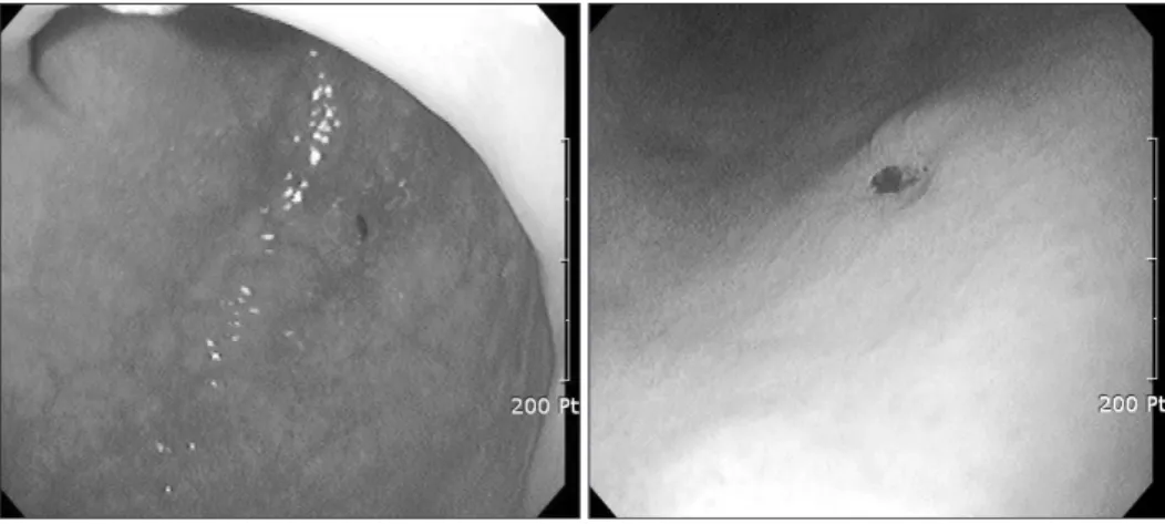
Fig. 1. Gastrofiberscopic findings.
A type IIc early gastric cancer in the greater curvature of the antrum.
Fig. 2. Abdominal computed tomography finding. There was neither specific lesion in the stomach nor regional lymph node enlargement.
Fig. 3. (A) H&E staining of regional lymph node (×100). A considerable amount of granulomatous inflammatory tissue (white arrows) was distrib- uted within the lymph node. (B) H&E staining of regional lymph node (×200). Tumor cells of the signet ring cell type are shown near the normal glands (black arrows).
Kang HJ, et al.
256
Discussion
More than two billion people are infected with tuberculosis worldwide, causing 1.7 million deaths in 2006 alone.(6) The inci- dence of extrapulmonary tuberculosis is increasing and accounts for 1 in 5 cases of the disease.(7) The most common site involved with intestinal tuberculosis is the ileocecal valve.(8) Additionally, cases have been documented involving sites such as the ascending colon, jejunum, duodenum, stomach, and sigmoid colon.(9) Among these sites, it is rare that the stomach is infected by tuberculosis.(10) In Korea, few cases of gastric tuberculosis have been documented, and gastric cancer with concomitant gastric tuberculosis is even more rarely seen.(11)
Gastric tuberculosis can be due to primary or secondary infec- tion. Infection of the stomach does not occur through intact mucous membranes, though it typically can spread to the stomach through ulcers, gastritis, erosions, ecchymoses, or cancer. There are four possible routes of infection; 1) direct infection through the mucous membrane, as occurs when bacilli-rich sputum is frequently swal- lowed, 2) hematogenous infection leading to miliary spread, 3) retrograde lymphatic spread, 4) and direct spread through contigu- ous organs.(6) Some symptoms are associated with gastric tuber- culosis infection such as abdominal pain, discomfort, and weight loss, but are fairly non-specific. Additionally, gastric tuberculosis may be easily confused with gastric cancer on initial studies such as computed tomography scan or endoscopy. In this case, there were no specific findings on initial presentation indicating tuberculosis infection. Gastric tuberculosis can be diagnosed by biopsy, so it is important to recognize that intestinal tuberculosis may involve any site along the gastrointestinal tract, particularly in patients with a history of pulmonary tuberculosis.(12) It is also important to estab- lish a quick and accurate diagnosis in order to prevent inappropriate treatment of the patient.
In this case, it is significant that gastric tuberculosis presents with gastric cancer. The specimen obtained through endoscopic biopsy of the lesion suspected to be early gastric cancer was con- firmed via slide review. Granulomatous lesion was located diffusely in whole stomach and main cancer lesion was tiny but located at the antrum of the stomach. Additionally, there was chronic granu- lomatous inflammation within the retrieved regional lymph nodes.
The cause of tuberculosis was unknown in this patient, though it may have been induced by an immune compromised state associ- ated with her gastric cancer. The inflammatory response triggered by infection precedes tumor development and is a part of normal
host defense, whose goal is pathogen elimination. However, tu- morigenic pathogens subvert host immunity and establish persistent infections associated with low grade but chronic inflammation.(13) The patient has history of pulmonary tuberculosis. I thought that tuberculosis was reactivated in immune compromised state caused by cancerous situation. Several authors have commented on the relationship between tuberculosis and cancer,(14) including Chow- dhary et al.(15) who reported that isolated cancer can develop in about 10% of cases of gastric tuberculosis. Thus it is important for clinicians to always consider this association between tuberculosis and neoplastic lesions.
References
1. Korea Centers for Disease Control and Prevention. Annual re- port on the notified tuberculosis cases patients in Korea 2010.
Seoul: Korea Centers for Disease Control and Prevention, 2011.
2. Wexner SD, Rosen L, Lowry A, Roberts PL, Burnstein M, Hicks T, et al. Practice parameters for the treatment of mucosal ulcerative colitis--supporting documentation. The Standards Practice Task Force. The American Society of Colon and Rectal Surgeons. Dis Colon Rectum 1997;40:1277-1285.
3. Chakravartty S, Chattopadhyay G, Ray D, Choudhury CR, Mandal S. Concomitant tuberculosis and carcinoma colon: co- incidence or causal nexus? Saudi J Gastroenterol 2010;16:292- 294.
4. Geo SK, Harikumar R, Varghese T, Rajan P, Aravindan KP.
Isolated tuberculosis of gastric cardia presenting as perforation peritonitis. Indian J Gastroenterol 2005;24:227-228.
5. Kim SH, Park JH, Kang KH, Lee JH, Park CK, Cho CM, et al.
Gastric tuberculosis presenting as a submucosal tumor. Gas- trointest Endosc 2005;61:319-322.
6. Donoghue HD, Holton J. Intestinal tuberculosis. Curr Opin Infect Dis 2009;22:490-496.
7. World Health Organization. Improving the diagnosis and treatment of smear-negative pulmonary and extrapulmonary tuberculosis among adults and adolescents. Geneva: World Health Organization, 2007.
8. Marshall JB. Tuberculosis of the gastrointestinal tract and peri- toneum. Am J Gastroenterol 1993;88:989-999.
9. Makharia GK, Srivastava S, Das P, Goswami P, Singh U, Tripa- thi M, et al. Clinical, endoscopic, and histological differentia- tions between Crohn's disease and intestinal tuberculosis. Am
Gastric Cancer Accompanied by Tuberculosis
257
J Gastroenterol 2010;105:642-651.
10. Mukhopadhyay M, Rahaman QM, Mallick NR, Khan D, Roy S, Biswas N. Isolated gastric tuberculosis: a case report and re- view of literature. Indian J Surg 2010;72:412-413.
11. Kim JH, Jeon YC, Kim TY, Eun CS, Sohn JH, Han DS, et al. A case of synchronous intestinal tuberculosis involving the stom- ach and colon. Korean J Gastroenterol 2008;52:320-324.
12. Giouleme O, Paschos P, Katsaros M, Papalexi F, Karabatsou S, Masmanidou M, et al. Intestinal tuberculosis: a diagnostic challenge--case report and review of the literature. Eur J Gas-
troenterol Hepatol 2011;23:1074-1077.
13. Grivennikov SI, Greten FR, Karin M. Immunity, inflammation, and cancer. Cell 2010;140:883-899.
14. Guntani A, Kakizoe S, Kakizoe H, Kakizoe Y, Kakizoe T, Kak- izoe K, et al. Peritoneal tuberculosis with gastric cancer: a case report. West Indian Med J 2006;55:358-359.
15. Chowdhary GN, Dawar R, Misra MC. Coexisting carci- noma and tuberculosis of stomach. Indian J Gastroenterol 1999;18:179-180.