Introduction
Cardiovascular disease accounts for 35-50% of all cause mortality in kidney transplant recipients. Among transplant- ed kidney patients, cardiovascular disease mortality rates are at least two-fold higher than an age stratified sample of the gen- eral population but significantly lower than an age stratified dialysis population.1) Detection and follow-up of cardiac ab- normalities in patients with end stage renal disease therefore plays an important role in clinical practice.
It has become apparent that torsion or twisting motion of the left ventricle (LV), which results from rotation of the apex and base of the heart in different directions, is integral to nor-
mal cardiac function. LV rotation plays an important role in maintaining efficient myocardial contraction during systole and aids in generating early suction power during the isovolu- mic relaxation period.2)3) Assessment of rotation may provide important insights into different types of myocardial dysfunc- tion and the effect of different treatment strategies.4-9) Recent technological advances in echocardiography such as velocity vector imaging allows for the quantification of myocardial mechanics including rotation, twist and torsion.
Prior studies have reported anatomic and functional abnor- malities in kidney transplant recipients,10)11) but the effects of kidney transplant on LV rotation, twist and torsion has never ORIGINAL ARTICLE J Cardiovasc Ultrasound 2013;21(4):171-176
Left Ventricular Torsion Changes Post Kidney Transplantation
Yan Deng, MD1, Anil Pandit, MD2, Raymond L. Heilman, MD2, Harini A. Chakkera, MD2, Marek J. Mazur, MD2 and Farouk Mookadam, MD2
1Department of Cardiovascular Ultrasound and Non-invasive Cardiology, Sichuan Academy of Medical Science &
Sichuan Provincial People’s Hospital, Sichuan, China
2Department of Internal Medicine, Mayo Clinic, Scottsdale, AZ, USA
Background: To quantify changes of left ventricular (LV) torsion in patients’ pre and post kidney transplantation.
Methods: A prospective study was conducted on 48 patients who received kidney transplantation for end stage renal disease and without myocardial infarction. The rotation, twist and torsion of LV were studied pre and post kidney transplantation (6 months post transplantation) using velocity vector imaging by echocardiography. The data is expressed as mean ± standard deviation and compared by paired t-test at the p < 0.05 significance level.
Results: Six months post kidney transplantation, left ventricular ejection fraction (from 40.33 ± 11.42 to 61.00 ± 13.68%), ratio of mitral early and late diastolic filling velocity (from 1.04 ± 0.57 to 1.21 ± 0.52), rotation of basal LV (from 4.48 ± 2.66 to 5.65 ± 2.64 degree), rotation of apical LV (from 4.27 ± 3.08 to 5.50 ± 4.25 degree), LV twist (8.75 ± 4.45 to 11.14 ± 5.25 degree) and torsion (from 1.06 ± 0.54 to 1.33 ± 0.61 degree/cm) were increased significantly (p < 0.05). Interventricular septum thickness (from 11.67 ± 2.39 to 9.67 ± 0.48 mm), left ventricular mass index (from 104.00 ± 16.47 to 95.50 ± 21.44 g/m2), systolic blood pressure (from 143.50 ± 34.99 to 121.50 ± 7.09 mmHg), serum blood urea nitrogen (from 42.40 ± 7.98 to 30.43
± 13.85 mg/dL) and creatinine (from 4.53 ± 1.96 to 2.73 ± 2.57 mg/dL) were decreased significantly (p < 0.05).
Conclusion: Kidney transplantation in end stage renal disease without myocardial infarction results in improvement in left ventricular structure, function and myocardial mechanics as detected by echocardiography and velocity vector imaging. Velocity vector imaging provided valuable information for detection and follow-up of cardiac abnormalities in patients with end stage renal disease.
KEY WORDS: Cardiac mechanics · Kidney transplant · Echocardiography.
• Received: August 10, 2013 • Revised: October 4, 2013 • Accepted: November 12, 2013
• Address for Correspondence: Farouk Mookadam, Department of Internal Medicine, Mayo Clinic College of Medicine, 13400 E Shea Blvd, Scottsdale, AZ 85259, USA Tel: +1-480-301-6801, Fax: +1-480-301-8018, E-mail: mookadam.farouk@mayo.edu
• This is an Open Access article distributed under the terms of the Creative Commons Attribution Non-Commercial License (http://creativecommons.org/licenses/by-nc/3.0) which permits unrestricted non-commercial use, distribution, and reproduction in any medium, provided the original work is properly cited.
been investigated. Therefore, we employed velocity vector im- aging to assess LV rotation, twist and torsion pre and post kid- ney transplant in end stage renal disease patients without myocardial infarction.
Methods Subjects
Sixty end stage renal disease Caucasian patients (12 female) aged 36-67 years who had undergone a renal transplantation were prospectively enrolled. Repeat echocardiography was performed 6 months after transplant surgery. Exclusion crite- ria were: 1) lack of immediate graft function; 2) early graft loss within the first three months of renal transplantation; 3) known cardiac infarction, valvular, ischemic or nonischemic cardiomyopathy, congestive heart failure and arrhythmias; and 4) previously diagnosed sleep-apnea syndrome. We excluded subjects with any known co-morbidity that may influence myocardial function. Forty-eight patients met inclusion crite- ria. Comorbidities among the group included: hypertension (n = 36), diabetes mellitus (n = 20), and treated coronary ar- tery disease (n = 24). Demographic, anthropometric and bio- chemical data included height, weight, blood pressure, blood urea nitrogen (BUN), creatinine, hemoglobin, electrocardio- gram and echocardiography prior to and six months post kid- ney transplantation. The study protocol was approved by the Mayo Clinic Institutional Review Board and the subjects pro- vided the informed consent.
Echocardiography
All subjects underwent a standard complete 2-dimensional, Doppler echocardiography and tissue Doppler imaging with an Acuson Sequoia C512 ultrasound system (Siemens Medical Solutions, Inc., Mountain View, CA, USA) with a 3.5 MHz transducer. Scans included: 1) apical 2-chamber and 4-cham- ber views for measurements of LV ejection fraction (LVEF); 2) short-axis apical and basal views for analysis of rotation by ve- locity vector imaging and calculation of torsion magnitude; 3) flow Doppler spectra with early (E-wave) and late (A-wave) component of LV filling measured by positioning the sample volume at the level of the tips of mitral leaflets in the apical 4-chamber view; and 4) tissue Doppler of early peak diastolic mitral annulus velocity (e’) measured at the basal medial annu- lus. Images of three cardiac cycles were acquired with a frame rate > 70 Hz. The LVEF was calculated by the modified Simp- son’s method.12) Early diastolic myocardial velocity was mea- sured at the medial mitral annulus. LV mass was estimated from LV linear dimensions as: LV mass = 0.8 × {1.04 [(LVIDd + PWTd + SWTd) - (LVIDd)]} + 0.6 g. Where LVIDd is LV internal dimension at end diastole, PWTd and SWTd are posterior wall thickness at end diastole and septal wall thick- ness at end diastole, respectively. LV mass was indexed by body surface area. Relative wall thickness at end of diastole
(RWtd) was calculated by the formula (2 × PWTd) / LVIDd.
All measurements were performed according to the guidelines of the American Society of Echocardiography.12)13)
Velocity vector imaging
Syngo velocity vector imaging technology software (Siemens Medical Solutions, Inc., Mountain View, CA, USA) was used offline to track endocardial motion. The apical and basal short-axis images were converted into uncompressed Digital Imaging and Communications in Medicine format for subse- quent velocity vector imaging analysis. To minimize inter-ob- server variability, a single experienced observer, blinded to the subject’s data, performed all tracing. An optimal frame was se- lected where the basal or apical endocardial definition was well seen, to allow clear endocardial border tracing. The best endo- cardial definition is usually in the mid to late systolic frames.
Where good endocardial definition is noted, points are placed on the endocardium close to the myocardium. An average of 7 points per trace was defined. In the basal short axis views, a frame is selected that clearly separates the mitral valve from the endocardial border, to avoid tracking the valve. The trace is started at the 12 o’clock position. Tracking results are se- lected only when the tissue is followed accurately. LV rotations at the basal or apical short-axis views were determined as aver- age angular displacement of 6 myocardial segments. The posi- tive peak of apical LV rotation and negative peak of basal LV rotation were automatically measured. Data points depicting the basal and apical LV rotation and rotational velocities were exported to Excel (Microsoft Corporation, Redmond, WA, USA) to calculate LV twist and torsion. LV twist is defined as the maximal instantaneous difference between the apical and basal rotations. LV torsion is defined for the purpose of this study as LV twist magnitude normalized to LV length.4)14)
All measurements are the averages derived from three con- secutive cardiac cycles. The velocity vector imaging measure- ments were repeated in 10 subjects by the same observer and a second observer to determine the intra-observer and inter-ob- server correlations. Both of the observers were blinded to sub- jects data.
Statistical analysis
The SPSS version 13.0 software (SPSS Inc., Chicago, IL, USA) was used for the statistical analyses. Categorical data were expressed as frequencies. Continuous data were expressed as mean ± standard deviation. Comparison of continuous vari- ables was performed by paired t-test. Comparison of categori- cal variables was performed by the chi-square test. Correla- tion between velocity vector imaging parameters and other data was tested by Spearman correlation coefficients. Linear stepwise regression was performed to assess the adjusted asso- ciation. Statistical significance was defined as p <0.05. Inter- class correlation coefficient was used for evaluation of repro- ducibility.
Results
Patient characteristics
The basic characteristics of the groups are shown in Table 1.
Systolic blood pressure, heart rate, BUN and creatinine post kidney transplantation were significantly lower than pre kid- ney transplantation (p < 0.05). Glucose was higher than pre kidney transplantation (p < 0.05). The distribution of cardio- active drugs was not statistically significant different pre- and post kidney transplantation. The noted change were that only two patients were on erythropoiesis stimulating agent after transplantation, compared with 52% of those before transplan- tation and all the patients were on immueosuppressive agents after transplantation. There was no significant difference in he- moglobin pre and post kidney transplantation (p > 0.05).
Conventional echocardiography
LVEF, ratio of mitral early and late diastolic filling velocity, and LV rotation, twist and torsion were increased significantly post kidney transplantation compared to pre kidney trans- plantation (p < 0.05) (Table 2). Interventricular septum thick- ness, left ventricular mass index, systolic blood pressure, serum blood urea nitrogen and creatinine were decreased significantly post kidney transplantation compared to pre kidney trans- plantation (p < 0.05). There was no significant difference pre and post kidney transplantation in RWtd (p > 0.05) (Table 2).
Based on data in Table 2, LV displayed concentric hypertro- phy pre and post kidney transplantation in end stage renal disease.
Velocity vector imaging
Post kidney transplantation peak rotation of apical LV (ROT-API), peak rotation of basal LV (ROT-BAS), peak twist (TW) and peak torsion of LV (TOR) were significantly higher
than pre kidney transplantation (p < 0.05) (Fig. 1, Table 2).
There was no significant difference between absolute value of ROT-BAS and ROT-API (p > 0.05). Peak TOR was positive correlation with E, A, E/A, e, LVEF, ROT-API, ROT-BAS, TW (0.65, 0.25, 0.6, 0.4, 0.49, 0.83, 0.77, 0.83, respectively, p < 0.05). Peak TOR was negative correlation with mitral E wave deceleration time (DT), interventricular septum (IVS), LV mass index (LVMI) (-0.31, -0.34, -0.77, respectively, p <
0.05). There was no independent predictor for improvement before kidney transplantation.
Reproducibility
The inter observer variability was 0.94 and 0.92 for ROT- API and ROT-BAS respectively. The intra observer variability was 0.96 and 0.70 for ROT-API and ROT-BAS respectively.
Discussion
This study demonstrated that kidney transplantation result- ed in improvement of left ventricular structure, function and torsion after 6 months transplantation. To our knowledge, this was the first study to focus on left ventricular torsion pre and post kidney transplantation.
This study showed that conventional echocardiographic in- dices of LV function, including LVEF and E/A were improved within 6 months after kidney transplantation. In our study, E/
A ratio was significantly increased after kidney transplanta- tion, but E/e’ was not changed significantly. And there was e’
< 0.08 m/s, which indicated left ventricular diastolic dysfunc- tion. Our results were in accordance with prior studies, and indicated the left ventricular diastolic function was improved, but still abnormal. LV structure also showed improvement of interventricular septum thickness and left ventricular mass. In general, correction of the uremic state by renal transplantation leads to improvement of LV structure and function. Prior
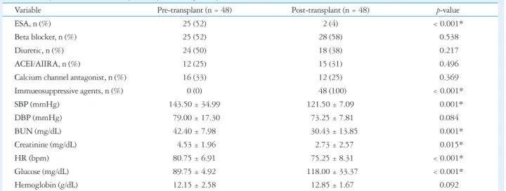
Table 1. Comparison of clinical data pre and post kidney transplantation
Variable Pre-transplant (n = 48) Post-transplant (n = 48) p-value
ESA, n (%) 25 (52) 2 (4) < 0.001*
Beta blocker, n (%) 25 (52) 28 (58) 0.538
Diuretic, n (%) 24 (50) 18 (38) 0.217
ACEI/AIIRA, n (%) 12 (25) 15 (31) 0.496
Calcium channel antagonist, n (%) 16 (33) 12 (25) 0.369
Immueosuppressive agents, n (%) 0 (0) 48 (100) < 0.001*
SBP (mmHg) 143.50 ± 34.99 121.50 ± 7.09 0.001*
DBP (mmHg) 79.00 ± 17.30 73.25 ± 7.81 0.084
BUN (mg/dL) 42.40 ± 7.98 30.43 ± 13.85 0.001*
Creatinine (mg/dL) 4.53 ± 1.96 2.73 ± 2.57 0.015*
HR (bpm) 80.75 ± 6.91 75.25 ± 8.31 < 0.001*
Glucose (mg/dL) 89.75 ± 4.92 118.00 ± 33.37 < 0.001*
Hemoglobin (g/dL) 12.15 ± 2.58 12.85 ± 1.67 0.092
Mean ± standard deviation. *p < 0.05. ESA: erythropoiesis stimulating agent, ACEI: angiotensin-converting enzyme inhibitor, AIIRA: angiotensin II receptor antagonist, SBP: systolic blood pressure, DBP: diastolic blood pressure, BUN: blood urea nitrogen, HR: heart rate
studies, not using VVI technology, demonstrated that there were structural and functional improvements in cardiac indi- ces post kidney transplantation.15)16) Wali et al.15) reported that kidney transplantation in end stage renal disease patients with advanced systolic heart failure resulted in an increase in LVEF.
Compared to pre kidney transplantation, ROT-BAS, ROT- API, TW and TOR were significantly higher post kidney transplantation, indicating an improvement in overall myo- cardial mechanical function, but there was no significant dif- ference between absolute value of ROT-BAS and ROT-API. It has been established that in healthy subjects rotation of the LV base is opposite to that of the apex but is significantly lower in its magnitude. In the normal heart, the counterdirectional rota- tion of the LV apex with respect to the base results in a wring- ing movement during ejection. The rotation angle increased with distance from base to apex, and subendocardial rotation was found to be higher than subepicardial rotation.17)18) The
pattern of net LV twist in which the apex and the base rotate in different directions has been explained on the basis of vary- ing spiral myofiber architecture of the apical and basal region and apex-to-base and transmural gradients in myosin phos- phorylation.19-21) For optimal cardiac mechanical function it is important to maintain rotation of the LV apex in a direction opposite to and higher than the base. Our study shows an im- provement of rotation, twist and torsion post kidney trans- plantation, rotation of the LV base was opposite to that of the apex but was higher in its magnitude, and the left ventricular rotation pattern was still different from normal subjects.
Physiological variables such as preload, afterload, contractil- ity, exercise and age may influence the extent of LV rotation.
The most important factors predisposing to abnormal cardiac performance and morphology in end stage renal diseases are sys- temic hypertension, anemia, volume overload, and in patients on hemodialysis, the arteriovenous fistula. Prolonged exposure
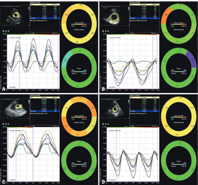
Fig. 1. Left ventricular rotation pre and post kidney transplantation. A: Left ventricular rotation at apex pre kidney transplantation. B:
Left ventricular rotation at base pre kidney transplantation. C: Left ventricular rotation at apex post kidney transplantation. D: Left ventricular rotation at base post kidney transplantation.
A
C
B
D
to uremic toxins can result in myocyte fibrosis and cell death.
Metabolic factors such as acidosis, hypoxia, hypocalcemia, and possibly high levels of parathyroid hormone, may impair LV function.22)23) Kidney transplantation is associated with a re- versal of the biochemical toxins and conditions associated with uremia, which resulted in the improvement of LV structure, function and torsion.
In this study, peak TOR was positive correlation with E, A, E/A, e, LVEF, ROT-API, ROT-BAS, TW (0.65, 0.25, 0.6, 0.4, 0.49, 0.83, 0.77, 0.83, respectively, p < 0.05) and peak TOR was negative correlation with DT, IVS, LVMI (-0.31, -0.34, -0.77, respectively, p < 0.05). Studies of Kim et al.24) and Takeuchi et al.25) demonstrated similar results to confirm that left ventricular rotation varied with changes in preload, afterload, and contractility.
In our study, post kidney transplantation fasting glucose was higher than pre transplantation. Evidence suggested that immunosuppressive drugs accounted this, the association be- tween immunosuppressive drugs and glucose increase had been established clearly and was related to cumulative dosages and therapy duration.26)
In this study, we performed myocardial rotation analysis by velocity vector imaging. Velocity vector imaging is not a sim- ple speckle-tracking technique, as it uses a more sophisticated approach that involves endocardial border tracking performed with Fourier techniques that ensure higher accuracy using the periodicity of the heart’s motion, which allows myocardial ro- tation to be accurately quantified for global and regional myo-
cardial functional assessment.4)27)
However, this was a small number and single center study, and the effects of kidney transplantation on LV structure and function need long-term follow-up.
In conclusion, kidney transplantation in end stage renal dis- ease without myocardial infarction results in improvement of LV structure, function, myocardial rotation, twist and torsion as detected by echocardiography and velocity vector imaging.
Assessment of LV rotation, twist and torsion provided impor- tant insights into different types of myocardial dysfunction and the effect of different treatment strategies. Velocity vector imaging provided valuable information for detection and fol- low-up of cardiac abnormalities in patients with end stage re- nal disease.
References
1. Sarnak MJ, Levey AS, Schoolwerth AC, Coresh J, Culleton B, Hamm LL, McCullough PA, Kasiske BL, Kelepouris E, Klag MJ, Parfrey P, Pfeffer M, Raij L, Spinosa DJ, Wilson PW; American Heart Association Councils on Kidney in Cardiovascular Disease, High Blood Pressure Research, Clinical Cardiology, and Epidemiol- ogy and Prevention. Kidney disease as a risk factor for development of cardiovascular disease: a statement from the American Heart Association Councils on Kidney in Cardiovascular Disease, High Blood Pressure Re- search, Clinical Cardiology, and Epidemiology and Prevention. Circulation 2003;108:2154-69.
2. Beyar R, Sideman S. Left ventricular mechanics related to the local distri- bution of oxygen demand throughout the wall. Circ Res 1986;58:664-77.
3. Ashikaga H, Criscione JC, Omens JH, Covell JW, Ingels NB Jr.
Transmural left ventricular mechanics underlying torsional recoil during re- Table 2. Anatomical and functional indexes of left ventricle pre and post kidney transplantation
Variable Pre-transplant Post-transplant p-value
IVS (mm) 11.67 ± 2.39 9.67 ± 0.48 < 0.001*
LVPW (mm) 9.67 ± 1.72 9.67 ± 0.96 1.000
LV mass index (g/m2) 104.00 ± 16.47 95.50 ± 21.44 0.043*
RWtd 0.49 ± 0.13 0.50 ± 0.12 0.775
LVEF (%) 40.33 ± 11.42 61.00 ± 13.68 < 0.001*
E (m/s) 0.67 ± 0.21 0.87 ± 0.13 < 0.001*
A (m/s) 0.63 ± 0.15 0.73 ± 0.11 < 0.001*
E/A 1.04 ± 0.57 1.21 ± 0.52 < 0.001*
e’ (m/s) 0.05 ± 0.01 0.06 ± 0.01 < 0.001*
a’ (m/s) 0.06 ± 0.02 0.08 ± 0.02 < 0.001*
e’/a’ 0.88 ± 0.33 0.81 ± 0.35 0.003*
E/e’ 15.57 ± 6.69 14.33 ± 4.07 0.126
DT (ms) 199.33 ± 49.69 181.00 ± 36.07 < 0.001*
ROT-BAS (degree) -4.48 ± 2.66 -5.65 ± 2.64 0.022*
ROT-API (degree) 4.27 ± 3.08 5.50 ± 4.25 0.004*
TW (degree) 8.75 ± 4.45 11.14 ± 5.25 0.004*
TOR (degree/cm) 1.06 ± 0.54 1.33 ± 0.61 0.003*
Mean±standard deviation. *p < 0.05. IVS: interventricular septum, LVPW: left ventricular posterior wall, RWtd: relative wall thickness in diastole, LVEF: left ventricular ejection fraction, E: mitral early diastolic filling velocity, A: mitral late diastolic filling velocity, E/A: a ratio of mitral early and late diastolic filling velocity, e’: mitral early diastolic annuluar velocity, a’: mitral late diastolic annuluar velocity, e’/a’: a ratio of mitral early and late diastolic annuluar velocity, E/e’:
a ratio of mitral early diastolic filling velocity and early diastolic annular velocity, DT: mitral E wave deceleration time, ROT-BAS: average rotation of basal LV, ROT-API: average rotation of apical LV, TW: average twist, TOR: average torsion
laxation. Am J Physiol Heart Circ Physiol 2004;286:H640-7.
4. Alharthi MS, Jiamsripong P, Calleja A, Sengupta PP, McMahon EM, Khandheria B, Tajik AJ, Belohlavek M. Selective echocardiograph- ic analysis of epicardial and endocardial left ventricular rotational mechan- ics in an animal model of pericardial adhesions. Eur J Echocardiogr 2009;10:357-62.
5. Alharthi MS, Jiamsripong P, Calleja A, Sengupta PP, McMahon EM, Khandheria B, Tajik AJ, Belohlavek M. Selective echocardiograph- ic analysis of epicardial and endocardial left ventricular rotational mechan- ics in an animal model of pericardial adhesions. Eur J Echocardiogr 2009;10:357-62.
6. Gustafsson U, Lindqvist P, Mörner S, Waldenström A. Assessment of regional rotation patterns improves the understanding of the systolic and dia- stolic left ventricular function: an echocardiographic speckle-tracking study in healthy individuals. Eur J Echocardiogr 2009;10:56-61.
7. Esch BT, Warburton DE. Left ventricular torsion and recoil: implications for exercise performance and cardiovascular disease. J Appl Physiol (1985) 2009;106:362-9.
8. Kim WJ, Lee BH, Kim YJ, Kang JH, Jung YJ, Song JM, Kang DH, Song JK. Apical rotation assessed by speckle-tracking echocardiogra- phy as an index of global left ventricular contractility. Circ Cardiovasc Im- aging 2009;2:123-31.
9. Burns AT, McDonald IG, Thomas JD, Macisaac A, Prior D. Doin’
the twist: new tools for an old concept of myocardial function. Heart 2008;
94:978-83.
10. Borrows R, Loucaidou M, Chusney G, Borrows S, Tromp JV, Cairns T, Griffith M, Hakim N, McLean A, Palmer A, Papalois V, Taube D. Anaemia and congestive heart failure early post-renal transplantation.
Nephrol Dial Transplant 2008;23:1728-34.
11. Boschiero L, Fior F, Nacchia F. Bimodal distribution of major cardiovascu- lar events in kidney allograft recipients. Transplant Proc 2009;41:1183-6.
12. Lang RM, Bierig M, Devereux RB, Flachskampf FA, Foster E, Pel- likka PA, Picard MH, Roman MJ, Seward J, Shanewise JS, Solomon SD, Spencer KT, Sutton MS, Stewart WJ; Chamber Quantification Writing Group; American Society of Echocardiography’s Guidelines and Standards Committee; European Association of Echocardiogra- phy. Echocardiography’s Guidelines and Standards Committee and the Chamber Quantification Writing Group, developed in conjunction with the European Association of Echocardiography, a branch of the European Society of Cardiology. J Am Soc Echocardiogr 2005;18:1440-63.
13. Quiñones MA, Otto CM, Stoddard M, Waggoner A, Zoghbi WA;
Doppler Quantification Task Force of the Nomenclature and Stan- dards Committee of the American Society of Echocardiography.
Recommendations for quantification of Doppler echocardiography: a report from the Doppler Quantification Task Force of the Nomenclature and Stan- dards Committee of the American Society of Echocardiography. J Am Soc Echocardiogr 2002;15:167-84.
14. van Dalen BM, Soliman OI, Vletter WB, ten Cate FJ, Geleijnse
ML. Age-related changes in the biomechanics of left ventricular twist mea- sured by speckle tracking echocardiography. Am J Physiol Heart Circ Physiol 2008;295:H1705-11.
15. Wali RK, Wang GS, Gottlieb SS, Bellumkonda L, Hansalia R, Ra- mos E, Drachenberg C, Papadimitriou J, Brisco MA, Blahut S, Fink JC, Fisher ML, Bartlett ST, Weir MR. Effect of kidney transplantation on left ventricular systolic dysfunction and congestive heart failure in patients with end-stage renal disease. J Am Coll Cardiol 2005;45:1051-60.
16. Sahagún-Sánchez G, Espinola-Zavaleta N, Lafragua-Contreras M, Chávez PY, Gómez-Núñez N, Keirns C, Romero-Cardenas A, Pérez-Grovas H, Acosta JH, Vargas-Barrón J. The effect of kidney transplant on cardiac function: an echocardiographic perspective. Echocar- diography 2001;18:457-62.
17. Rüssel IK, Götte MJ, Bronzwaer JG, Knaapen P, Paulus WJ, van Rossum AC. Left ventricular torsion: an expanding role in the analysis of myocardial dysfunction. JACC Cardiovasc Imaging 2009;2:648-55.
18. Helle-Valle T, Crosby J, Edvardsen T, Lyseggen E, Amundsen BH, Smith HJ, Rosen BD, Lima JA, Torp H, Ihlen H, Smiseth OA.
New noninvasive method for assessment of left ventricular rotation: speckle tracking echocardiography. Circulation 2005;112:3149-56.
19. Sengupta PP, Korinek J, Belohlavek M, Narula J, Vannan MA, Jah- angir A, Khandheria BK. Left ventricular structure and function: basic science for cardiac imaging. J Am Coll Cardiol 2006;48:1988-2001.
20. Davis JS, Hassanzadeh S, Winitsky S, Lin H, Satorius C, Vemuri R, Aletras AH, Wen H, Epstein ND. The overall pattern of cardiac con- traction depends on a spatial gradient of myosin regulatory light chain phos- phorylation. Cell 2001;107:631-41.
21. Taber LA, Yang M, Podszus WW. Mechanics of ventricular torsion. J Biomech 1996;29:745-52.
22. Alpert MA. Cardiac performance and morphology in end-stage renal dis- ease. Am J Med Sci 2003;325:168-78.
23. Amann K, Breitbach M, Ritz E, Mall G. Myocyte/capillary mismatch in the heart of uremic patients. J Am Soc Nephrol 1998;9:1018-22.
24. Kim HK, Sohn DW, Lee SE, Choi SY, Park JS, Kim YJ, Oh BH, Park YB, Choi YS. Assessment of left ventricular rotation and torsion with two-dimensional speckle tracking echocardiography. J Am Soc Echocar- diogr 2007;20:45-53.
25. Takeuchi M, Otsuji Y, Lang RM. Evaluation of left ventricular function using left ventricular twist and torsion parameters. Curr Cardiol Rep 2009;11:225-30.
26. Rodrigo E, Fernández-Fresnedo G, Valero R, Ruiz JC, Piñera C, Palomar R, González-Cotorruelo J, Gómez-Alamillo C, Arias M.
New-onset diabetes after kidney transplantation: risk factors. J Am Soc Nephrol 2006;17(12 Suppl 3):S291-5.
27. Jiamsripong P, Alharthi MS, Calleja AM, McMahon EM, Mookad- am F, Khandheria BK, Belohlavek M. Quantification of left ventricular twisting mechanics by velocity vector imaging in an animal model of pericar- dial adhesions. Ultrasound Med Biol 2009;35:1963-72.