http://dx.doi.org/10.4174/astr.2016.90.5.265 Annals of Surgical Treatment and Research
Early results with the Mutaf technique: a novel off- midline approach in pilonidal sinus surgery
Musa Zorlu, İbrahim Tayfun Şahiner, Ethem Zobacı1, Cem Kocak2, Ahmet Çınar Yastı, Mete Dolapçı
Department of General Surgery, Medical Faculty of Hitit University, Çorum, 1Department of General Surgery, Çorum Research and Training Hospital, Medical Faculty of Hitit University, Çorum, 2Department of Statistics, School of Health, Hitit University, Çorum, Turkey
INTRODUCTION
Pilonidal sinus (PS) is an acute, chronic, symptomatic or asymptomatic clinical condition occurring at the natal cleft in the sacrococcygeal area. Untreated subjects experience a vicious cycle consisting of the development of subcutaneous infection, abscess formation, spontaneous or surgical drainage of the abscess, and serous discharge. Therefore, although it is not a lifethreatening condition, it may adversely affect the quality of life with restriction of the activities of daily living.
PS is more prevalent in the young working population, with a male to female ratio of 2.24 to 1 (males prevalent
is 2.2-4 times more than females). The reported incidence rate in the United States is 26/100 [1]. Surgery represents a therapeutic option for patients with PS and despite many different techniques described in the literature for the surgical treatment of this condition, no consensus has been reached regarding the best strategy. Low recurrence rates reported in recent publications for offmidline closure techniques has led to the routine recommendation of this management strategy [2]. Previously described techniques all have their pros and cons. The primary prerequisite in an ideal surgical technique involves low recurrence and surgical complication rates, as well as other attributes such as the simplicity of the technique, Purpose: The objective of the present study was to compare different off-midline techniques in terms of their advantages and disadvantages.
Methods: A total of 81 patients were included in this prospective, controlled, randomized study. Patients in group 1 were treated with the Limberg flap, and patients in group 2 were treated with Mutaf technique. Patients were followed up for 9 months postsurgically and assessed at regular intervals.
Results: A total of 41 and 40 patients received surgical treatment with Limberg or Mutaf techniques, respectively. The 2 groups were similar in terms of age, gender, body mass index, and Tezel pilonidal sinus classification. Also, the 2 groups were comparable with regard to the frequency of preoperative discharge from the wound site, history of abscess formation, and the resultant antibiotic use. Early results showed similar recurrence rates and surgical-site complications between the 2 groups. Although a lower visual analogue scale score was found in group 2 at postoperative day 1, seroma persistence, time to withdrawal of surgical drains, and wound healing were more prolonged.
Conclusion: In this study, Mutaf technique was comparable to Limberg flap in the treatment of pilonidal sinus. Therefore, Mutaf technique may be offered as a viable surgical therapeutic option among off-midline closure approaches.
[Ann Surg Treat Res 2016;90(5):265-271]
Key Words: Pilonidal sinus, Surgical Flaps, Surgical treatment
Reviewed January February March April May June July August September October November December
Received November 17, 2015, Revised January 26, 2016, Accepted February 15, 2016
Corresponding Author: Ibrahim Tayfun Şahiner
Department of General Surgery, Hitit University School of Medicine, Bahçelievler Mahallesi , Çamlık Caddesi No:2, Çorum,19030, Turkey Tel: +90-364-223-0300, Fax: +90-364-223-0323
E-mail: tayfunsahiner@gmail.com
Copyright ⓒ 2016, the Korean Surgical Society
cc Annals of Surgical Treatment and Research is an Open Access Journal. All articles are distributed under the terms of the Creative Commons Attribution Non- Commercial License (http://creativecommons.org/licenses/by-nc/4.0/) which permits unrestricted non-commercial use, distribution, and reproduction in any medium, provided the original work is properly cited.
lowcost, short duration of hospital stay, quick recovery and return to daily activities, and minimum adverse cosmetic and psychological effects.
In this regard, Limberg flap technique, an offmidline clo
sure method, has long been considered a standard surgical approach by many experts and is commonly recommended for the surgical treatment of this condition. On the other hand, the Mutaf triangle closure technique, originally described by Mutaf, was first used in patients with large myelomeningocele defects [3]. Thanks to unequal zplasty, two different flaps can be formed that have been reported to provide better tissue relaxation, which allows both the primary closure of the donor area and better vascularization of the flap tissue and tension
free closure. [3]. Thus, in this study our aim was to compare this novel technique with Limberg flap technique.
METHODS
This was a prospective, controlled, randomized, single
center study. The study protocol was approved by the Ethics Committee, Ankara Numune Training and Research Hospital (approval number: 20796219724.0849). A total of 81 patients who were diagnosed with PS between 01 January 2014 and
Table 1. Tezel classification
Type Tezel classification
I Asymptomatic sinus II Acute pilonidal abscess
III Chronic (symptomatic) restricted in navicular area IV Chronic (symptomatic) exceeded navicular area
V Recurrent pilonidal sinus
A B
C D
Fig. 1. Mutaf triangle closure technique: (A) drawing flap, (B) excision of natal cleft, (C) preparation of flap, and (D) post- operative day 60.
31 December 2014 at Çorum Research and Training Hospital, Hitit University Medical Faculty and who met the inclusion criteria were included in this study. The study was conducted in accordance with the principles set forth in the Helsinki Declaration. All patients provided written informed consent before study participation. The randomization scheme was based on the outpatient attendance number such that those with an odd number were assigned into group 1 and those with an even number into group 2, who were treated with Limberg flap technique and Mutaf triangular defect closure technique, respectively.
Patient selection
Female or male PS patients over 18 years of age were included and classified according to the navicular area concept proposed by Tezel [4] (Table 1).
Preoperatively age, gender, body mass index (BMI), previous treatments (discharge, antibiotics, use of antibiotics, etc.) were recorded. Also, the duration of surgery, defined as the time from initiation of incision to the placement of last skin suture, was recorded.
Postoperative followup controls were performed at bedside following surgery, at 30 days and at 6 months at the outpatient unit. The visual analogue scale (VAS) scores (between 0 and 10) were recorded at day 1, 10, and 30.
Surgicalsite related complications including induration, wound dehiscence, seroma formation, hematoma, sensory loss at the wound site, and infections were recorded.
Duration of hospital stay was defined as the time from the termination of surgery to discharge. Healing time was defined as the duration of time from the day of surgery to the removal of stitches (or to complete healing when complications were present). The incidence of early recurrence was determined with 6 months of followup data.
Technical descriptions
Patients in group 1 were operated on using Limberg flap technique according to the descriptions by Mentes et al. [5], while those in group 2 were operated on using Mutaf triagular closure technique as described by Mutaf et al. [3] (Fig. 1).
In both techniques, a 12F suction drain tube was placed subcutaneously and anatomical layers were closed with 2 layers. The 2/0 absorbable polyglactin sutures were used for the subcutaneous layers, while the skin was closed using 3/0 unabsorbable polypropylene sutures with mattress stitches.
Prior to surgery all patients received a single dose of 1g cefazolin intravenously for infection prophylaxis.
No further antibiotic treatments were given postoperatively or at discharge. All surgical procedures were performed under standardized spinal anesthesia. Diclofenac intramuscular was given at postoperative hour 8 for analgesia, followed by
diclofenac oral tablets twice a day. Drains were removed when the output was below 20 mL/day. Sutures were removed at postoperative day 10.
Statistical analyses
IBM SPSS Statistics ver. 20.0 (IBM Co., Armonk, NY, USA) was used for statistical analyses. For statistical analysis, frequency distributions according to dependent variables and descriptive statistics were obtained for both surgical techniques. For examination of the effect of surgical technique on dependent variables, chisquare and Fisher exact chisquare analyses were used for the qualitative variables, while tests of normality were performed for the quantitative analyses for the association between the surgical technique and the quantitative dependent variables. Pairwise group comparisons were carried out using the twosample ttest and MannWhitney Utest depending on the normal distribution of the data. Ninetyfive percent confidence intervals were provided and a Pvalue of less than 0.05 was considered statistically significant.
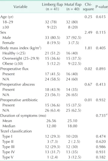
Table 2. Basics characteristics of patients Variable Limberg flap
(n = 41) Mutaf flap (n = 40) Chi-
square P-value
Age (yr) 0.25 0.615
18–29 32 (78) 32 (80)
≥30 9 (22) 8 (20)
Sex 2.49 0.115
Male 33 (80.5) 37 (92.5)
Female 8 (19.5) 3 (7.5)
Body mass index (kg/m2) 1.81 0.405
Healthy (<25) 21 (51.2) 16 (40) Overweight (25–29.9) 15 (36.6) 15 (37.5)
Obese (≥30) 5 (12.2) 9 (22.5)
Preoperative flux 0.02 0.893
Present 17 (41.5) 16 (40)
N/A 24 (58.5) 24 (60)
Preoperative abscess 0.67 0.413
Present 18 (43.9) 14 (35)
N/A 23 (56.1) 26 (65)
Preoperative antibiotic 0.01 0.932
Present 15 (36.6) 15 (37.5)
N/A 26 (63.4) 25 (62.5)
Duration of symptoms (mo) 0.735a)
Mean 26.56 25.10
Median 12.00 18.00
Tezel classification
Type I 12 (29.3) 10 (20) 0.474
Type II 3 (7.3) 2 ( 2.5) 0.620
Type III 12 (29.3) 12 (30) 0.986
Type IV 13 (31.7) 13 (35) 0.931
Type V 1 (2.4) 3 (12.5) 0.182
Values are presented as number (%) unless otherwise indicated.
N/A, absent.
a)Mann-Whitney test.
RESULTS
The demographic data for the study subjects (n = 81) is summarized in Table 2. Groups 1 and 2 consisted of 41 and 40 patients, respectively, with an overall maletofemale ratio of 70:11. The 2 groups were comparable in terms of age, gender, BMI, and Tezel PS classification as well as preoperative history of wound site discharge, and abscess formation and related antibiotic use. The mean follow up was 9.2 and 8.97 months, respectively.
In both groups, the majority of the patients were between 18 and 29 years of age and had a BMI < 25 kg/m2. Preoperatively, the average duration of symptoms was 26.56 and 25.1 months in groups 1 and 2, respectively.
Intra and postoperative patient data are shown in Table 3.
The duration of surgery was similar across the study groups.
Rate of surgical site complications such as induration at the surgical wound, wound dehiscence, hematoma formation, and sensory loss were comparable between the 2 groups. However, significant differences were found between the 2 groups in terms of seroma formation, surgical drain removal time, and healing time.
A significant difference in favor of Mutaf technique was found with regard to VAS scores at postoperative day 1, while no difference in VAS scores were observed at postoperative days 10 and 30.
The duration of hospital stay was similar between the 2 groups, as were the rates of recurrence at 6 months post
operative.
DISCUSSION
The word pilonidal derives from the Latin words pilus ("hair") and nidus (“nest”) and PS was originally described by Andersson in 1847. It is an infectious disease arising from the hair follicles on the natal cleft in the sacrococcygeal region that may have an acute, chronic, symptomatic or asymptomatic course. It is generally considered as an acquired condition rather than a hereditary one [6]. The treatment options vary from simpler surgical approaches such as fistulotomy + curettage to those involving more advanced flap repair techniques. Defects formed after total excision of PS have been traditionally treated with primary midline repair, marsupialization, leaving the wound open, or MacFee technique. Also, offmidline techniques have been utilized for defect reconstruction such as Rhomboid excision plus Limberg flap, Karyadakis flap, Bascom (cleft lift), VY advancement flap, Zplasty, elliptical rotation flap, perforator flaps, and gluteus maximus muscleskin flaps, with certain pros and cons associated with each of these techniques.
An ideal surgical technique should have low recurrence and Table 3. Preoperative and postoperative evaluation results
Variable Limberg
group (n = 41)
Mutaf group (n = 40)
Chi-
square P-value
Operation time (min) 34.27 ± 8.0 31.60 ± 9.2 0.170
Induration 0.201a)
N/A 40 (97.6) 36 (90.0)
Present 1 (2.4) 4 (10.0)
Wound separation 0.494a)
N/A 41 (100) 39 (97.5)
Present 0 (0) 1 (2.5)
Seroma 0.026a),*
N/A 41(100) 35(87.5)
Present 0 (0) 5 (12.5)
Hematoma 0.494a)
N/A 41 (100) 39 (97.5)
Present 0 (0) 1 (2.5)
Loss of sensation 2.17 0.141
N/A 21 (51.2) 14 (35.0)
Present 20 (48.8) 26 (65.0)
Infection 2.76 0.096
N/A 38 (92.7) 32 (80.0)
Present 3 (7.3) 8 (20.0)
Follow-up time (mo) >0.05
Mean 9.2 8.9
Range 6–13 6–13
Recurrence (control 6 mo) >0.616a)
N/A 40 (97.6) 38 (95.0)
Present 1 (2.4) 2 (5.0)
VAS score
Postop day 1 0.048*
Mean 1.68 1.05
Median 2.00 1.00
Postop day 10 0.796
Mean 0.37 0.35
Median 0 0
Postop day 30 0.975
Mean 0.17 0.07
Median 0 0
Duration of hospitalization (day) 0.600
Mean 2.49 3.38
Median 2.00 2.00
Drain removal time (day) <0.001**
Mean 4.07 9.42
Median 0 3.00
Healing time (day) <0.001**
Mean 11.95 15.03
Median 11.00 15.00
Values are presented as mean±standard deviation or number (%) unless otherwise indeciated.
N/A, absent; VAS, visual analogue scale.
a)Fisher exact test. *P < 0.05 significant level of importance. **P <
0.01 significant level of importance.
surgical complication rates, as well as other attributes such as the simplicity of the technique, lowcost, short duration of hospital stay, quick recovery and return to daily activities, and minimum adverse aesthetic and psychological effects.
In a 2008 metaanalysis by McCallum et al. [2] involving a number of randomized controlled studies found low recurrence rates with offmidline closure techniques, hence the authors recommended that offmidline techniques be the standard approach in the surgical treatment of patients with PS. In another metaanalysis by Horwood et al. [7] in 2012, only 8 studies could be included in the data pool, while only 2 studies compared Karydakis and Limberg flap techniques. Therefore, the authors concluded that more highquality comparative studies are warranted to reach a definitive conclusion [7].
Orhalmi et al. [8] compared Limberg flap and Karydakis flap in their study in 2014 and found similarly good results with both techniques. Guner et al. [9] in their study comparing Bascom and Limberg flap techniques, reported on the specific advantages of each approach. Ozdemir et al. [10] used off
midline closure using the whole natal cleft excision technique in 234 patients in 2014 with successful results. Although off
midline surgery is recommended as the standard treatment for PS in the metaanalysis by McCallum et al. [2], it should also be pointed out that no adequate prospective, randomized studies have been performed that directly compared different offmidline approaches and that such studies are warranted.
Thus, our study involved the comparison of two offmidline techniques, i.e., Limberg flap technique and Mutaf triangular closure technique.
In Mutaf triangular closure method, unequal zplasty allows extra tissue relaxation and the donor area may be closed with primary closure. In the study where Mutaf describes his technique, a good vascularization was reported in the flap tissue in 5 patients with large myelomeningocele defects [3]. On the other hand, Limberg flap technique was reported to provide adequate closure for small or mediumsized myelomeningocele defects, while it was shown to fail in providing adequate tensionfree closure in larger defects [11]. Also, in a study by Mutaf involving PS patients, a first flap for the closure of the defect area and a second flap covering the donor area, together with an unequal zplasty, allowed better tissue relaxation and was able to provide tensionfree closure even in large defects [3].
PS occurs more commonly in the second or third decades of life with a male to female ratio of 2.2-4 to 1 (males prevalent is 2.2-4 times more than females) [1]. More frequent occurrence of this condition in the young and working population places more significance on PS due to its influence on professional and social life. In our study, the male to female ratio was 70:11 (i.e., 6.36), once more confirming a higher frequency among male subjects. Similar to previous reports, patients between 18 and 29 years of age comprised the majority of our study participants
[1].
Ideal PS surgery should be associated with low risk of sur
gicalsite complications. In a study by Petersen et al., [12] the incidence of wound site infections was 12% with the Karydakis flap technique. In two different studies, the reported rates of infection for Limberg flap varied between 0.8% and 3% [11,13].
Guner et al. [9] performed a Limberg flap in 61 patients and found an overall infection rate of 9.7% including both superficial and deep infections. In our study, infection occurred in 7.3% of patients in Limberg flap group and in 20% of patients in Mutaf technique group, with no significant differences between the 2 groups (P = 0.096).
The higher rate of infection despite low occurrence of seroma formation was explained on the basis of inadequate personal hygiene and use of surgical drains in two different studies [14,15]. In the present study, the time to drain removal was 4.07 and 9.42 days in groups 1 and 2, respectively, with a significant difference in this regard (P < 0.001). Also, seroma formation was not observed in any patients in group 1 vs. in 12% of patients in group 2, again with a significant difference (P = 0.026). Due to the use of two different flaps in Mutaf technique, more frequent formation of seromas and more prolonged need for surgical drains were not considered unexpected. Similarly, higher rate of infections in patients treated with Mutaf technique was probably associated with these two factors, i.e., seromas and prolonged time to drain removal. Of the 8 patients with infections in Mutaf technique group, only 1 had deep surgical site infection, while 7 were found to have superficial skin infections.
In a study examining wound dehiscence, classical Limberg flaps were associated with higher rates of wound dehiscence as compared to a modified Limberg flap technique (23%, 3%, P
= 0.03) [2]. In the present study, the dehiscence rates were 0%
and 2.5% in groups 1 and 2, respectively (P > 0.05). A previous study reported no patients developing hematoma after PS surgery with Limberg flap or Bascom techniques [9]. In our study, 0% and 2.5% of patients in groups 1 and 2 had hematoma formation, respectively (P > 0.05).
A determinant of postoperative success in patients under
going PS surgery is local sensory loss at the site of surgery, which occurred in 48.8% and 65% of our patients in groups 1 and 2, respectively (P = 0.141). Preparation of 2 flaps with the Mutaf technique was associated with increased rates of local sensory loss, although this was not considered a disadvantage as reflected by the absence of significant differences between the 2 groups.
Earlier completion of postoperative wound healing allows earlier return to normal daily activities. In a study examining the healing time in Limberg flap and Bascom techniques, the respective healing times of 11.55 and 11.50 days were reported [9]. In another study, the wound healing time reported for a
modified Limberg technique was 14 days [16,17]. In our study, the corresponding values in groups 1 and 2 were 11.56 and 15.4 days, respectively (P < 0.001). The more prolonged healing process in the group treated with Mutaf technique was thought to be associated with the longer time to removal of the drain, which probably caused a relative increase in the time to healing and a statistically significant difference.
Postoperative pain is an important factor for patients’ com
fort and an ideal surgical technique should be associated with minimum pain. In three different studies, Limberg flap technique was compared with primary closure and secondary healing, and was found to cause less pain than other groups [1820]. In a prospective study with 122 patients, Limberg flap was compared with Bascom technique, with no significant differences in VAS scores between the two groups [9]. In our study, VAS scores at 1, 10 and 30 days after surgery, the mean scores on day 1 in groups 1 and 2 were 1.68 and 1.05, respec
tively, with lesser intensity of pain in patients in Mutaf tech
nique group (P = 0.048); VAS scores at postoperative day 10 and 30 were not significantly different (P > 0.05). This difference on day 1 was probably due to the presence of a tensionfree flap, which was prepared using 2 separate flaps.
Duration of hospital stay after surgery is linked with cost
effectiveness and patient satisfaction of a specific surgical tech
nique. In three different studies, the length of hospital stay in patients undergoing PS surgery with Limberg technique were 2, 4, and 6 days respectively [16,21,22]. In our study, the length of hospital stay in groups 1 and 2 were 2.49 and 3.38 days, respectively, with no significant difference in this regard (P = 0.06). Thus, neither technique was associated with significantly prolonged hospital stay.
In the study by Guner et al., [9] the duration of surgery was 36 minutes (range, 22–50 minutes) and 29 minutes (range, 20–
42 minutes) for Limberg and Bascom techniques, respectively (P < 0.0001). In our study, this value in groups 1 and 2 were 34.27 ± 8 and 31.60 ± 9.2 minutes. The surgery time observed in our patient groups is consistent with previous reports and no significant differences were detected between the 2 groups.
Previously, BMI has been found to bear clinical significance in terms of the risk of surgicalsite related complications and recurrence. For instance, a direct link between BMI and wound infection, wound dehiscence, and recurrence rate was found in two previous studies [23,24]. In a study by Sievert et al. [25]
examining the effect of lifestyle on longterm recurrence risk in a total of 534 patients and in another study by Poorghasem and Mahoori [26] again looking at the effect of BMI on recurrence rates, recurrent PS was found to occur primarily in those with a BMI of greater than 30 kg/m2 (obese) or in those with a BMI between 25–30 kg/m2 (overweight), with reduced recurrence in those having a BMI below 25 kg/m2 [26]. In another study Alptekin et al. [27] found higher recurrence rates in patients with a high volume of specimen regardless of the operation method. In our study, a patient who had wound dehiscence and recurrence had a BMI between 25 and 29.9 kg/m2 (i.e., overweight). Another patient with recurrence had a BMI > 30 kg/m2, again consistent with previous reports. All 5 patients (45%) with wound site infection had a BMI greater than 30 (i.e., obese).
The primary priority in PS surgery is to prevent recurrences.
In a 7,471 patient series reported by Karydakis [28], the reported rate of recurrence was 1%. In a study by Tezel et al. [29], the reported recurrence rate with Bascom cleft lift technique was 1.3%. In a review by Petersen et al. [16] involving 16 studies of Limberg flap technique, a recurrence rate of 1.5% (range, 0.8%–
2.7%) was found. In our study the recurrence rates in groups 1 and 2 were 2.4% and 5%, respectively, with no significant differences (P > 0.616). Therefore, Limberg flap and Mutaf techniques had a comparable early recurrence rate. Although Mutaf technique was associated with more frequent seroma formation, pro longed time to drain removal, and prolonged healing time, the 2 groups were similar in terms of early recurrence rate, wound site induration, wound dehiscence, hematoma formation, local sensory loss, and infection rate.
Also, a lower VAS score at day 1 in Mutaf technique group was considered as an advantage. Therefore, we believe that Mutaf triangular closure technique may represent a viable surgical therapeutic alternative among offmidline approaches. This view should be supported with larger clinical samples involving the use of this technique in larger defects and involving longer
term followup.
CONFLICTS OF INTEREST
No potential conflict of interest relevant to this article was reported.
1. Keskin AI, Polat Y, Duran E, Cetinkünar S, Zorlu M. Comparison of four different sur
gical techniques in cases with pilonidal
sinus. Dicle Med J 2014;41:55863.
2. McCallum IJ, King PM, Bruce J. Healing by primary closure versus open healing after
surgery for pilonidal sinus: systematic review and metaanalysis. BMJ 2008;336:
86871.
REFERENCES
3. Mutaf M, Bekerecioglu M, Erkutlu I, Bulut O. A new technique for closure of large meningomyelocele defects. Ann Plast Surg 2007;59:53843.
4. Tezel E. A new classification according to navicular area concept for sacrococcygeal pilonidal disease. Colorectal Dis 2007;9:
5756.
5. Mentes O, Bagci M, Bilgin T, Ozgul O, Ozdemir M. Limberg flap procedure for pilonidal sinus disease: results of 353 patients. Langenbecks Arch Surg 2008;
393:1859.
6. Akinci OF, Bozer M, Uzunkoy A, Duzgun SA, Coskun A. Incidence and aetiological factors in pilonidal sinus among Turkish soldiers. Eur J Surg 1999;165:33942.
7. Horwood J, Hanratty D, Chandran P, Billings P. Primary closure or rhomboid exci sion and Limberg flap for the manage
ment of primary sacrococcygeal pilonidal disease? A metaanalysis of ran domized controlled trials. Colorectal Dis 2012;14:
14351.
8. Orhalmi J, Sotona O, Dusek T, Ferko A.
Pilonidal sinus: possibilities surgical treat
ment. Rozhl Chir 2014;93:4915.
9. Guner A, Boz A, Ozkan OF, Ileli O, Kece C, Reis E. Limberg flap versus Bascom cleft lift techniques for sacrococcygeal pilonidal sinus: prospective, randomized trial. World J Surg 2013;37:207480.
10. Ozdemir H, Unal Ozdemir Z, Tayfun Sahiner I, Senol M. Whole natal cleft excision and flap : an alternative surgical method in extensive sacrococcygeal pilo
nidal sinus disease. Acta Chir Belg 2014;
114:26670.
11. Lapid O, Rosenberg L, Cohen A. Meningo
myelocele reconstruction with bilobed flaps. Br J Plast Surg 2001;54:5702.
12. Petersen S, Aumann G, Kramer A, Doll D, Sailer M, Hellmich G. Shortterm results of Karydakis flap for pilonidal sinus dis
ease. Tech Coloproctol 2007;11:23540.
13. Mentes BB, Leventoglu S, Cihan A, Tatlicioglu E, Akin M, Oguz M. Modified Limberg transposition flap for sacroco
ccygeal pilonidal sinus. Surg Today 2004;
34:41923.
14. Topgul K, Ozdemir E, Kilic K, Gokbayir H, Ferahkose Z. Longterm results of limberg flap procedure for treatment of pilonidal sinus: a report of 200 cases. Dis Colon Rectum 2003;46:15458.
15. Petersen S, Koch R, Stelzner S, Wendlandt TP, Ludwig K. Primary closure techniques in chronic pilonidal sinus: a survey of the results of different surgical approaches.
Dis Colon Rectum 2002;45:145867.
16. Cihan A, Mentes BB, Tatlicioglu E, Ozmen S, Leventoglu S, Ucan BH. Modified Lim
berg flap reconstruction compares favou
rably with primary repair for pilonidal sinus surgery. ANZ J Surg 2004;74:23842.
17. Akin M, Leventoglu S, Mentes BB, Bostanci H, Gokbayir H, Kilic K, et al.
Comparison of the classic Limberg flap and modified Limberg flap in the treat
ment of pilonidal sinus disease: a retro
spective analysis of 416 patients. Surg Today 2010;40:75762.
18. Ertan T, Koc M, Gocmen E, Aslar AK, Keskek M, Kilic M. Does technique alter quality of life after pilonidal sinus sur
gery? Am J Surg 2005;190:38892.
19. Jamal A, Shamim M, Hashmi F, Qureshi MI. Open excision with secondary healing versus rhomboid excision with Limberg transposition flap in the management of sacrococcygeal pilonidal disease. J Pak Med Assoc 2009;59:15760.
20. Mahdy T. Surgical treatment of the pilonidal disease: primary closure or flap reconstruction after excision. Dis Colon Rectum 2008;51:181622.
21. Daphan C, Tekelioglu MH, Sayilgan C.
Limberg flap repair for pilonidal sinus disease. Dis Colon Rectum 2004;47:2337.
22. Tekin A. A simple modification with the Limberg flap for chronic pilonidal disease.
Surgery 2005;138:9513.
23. Maghsoudi H, Nezami N, Ghamari AA.
Ambulatory treatment of chronic piloni
dal sinuses with lateral incision and pri
mary suture. Can J Surg 2011;54:7882.
24. Yeghaneh RA, Faegheh A, Mina A. Body mass ındex and recurrence of pılonıdal sınus dısease. Iran J Surg 2008;16:85-90.
25. Sievert H, Evers T, Matevossian E, Hoene
mann C, Hoffmann S, Doll D. The in
fluence of lifestyle (smoking and body mass index) on wound healing and long
term recurrence rate in 534 primary pilo
nidal sinus patients. Int J Colorectal Dis 2013;28:155562.
26. Poorghasem J, Mahoori A. The effect of body mass ındex on relapse of pilonidal sinus disease in adult patients. ZJRMS 2012;14:235.
27. Alptekin H, Acar F, Sahın M, Yilmaz H, Kafali ME, Beyhan S. Specimen index may be a predictive factor for recurrence after primary closure of pilonidal disease.
J Korean Surg Soc 2012;83:36773.
28. Karydakis GE. Easy and successful treat
ment of pilonidal sinus after explanation of its causative process. Aust N Z J Surg 1992;62:3859.
29. Tezel E, Bostanci H, Anadol AZ, Kurukah
vecioglu O. Cleft lift procedure for sacro
coccygeal pilonidal disease. Dis Colon Rectum 2009;52:1359.