www.krspine.org
A Rare Extradural Spinal Meningioma with Nocturnal Chest Pain - A Case Report -
Sang Bum Kim, M.D., Ph.D., Yougun Won, M.D., Min Gu Jang, M.D., Young Ki Min, M.D., Andreas Gutzeit, M.D., Fabio Casari, M.D., Oliver Nic Hausmann, M.D.
J Korean Soc Spine Surg 2019 Sep;26(3):100-104.
Originally published online September 30, 2019;
https://doi.org/10.4184/jkss.2019.26.3.100
Korean Society of Spine Surgery
SMG-SNU Boramae Medical Center, 20, Boramae-ro 5-gil, Dongjak-gu, Seoul 07061, Korea Tel: +82-2-831-3413 Fax: +82-2-831-3414
©Copyright 2017 Korean Society of Spine Surgery pISSN 2093-4378 eISSN 2093-4386
The online version of this article, along with updated information and services, is located on the World Wide Web at:
http://www.krspine.org/DOIx.php?id=10.4184/jkss.2019.26.3.100
This is an Open Access article distributed under the terms of the Creative Commons Attribution Non-Commercial License (http://
creativecommons.org/licenses/by-nc/4.0) which permits unrestricted non-commercial use, distribution, and reproduction in any medium, provided the original work is properly cited.
Journal of Korean Society of
Spine Surgery
A Rare Extradural Spinal Meningioma with Nocturnal Chest Pain - A Case Report -
Sang Bum Kim, M.D., Ph.D.
*, Yougun Won, M.D.
†, Min Gu Jang, M.D.
†, Young Ki Min, M.D.
†, Andreas Gutzeit, M.D.
‡, Fabio Casari, M.D.
∮, Oliver Nic Hausmann, M.D.
∥*Department of Orthopaedic Surgery, Chungnam National University School of Medicine, Korea
†Department of Orthopedic Surgery, Konyang University College of Medicine, Korea
‡Department of Radiology, Klinik St. Anna, Luzern, Switzerland and Department of Radiology, Salzburg University Hospital, Austria
∮University of Salzburg, Austria
∥Department of Neurosurgery, Klinik St. Anna, Luzern, Switzerland
Study Design: Case report.
Objectives: To report a rare case of a spinal extradural meningioma in a patient with longstanding nonspecific thoracic nocturnal pain Summary of Literature Review: Meningioma is a frequent intradural extramedullary tumor that is associated with pain, sensory/motor deficits, and sphincter weakness. Spinal meningiomas most commonly occur in the thoracic spine, although they can also be found at other locations.
Materials and Methods: A 65-year-old woman first visited the cardiac and gastrointestinal departments of our institution due to chest pain 2 years previously. No explanation for the complaint could be found in the heart or other organs. On a computed tomography scan of the thorax, a spinal mass was found a few months before the diagnosis. On magnetic resonance imaging, an extramedullary and extradural mass was observed at T7/8.
Results: We performed surgery and found an extradural spinal meningioma upon the histological diagnosis. Postoperatively, the patient could adequately move both legs and feet and the nocturnal chest pain disappeared after surgery without any complications.
Conclusions: Awareness of the rarity and nonspecific symptoms of extradural spinal meningiomas will be beneficial for their accurate diagnosis and proper treatment.
Key Words: Extradural space, Thoracic pain, Iatrogenic dural injury, Spinal meningioma, Spine tumors
Received: November 16, 2018 Revised: February 1, 2019 Accepted: July 15, 2019
Published Online: September 30, 2019 Corresponding author: Yougun Won, M.D.
ORCID ID: Sang Bum Kim: https://orcid.org/0000-0002-7497-9077 Yougun Won: https://orcid.org/0000-0002-0874-5248 Young Ki Min: https://orcid.org/0000-0001-8073-5434
Department of Orthopedic Surgery, College of Medicine, Konyang University 685, Gasoowon-dong, Seo-Gu, Daejeon 35365, Korea (R.O.K)
TEL: +82-42-600-9862, FAX: +82-42-545-2373
E-mail: yougunwon@gmail.com, yougunwon@kyuh.ac.kr Spinal Meningioma is the most frequent type of intradural
extramedullary tumor.1) The meningioma entirely originated from extradural space is very rare. Only 5 to 14% of spinal meningioma was reported as it has an extradural component.2) Spinal meningiomas most commonly occur in the thoracic spine.2-5) Meningiomas occur more frequently in women, and they occur in a slightly older age group compared with nerve sheath tumors.4) The common presentations of spinal meningioma are a pain, sensory/motor deficits, and sphincter weakness. Usually, back pain or neck pain appears before the motor and sensory changes. The present patient had longstanding nocturnal pain that was first managed by cardiac and gastrointestinal departments for many years. We report on a rare extradural spinal meningioma detected as an intradural meningioma on magnetic resonance imaging (MRI) in a
patient with nonspecific nocturnal chest pain.
Case Report
The patient was a 65-year-old woman with a history of
A Long Unrecognized Symptom of a Rare Extradural Spinal Meningioma Journal of Korean Society of Spine Surgery
www.krspine.org 101 thyroidectomy who complained of nocturnal chest pain. The
radiating pain was progressively increased to the right chest wall under nipple for two years. She first visited the cardiac and gastrointestinal departments for the evaluation of chest pain for 2 years. Despite intensive multidisciplinary evaluation, no explanations for the complaint could be found in the heart or other organs. On the computed tomography (CT) scan of the thorax, obtained a few months before the diagnosis, a spinal mass could be seen retrospectively; however, as it was very discrete, it was not described. There were no signs of back pain and neurologic deficits, including motor deficits or sensory changes. The deep tendon reflexes were exaggerated in lower limbs.
On MRI, an extramedullary and extradural mass was observed at T7/8 which had infiltrated the neuroforamen on the right (Fig. 1, 2). The tumor absorbed a gadolinium- containing contrast agent homogeneously and showed a maximum extension of 2 cm. The myelon was significantly shifted to the left (Fig. 3).
The patient was taken in a prone position after installation of neuromonitoring. Intervertebral disc area T7/8 and T8/9 were localized with lateral fluoroscopy. The median skin incision was made with the cutting of subcutaneous fat. Pushing off the thoracolumbar fascia from T7 to T9 was done in. Dissection of the paraspinal muscles from the spinous process and lamina
to the facet joint was done. Despite normal blood-lab values, this area had a markedly intensified bleeding tendency. A Laminectomy on T8 was performed. Careful inspection of the extradural intraspinal area was done. Caudally a fleshy, partly translucent, partly red mass was seen, which is located dorso- lateral to the dura sac. The latter exceeds the caudal part of the lamina of T9 was undermined caudally on the right side.
Fig. 2. Sagittal T1-weighted image showing an isointense extradural lesion on T8, mainly on the right side. The lesion extended along the fora- men.
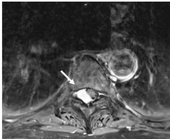
Fig. 3. Axial T1-weighted fat-suppressed gadolinium-containing contrast- enhanced magnetic resonance imaging scan showing an extradural lesion with some heterogeneity, but mostly a hyperintense signal at T8. The mass on the right side was pushing the cord to the left.
Fig. 1. Sagittal T1-weighted (T1W), T2-weighted (T2W), and T1 fat- suppressed and gadolinium-containing contrast-enhanced magnetic reso- nance imaging scan. (A) T1W image showing an isointense extradural le- sion at the T8 level. (B) T2 fat-suppressed image showing a hypointense lesion. (C) T1 gadolinium-containing contrast-enhanced image showing an extradural lesion with some heterogeneity, but mostly a hyperintense signal.
A B C
Under microscopic view, the blunt release of the tumor mass from the dura sac was done. The histologic specimen was sent for pathologic confirmation. As expected, along the root which was totally atrophic and agglutinated, the mass was running on extraspinal space, and it was extensively overgrowth between tumor and root. Gradually the tumor mass could be completely removed from the intraspinal chamber without dura injury.
We made the median opening of the dura to explore of intradural mass. We carefully inspected the intradural area under microscopic observation. The spinal cord seems free and no relevant mass could be found. The exiting root appears massively edematous and red colored. To proximal, no tumor mass could be seen. The latter is resected respectively also coagulated. We could remove the whole tumor. We repaired the dura with silk 4.0. It was covered with the stitching application of fibrin glue. The wound was closed layer by layer. The intraoperative SSEP and MEP’s were constantly unsuspicious. Postoperatively, the patient could move both legs and feet with good strength. Histological diagnosis was meningioma (meningotheliomatous type, grade 1, according to the World Health Organization classification). The nocturnal chest pain was disappeared after surgery, and the patient was recovered without any complication (IRB KYUH 2019-01- 007).
Discussion
The most frequent location of spinal meningiomas is the intradural space, and purely extradural spinal meningiomas are extremely rare, and information about these tumors is limited. In our case, we were not aware that meningiomas could occur in the extradural space exclusively. On MRI, the extramedullary mass was located posterior to 8 vertebral bodies with infiltration and displacement into the neuroforamen between the 8 and 9 vertebral bodies (Fig.
1, 2). However, the dura tail sign on figure 3 represents the possibility of intradural mass which could be related to common intradural mass like meningioma. In this case, clinical signs and image signs were very extra-ordinary and made the surgeon explore intra and extra dura space. The initial radiological differential diagnosis was a meningioma. The unique feature of our case is that the mass seemed to have an intradural extension or to originate
from the intradural extramedullary space on MRI. Spinal meningiomas frequently occur as ventral/ventro-lateral intradural extramedullary tumors, growing from arachnoid cap cells around the nerve root.6) On MRI, the location of the mass was not a frequent site, and it was not helpful in avoiding intradural invasive exploration. In the operating field, through the median opening of the dura, with careful inspection of the intradural area under microscopy, an extradural mass was found compressing the dural sac and exiting root. As the mass showed intradural extramedullary features on MRI, although it looked extradurally located in the operating field, we decided to explore the intradural space. To remove the intradural meningioma, intradural exploration was inevitable. Dural injury during exploration has a risk of complications including meningitis, neural damage, and embolism.2,4,7) As foraminal widening is a sign of an extradural mass effect (Fig. 2), the widening of the nerve root foramen, in this case, can be a clue that the tumor was an extradural mass. However, foraminal widening is not a typical sign of meningiomas, but rather of fibromas or schwannomas.
Because they are slow growing, meningiomas induce a slow, indolent course of symptoms. The symptoms largely depend on the tumor location and duration. The most common symptoms are motor deficits and spinal pain.3,7) In the first stage, spinal pain occurs because of nerve root irritation. In the second stage, the motor deficit develops owing to spinal cord compression. In the first stage, the most notable symptom of nerve root involvement is a pain, which predominates and may precede all other symptoms by months or years. As the tumor gradually enlarges, the spinal cord and nerve roots are first displaced and then compressed within the bony confines of the spinal canal.
This is compounded by microtrauma to neural tissues resulting from repetitive motion or stretching of the cord relative to the tumor. The pain may assume a radicular, radiating character or may present as localized back pain.
The known pain character caused by spinal neoplasia is typically persistent and progressive and is not alleviated by rest. Often the pain is worse at night, waking the patient from sleep.9) The other common symptoms of this stage are partial weakness and sensory loss (hypoesthesia, paresthesia). As the tumor begins to compress the spinal
A Long Unrecognized Symptom of a Rare Extradural Spinal Meningioma Journal of Korean Society of Spine Surgery
www.krspine.org 103 cord, motor deficits including gait disturbance occur. A
substantially larger subarachnoid space in the upper cervical spine and the lumbosacral spine permits larger tumor growth before the onset of symptoms; however, once the spinal cord and roots become compressed, the progression of symptoms is unrelated to the tumor location along the spine. In our case, the first symptom was nonspecific nocturnal chest pain and the patient had to undergo a full cardiac evaluation and then visited the gastrointestinal department to determine which internal organ was involved. No pathology of the heart and internal organs were found. For cardiac evaluation, the patient underwent CT, which led to the incidental diagnosis of a mass in the spinal canal. Owing to advances in diagnostic techniques, the average time to the diagnosis of a spinal mass was shortened by about 6 months; consequently, patients experience less severe neurologic deficits before surgery.2,7,8) These advancements have made diagnosis simpler and more accurate; however, they also tend to omit the classical consideration of the possibility of rare diseases.
Conclusions
Awareness of the rarity and nonspecific symptoms of extradural spinal meningiomas will be beneficial for their accurate diagnosis and understanding the image findings of extradural meningioma can provide suitable treatment options.
REFERENCES
1. Calogero JA, Moossy J. Extradural spinal meningiomas:
report of four cases. J Neurosurg. 1972 Oct;37(4):442-7.
DOI: 10.3171/jns.1972.37.4.0442.
2. Gottfried ON, Gluf W, Quinones-Hinojosa A, et al. Spi- nal meningiomas: surgical management and outcome.
Neurosurg Focus. 2003 Jun 15;14(6):e2. DOI: 10.3171/
foc.2003.14.6.2.
3. Guidetti B. Removal of extramedullary benign spinal cord tumors. Advances and Technical Standards in Neurosur- gery, 1974, pp 173-197. DOI: 10.1007/978-3-7091- 7099-1_6.
4. Levy WJ Jr, Bay J, Dohn D. Spinal cord meningioma.
J Neurosurg. 1982 Dec;57(6):804-12. DOI: 10.3171/
jns.1982.57.6.0804.
5. Powell M. Sir Victor Horsley—an inspiration. BMJ.
2006 Dec 23;333(7582):1317-9. DOI: 10.1136/
bmj.39056.527407.55.
6. Santiago BM, Rodeia P, Cunha E Sa M. Extradural thoracic spinal meningioma. Neurol India. 2009 Jan-Feb;57(1):98.
DOI: 10.4103/0028-3886.48802.
7. Klekamp J, Samii M. Surgical results for spinal meningio- mas. Surg Neurol. 1999 Dec;52(6):552-62. DOI: 10.1016/
S0090-3019(99)00153-6.
8. King AT, Sharr MM, Gullan RW, Bartlett JR. Spinal meningiomas: a 20-year review. Br J Neurosurg. 1998 Dec;12(6):521-6. DOI: 10.1080/02688699844367.
9. Deyo, Richard A. Diehl, Andrew K. Cancer as a cause of back pain. Journal of General Internal Medicine. 1988.
3(3): 230-238. DOI: 10.1007/bf02596337.
10. Herkowitz HN, Garfin SR, Eismont FJ, et al. Rothman- Simeone The Spine E-Book: Expert Consulted: Elsevier Health Sciences; 2011.
야간 흉통을 호소하는 드문 흉추 경막외 수막종 - 증례 보고-
김상범* • 원유건† • 장민구† • 민영기† • Andreas Gutzeit‡ • Fabio Casari∮ • Oliver Nic Hausmann∥
*충남대학교병원 정형외과학교실, †건양대학교병원 정형외과학교실
‡Department of Radiology, Klinik St. Anna, Luzern, Switzerland and Department of Radiology, Salzburg University Hospital, Austria
∮University of Salzburg, Austria
∥Department of Neurosurgery, Klinik St. Anna, Luzern, Switzerland
연구 계획: 증례 보고
목적: 저자들은 장기간 지속되는 비특이적인 흉추부의 야간통을 호소한 환자에게 발견된 경막외 척추 수막종 증례를 보고하고자 한다.
선행 연구문헌의 요약: 척추 수막종은 통증, 감각/운동의 저하, 항문 수축 기능 저하 등과 관련된 경막내 척수외 종양이다. 경막내 척수외 수막종은 흉추 부에서 흔히 발생한다.
대상 및 방법: 65세 여환은 2년 전부터 지속되는 흉통으로 심장내과와 소화기내과를 진료를 보았다. 시행한 검사에서 심장이나 다른 장기에서는 흉통을 설명할 소견은 보이지 않았다. 척추 종양이 진단되기 수 개월 전에 시행한 전산화 단층 촬영에서 흉추부의 종양이 확인되었고, 자기공명영상에서 흉추 제 7-8번에서 경막외 골수외 종양이 확인되었다.
결과: 본 환자에 대해서 수술적 치료를 시행하였으며 조직학적 진단상 척추 수막종이 확인되었다. 수술 후 환자의 야간통은 사라졌으며 보행상의 문제도 나타나지 않았고 특이 합병증은 없었다.
결론: 경막외 척추 수막종의 비특이적인 증상과 희귀성을 인지하는 것은 정확한 진단과 적절한 치료를 하는데 도움이 될 것이다.
색인 단어: 경막외 공간, 흉추부 통증, 의인성 경막 손상, 척추 수막종, 척추 종양 약칭 제목: 비특이적인 증상을 동반한 경막외 척추 수막종
접수일: 2018년 11월 16일 수정일: 2019년 2월 1일 게재확정일: 2019년 7월 15일 교신저자: 원유건
대전광역시 서구 가수원동 685 건양대학교병원 정형외과학교실
TEL: 042-600-9862 FAX: 042-545-2373 E-mail: yougunwon@gmail.com, yougunwon@kyuh.ac.kr