대한족부족관절학회지: 제8권 제2호 2004
J Korean Foot Ankle Soc. Vol 8. No. 2. pp.157-160, 2004
- 157 -
∙Address for correspondence Hyon Jeong Kim, M.D., Ph.D.
Department of Orthopaedic Surgery, Ajou University School of Medicine San 5 Wonchon-dong Youngtong-gu Suwon 443-721, Korea
Tel:+82-31-219-5221 Fax:+82-31-219-5229 Email:osgirl@korea.com
족관절 해면골의 미세 구조 분석
아주대학교 의과대학 정형외과학교실 김현정․안태선․백명현․최문권․원예연
Micro-Structural Profiles of Trabecular Bone at the Ankle Joint
Hyon Jeong Kim, M.D., Tae Sun An, M.D., Myong Hyun Baek, B.S., Mun Kwon Choi, M.D., Ye Yeon Won, M.D.
Department of Orthopaedic Surgery, Ajou University School of Medicine
=Abstract=
Purpose: This study aimed to investigate and compare the micro-structural profiles of trabecular bone from different facets at the ankle joint.
Materials and Methods: In a fresh cadaver ankle, four cored 10 mm of diameter cylindrical specimens of trabecular bone were harvested from the distal tibia, the talar dome, the medial malleolus, and the lateral malleolus. Using a micro-computed tomography, two-dimensional and three-dimensional micro-structural indices of the trabecular bone were analyzed.
Results: Each specimen from the tibia, talus, medial malleolus, and the lateral malleolus showed unique micro-structural pattern. Tibia versus talus, the talus was seen a higher bone volume fraction and a wider supporting zone subchondrally whereas the tibia was seen a relatively lower bone volume fraction and a much narrower supporting zone subchondrally. Lateral malleolus versus medial malleolus, the lateral malleolus was seen the thicker but sparse trabeculae pattern whereas the medial malleolus was seen the thinner but more compact trabecular pattern.
Conclusion: Each four locations from the different facet at the ankle joint have distinct own micro-structural patterns of the trabecular bone, suggesting different mechanical properties.
Key Words: Ankle, Trabecular bone, Micro-computed tomography
서 론
최근 족관절 인공관절 성형술의 화두는 디자인 측면에선 보다 해부학적 모양에 가까운 기구를 만드는 것이고 기술적 측면에서는 관절 주변의 뼈를 최소한으로 깎아 최대한 보존
하는 것이다. 이러한 노력을 기울이는 바탕에는 족관절을 이 루는 주요 지지 골격인 경골 원위부와 거골에 대한 생역학적 연구 자료들이 이론적 기반이 되고 있다. 전반적으로 경골의 강도는 거골보다 약 40% 정도 약하고 후외방이 비교적 강하 다고 알려져 있다5). 역학 실험에 의하면 발목관절의 경골과 거골에서 2 mm 간격으로 관절면에 평행 절단하면 경골은 절 단이 진행할 때마다 크게 강도가 떨어지며 4 mm 이상 제거하 면 급속히 강도가 약화된다고 한다. 거골에서는 처음 2 mm를 절단에서는 강도가 증가하지만 그 다음 더 깊이 절단을 진행 하면 강도가 약화된다고 보고되었다5). 즉, 경골은 연골하골 의 지지를 잃으면 압박 저항이 30~50%로 감소하고 3 cm 이
김현정․안태선․백명현․최문권․원예연
- 158 -
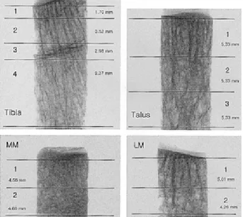
Figure 1. Scout X-ray transmission images of each specimen were seen.
Figure 2. Two-dimensional micro-computed tomography images were seen from each zone of the tibia specimen.
상 근위부에 이르면 압박 저항이 '0'이 된다1).
이러한 역학 실험은 관절을 이루는 구조들의 강도와 저항 등 기계적 성격에 대해 많은 정보를 주었으나 관절 주변을 지 지하고 있는 해면골의 micrometer 단위의 섬세한 미세구조 양상을 조사하는 데에는 부족하였다. 또한, 경골과 거골이 족관절의 체중부하에 주요한 기능을 하고 있다는 것은 잘 알 려져 있으나 양쪽 옆을 지지하고 있는 경골 내과와 비골 외과 에 대해서는 자료가 적다. 본 연구에서는 미세 단층 촬영기 (micro-computed tomography)를 사용하여 족관절을 이루 는 여러 관절면인 경골면, 거골면, 내과면, 외과면 각각에서 관절면을 지지하고 있는 해면골의 미세 구조의 형태학적 특 징을 조사하고 그 차이를 비교함으로써, 미세구조와 역학적 기능과의 상관 관계를 연역해 보고자 하였다.
대상 및 방법
1. 시편의 채취
급성 비브리오 패혈증과 괴사성 근막염으로 슬하 절단술 시행 받은 54세 남자환자의 신선 절단지의 족관절에서 원통 형 톱날을 이용하여 원위 경골, 거골, 비골 외과와 경골 내과 네 군데에서 지름 10 mm의 원통형 시편을 채취하였다.
2. 시편의 미세 단층 촬영
획득된 각 부위의 해면골 시편은 미세 단층 촬영기(model SkyScan-1072, SKYSCAN, Belgium)를 이용하여 시편 전 체를 단순 방사선 촬영한 후 200 um 간격으로 촬영하여 이 차원 미세 단면 영상을 각각의 시편에 대해 870장에서 1650 장씩 얻었다. 삼차원 영상은 이차원 영상을 바탕으로 해면골 의 미세구조를 재구성하여 얻었다.
3. 미세 구조 특성 지수
(micro-structural property indexes)의 분석
시편의 분석에는 CT-Analyser™ (SKYSCAN, Belgium) 소프트웨어를 이용하였고, 형태학적 지수(histomorpho- metric index) 분석을 위해서는 ANT™ (SKYSCAN, Belgium) 소프트웨어를 사용하였다. 이차원 영상으로부터 골소주 두께(Tb.Th: trabecular thickness), 골소주 거리 (Tb.Sp: trabecular spacing), 골소주 개수(Tb.N: trabe- cular number)의 지수를 구했고, 삼차원 영상으로부터 골 체적(BV: bone volume), 골 체적비(BV/TV: bone volume
fraction), 이방성 정도(DOA: degree of anisotrophy), 구 조 모델 지수 (SMI: structure model index)의 지수를 구했 다.
결 과
1. 골 체적비
경골, 거골, 내과, 외과에서 나온 각각의 시편은 독특한 미세구조 특징을 보였다. 이차원 미세영상에서 보이는 해면 골 유형의 변화에 따라 원위 경골은 네 개의 영역으로, 거골 은 세 개의 영역으로, 외과와 내과는 각각 두 개의 영역으로 구분하였다(Fig. 1~5, Table 1).
모든 시편에서 관절 연골 바로 아래 연골 하골에 해당하는 부위 즉 제1 영역이 가장 골량이 많고 골 체적비가 높았다.
비교적 균질한 연골 하골의 두께는 시편마다 달라서 경골은 약 1.7 mm, 거골은 약 5.3 mm, 내과는 약 4.5 mm, 외과는
족관절 해면골의 미세 구조 분석
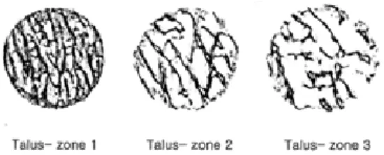
- 159 - Figure 3. Two-dimensional micro-computed tomography images were seen from each zone of the talar specimen.
*Tb.Th †Tb.Sp ‡Tb.N §BV (mm3) ∥BV/TV (%) ¶DOA **SMI
Talus Total 0.228 0.671 1.11235 139.514 30.186 0.241 0.874
Zone 1 0.247 0.424 1.490313 65.211 41.883 0.362 0.867
Zone 2 0.225 0.763 1.012146 42.743 27.453 0.285 0.839
Zone 3 0.2 0.832 0.968992 31.9 20.188 0.392 1.214
Tibia Total 0.166 0.657 1.21507 111.056 21.847 0.261 1.441
Zone 1 0.222 0.294 1.937984 23.739 47.247 0.346 0.926
Zone 2 0.203 0.621 1.213592 26.302 25.543 0.216 1.647
Zone 3 0.174 0.553 1.375516 27.792 31.775 0.296 1.55
Zone 4 0.119 0.738 1.166861 34.602 12.649 0.414 1.496
††LM Total 0.249 0.668 1.09051 73.826 27.297 0.209 0.468
Zone 1 0.264 0.595 1.164144 50.265 34.336 0.205 0.202
Zone 2 0.225 1.101 0.754148 22.082 18.638 0.044 0.987
‡‡MM Total 0.18 0.464 1.5528 82.518 30.753 0.398 0.699
Zone 1 0.193 0.464 1.52207 43.049 32.129 0.217 0.561
Zone 2 0.158 0.47 1.592357 39.622 28.902 0.461 0.923
*; Tb.Th, trabecular thickness, †; Tb.Sp, Trabecular spacing, ‡; Tb.N, trabecular number, §; BV, bone volume, ∥; BV/TV, bone volume/total volume (bone volume fraction), ¶; DOA, degree of anisotropy, **; SMI, structural model index, ††; LM, lateral malleolus, ‡‡; MM, medial malleolus.
Table 1. Micro-Structural Indexes of the Ankle Joint
Figure 5. Two-dimensional micro-computed tomography images were seen from each zone of the lateral malleolus specimen.
Figure 4. Two-dimensional micro-computed tomography images were seen from each zone of the medial malleolus specimen.
약 5.0 mm 를 보였다. 각 시편에서 연골 하골에 해당하는 제1영역만을 비교했을 때는 좁지만 경골의 연골 하골이 47.247%로 거골의 41.883%, 외과의 34.336%, 내과의 32.129%에 비하여 가장 높았다. 특징적으로 원위 경골에는
성장판이 있었던 곳에서 좀 더 두꺼워진 골소주들이 모여있 는 영역이 잠깐 나타나는데(경골 제3 영역) 이 영역에서 부분 적으로 골 체적비가 다소 상승하였다.
관절면을 마주하고 있는 경골과 거골을 비교할 때 경골은 거골에 비해 약 1/3 정도 골 체적비가 낮았다(21.85%:
30.186). 내과와 외과를 비교할 때 내과는 골소주가 가늘지 만(0.18) 보다 촘촘했고(0.464) 외과는 골소주가 다소 두껍 지만(0.249) 간격이 넓었다(0.668).
2. 이방성 정도와 구조 모델 지수(Table 1)
이방성 정도는 0~1의 숫자로 표현되며 0에 가까울수록 등 방성(isotropy), 1에 가까울수록 이방성(anisotropy)으로 정 의된다2). 경골과 거골은 정도와 분포에 있어서 서로 비슷한
김현정․안태선․백명현․최문권․원예연
- 160 - 양상의 이방성을 보였다. 외과와 내과를 비교하면 제2 영역 에서 외과는 0.044로 등방성 구조에 가까운 반면 내과는 0.461로 보다 이방체에 가까운 차이를 보였다.
구조 모델 지수는 해면골 구조의 특성을 나타내는 지수로 서 0~3의 숫자로 표현되며 0에 가까울수록 판 구조 (plate-like structure), 3에 가까울수록 막대 구조 (rod- like structure)로 정의된다2). 전체적으로 볼 때 거골은 0.874로 경골의 1.441에 비해 보다 판 구조에 가깝고 외과는 0.468로 내과의 0.699에 비해 보다 판 구조에 가까운 결과를 보였다.
고 찰
본 연구에서 얻은 미세 단층 촬영을 이용한 족관절의 경골 과 거골의 미세 구조 분석 결과는 이전의 다른 여러 역학 실 험의 결과들1,3,5)과 잘 맞는다. 경골은 거골에 비해 약 1/3 작 은 골 체적비를 보였고 연골 하골의 특성을 보이는 제1 영역 의 두께가 1.7 mm로 몹시 좁았다. Hvid 등5)의 2 mm 연속 평행 절단 실험에서 경골이 거골과는 대조적으로 첫 번 절단 에서부터 급속히 강도 저하를 보인 결과를 설명할 수 있다.
태생적으로 거골은 표면에 피질골의 비중이 아주 적고 많은 부분 관절면으로 둘러싸여 있어서 그 아래 해면골이 하중의 대부분을 비교적 균질하게 감당해내야 할 것으로 추정된다.
하지만 경골은 대표적 장관골로써 가해지는 하중의 많은 부 분이 변연부의 피질골로 편중되어 상대적으로 관절 중앙부는 약해질 것으로 설명해 볼 수 있다3,4,7).
비골 외과와 경골 내과의 경우 다른 역학실험 자료는 찾아 볼 수 없었다. 본 연구의 미세구조 분석 결과에 의하면 두 구 조는 전체적 골 체적비는 외과 27.297, 내과 30.753으로 비 슷하였으나, 대조적으로 외과는 골소주의 두께가 두꺼운 대 신 간격이 넓었으며 내과는 골소주의 두께가 가는 대신 간격 이 촘촘하였다. 또한 연골 바로 아래 연골 하골에 해당하는 제1 영역의 구조모델지수를 비교할 때 외과는 0.202로 상당 히 판구조에 가깝고 내과는 0.561로 막대구조 쪽으로 향해있 다. 비슷한 골 체적비의 두 구조라도 이렇듯 미세구조 특성 지수가 상당히 달라서 기계적 특성도 다를 것이며 골소주가 두껍고 판구조에 가까운 외과가 역학적으로는 내과보다 강할 것으로 예상된다. 이러한 결과는 족관절의 중립 위에서 내과 와 접하는 관절면의 방향은 지면에 대해 거의 수직이지만 외
과와 접하는 관절면은 비스듬한 각을 형성하고 있기 때문에 외과가 하지에 가해지는 하중의 약 1/6을 감당한다는 보고를 뒷받침할 수 있다6).
본 연구에 사용한 미세 단층 촬영은 일반 단층 촬영이나 자기 공명 촬영보다 훨씬 높은 해상도를 제공하여 해면골을 이루고 있는 골소주의 세세한 미세 구조 특성을 분석하는 데 에 아주 우수한 방법이다. 한걸음 더 나아가 이렇게 획득한 미세 구조의 삼차원 영상에 유한요소 모델을 적용하여 유한 요소해석을 실행함으로써 해면골의 기계적 특성을 연구하고 기계를 이용한 역학 실험을 시연해 볼 수 있다.
결 론
미세 단층 촬영을 통한 족관절 해면골의 미세구조 분석에 서 경골과 거골과 비골 외과와 경골 내과는 각각 독특한 미세 구조 특징을 보였다.
REFERENCES
1) Aitken GK, Bourne RB, Finlay JB, Rorabeck CH and Andreae PR: Indentation stiffness of the cancellous bone in the distal human tibia. Clin Orthop, 201: 264-270, 1985.
2) Day JS, Ding M, Odgaard A, Sumner DR, Hvid I and Weinans H: Parallel plate model for trabecular bone exhibits volume fraction-dependent bias. Bone, 27: 715- 720, 2000.
3) Goldstein SA, Wilson DL, Sonstegard DA and Matthews LS: The mechanical properties of human tibial trabecular bone as a function of metaphyseal location. J Biomech, 16:
965-969, 1983.
4) Hintermann B and Valderrabano V: Total ankle replace- ment. Foot Ankle Clin, 8: 375-405, 2003.
5) Hvid I, Rasmussen O, Jensen NC and Nielsen S:
Trabecular bone strength profiles at the ankle joint. Clin Orthop, 199: 306-312, 1985.
6) Lambert KL: The weight-bearing function of the fibula. A strain gauge study. J Bone Joint Surg, 53A: 507-13, 1971.
7) Trouillier H, Hansel L, Schaff P, Rosemeyer B and Refior HJ: Long-term results after ankle arthrodesis:
clinical, radiological, gait analytical aspects. Foot Ankle Int, 23: 1081-1090, 2002.