• 검색 결과가 없습니다.

Intravenous Single Dose Toxicity of Sweet Bee Venom in Sprague-Dawley Rats

N/A
N/A
Protected

Academic year: 2021

Share "Intravenous Single Dose Toxicity of Sweet Bee Venom in Sprague-Dawley Rats"

Copied!
8
0
0

로드 중.... (전체 텍스트 보기)

전체 글

(1)

Intravenous Single Dose Toxicity of Sweet Bee Venom in Sprague-Dawley Rats

Kwang-Ho Lee 1 , JunSang Yu 2 , Seungho Sun 3 , KiRok Kwon 4 *

Abstract

Objectives: Anaphylactic shock can be fatal to people who become hypersensitive when bee venom pharma- copuncture (BVP) is used. Thus, sweet bee venom (SBV) was developed to reduce these allergic responses. SBV is almost pure melittin, and SBV has been reported to have fewer allergic responses than BVP. BVP has been admin- istered only into acupoints or intramuscularly, but we thought that intravenous injection might be possible if SBV were shown to be a safe medium. The aim of this study is to evaluate the intravenous injection toxicity of SBV through a single-dose test in Sprague-Dawley (SD) rats.

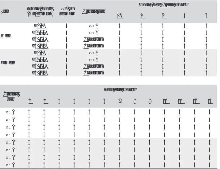
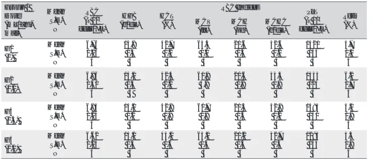
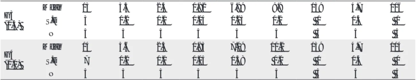
Methods: Male and female 6-week-old SD rats were in- jected intravenously with SBV (high dosage: 1.0 mL/an- imal; medium dosage: 0.5 mL/animal; low dosage: 0.1 mL/animal). Normal saline was injected into the con- trol group in a similar method. We conducted clinical observations, body weight measurements, and hema- tology, biochemistry, and histological observations.

Results: No death was observed in any of the experi- mental groups. Hyperemia was observed in the high and the medium dosage groups on the injection day, but from next day, no general symptoms were observed in

any of the experimental groups. No significant changes due to intravenous SBV injection were observed in the weights, in the hematology, biochemistry, and histo- logical observations, and in the local tolerance tests.

Conclusion: The results of this study confirm that the lethal dose of SBV is over 1.0 mL/animal in SD rats and that the intravenous injection of SBV is safe in SD rats.

1. Introduction

Bee venom pharmacopuncture (BVP) is a new type of treatment combining the efficacy of acupuncture and the pharmacological actions of the venom that is artificially extracted and refined from live honey bees (Apismellifera) [1]. BVP has been used to treat degen- erative and rheumatoid arthritis [2-5] and spine disor- ders [6-9]. However, several types of allergic respons- es can occur during the treatment period; especially, anaphylactic shock, which is fatal to people who are hypersensitive to bee venom, is an obstacle faced by Korean doctors who use BVP. Thus, sweet bee ven- om (SBV) was developed to reduce these allergic re- sponses. Enzymes, known as allergens, are eliminated through a protein separation technique, so only melit- tin is left in SBV. Melittin is the dominant component of bee venom; melittin constitutes 40% ─ 50% of bee venom’s dried weight and has strong anti-inflammato- ry and analgesic actions [10, 11]. SBV has been report- ed to have fewer allergic responses than BVP; for that reason, SBV treatment is considered to be as effective,

Original article

Key Words

bee venom, intravenous injection, melittin, toxicity test

This is an Open-Access article distributed under the terms of the Creative Commons Attribution Non-Commercial License (http://creativecommons.org/licenses/by-nc/3.0/) which permits unrestricted noncommercial use, distribution, and reproduction in any medium, provided the original work is properly cited.

This paper meets the requirements of KS X ISO 9706, ISO 9706-1994 and ANSI/NISO Z39.48-1992 (Permanence of Paper).

*

Corresponding Author

Ki-Rok Kwon. Korean Pharmacopuncture Institute, 4F, Association of Korean Oriental Medicine B/D, 26-27, Gayang-dong, Gangseo-gu, Seoul 157-200, Korea.

Tel: +82-33-744-9304 Fax: +82-33-744-9305 E-mail: [email protected]

ⓒ 2015 Korean Pharmacopuncture Institute http://www.journal.ac

1

Department of Acupuncture &Moxibustion Medicine, College of Korean Medicine, Sangji University, Wonju, Korea

2

Department of Sasang Constitutional Medicine, College of Korean Medicine, Sangji University, Wonju, Korea

3

Department of Internal Medicine, College of Korean Medicine, Sangji University, Wonju, Korea

4

Research Center of the Korean Pharmacopuncture Institute, Seoul, Korea

Received: Apr 9, 2015 Reviewed: Jun 5, 2015 Accepted: Jun 15, 2015

(2)

or better, than BVP treatment [12, 13]. Also studies using several toxicity tests have shown SBV to be safe [14-19].

Until now BVP has been administered intramuscularly, intracutaneously or into acupoints. However, in our opin- ion, intravenous injection should be possible if SBV is a safe medium. Thus, we conducted a single-dose toxicity test of SBV administered intravenously in Sprague-Dawley (SD) rats, and we report the results here.

2. Material and Methods

Twenty-four SD rats of each gender were obtained from a specific pathogen-free facility (ORIENTBIO Inc., Gyeo- ng-gi, Korea) at 5-weeks of age and were used after a week of quarantine and acclimatization. The animals were housed in a room maintained at 20.0 ─ 23.0℃ under a relative humidity of 42.8% ─ 68.9%. The room was illumi- nated with artificial lighting from 07:00 to 19:00 hours and 10 ─ 15 air changes per hour. Three animals were housed in suspended stainless-steel wire-mesh cages and were allowed sterilized tap water and commercial rodent chow (Teklad Certified Irradiated Global 18% Protein Rodent Diet 2918C, Harlan Laboratories, Inc., U.S.A.). The proto- col of this study was approved by the institutional Animal Care Committee of Biotoxtech, Co. (Oh Chang, Korea).

SBV was prepared using a freeze dryer (FD 8508, Ilshin Lab Co. Ltd., Korea) by Ginseng & Venom (G & V) Co.

(Wonju, Korea) and was stored at ─ 20℃. A high-perfor- mance liquid chromatography (HPLC, Agilent 1100 series, Agilent Technologies Ltd., U.S.A.) analysis was performed to confirm that SBV was pure melittin.

Twenty healthy male and 20 healthy female rats were se- lected by average weights, and 5 rats of each gender were assigned to 1 of 4 groups: control (normal saline), low dos- age (0.1 mL/animal), medium dosage (0.5 mL/animal), and high dosage (1.0 mL/animal). In a pilot study, no mor- talities had been observed at a dosage of 1.0 mL/animal of SBV in male and female SD rats; based on that results, we set the high dose at 1.0 mL/animal. SBV was administered to the rats by intravenous injection into the caudal vein.

The control group was administered 1.0 mL/animal of normal saline (Lot No. 12115, Choongwae Pharma Corp., Korea).

All animals were observed for clinical signs at 30 minutes, 1 hour, 2 hours, 4 hours and 6 hours after the injection of SBV. Clinical signs were observed daily from the injection day to 14 day after the first injection. The body weight of each rat was measured at the initiation of treatment and at 3 days, 7 days and 14 days after the injection.

The animals were fasted for 18 hours prior to necropsy and blood collection. Blood samples were drawn from the abdominal aorta by using a syringe needle under ether anesthesia. Blood samples were collected into tubes con- taining ethylenediaminetetraacetic acid (EDTA) and were analyzed by using a blood counting analyzer (ADVIA 120, SIEMENS, Germany) to determine the red blood cell count (RBC), hemoglobin concentration (HGB), hematocrits (HCT), mean corpuscular cell volume (MCV), mean cor- puscular hemoglobin (MCH), mean corpuscular cell he- moglobin concentration (MCHC), platelet count (PLT),

white blood cell count (WBC), WBC differential count, re- ticulocyte (Reti) count, prothrombin time (PT) and active partial thromboplastin time (APTT).

For the serum biochemistry analysis, blood samples were centrifuged at 3,000 rpm for 10 minutes and analyz- ed using an auto-analyzer (7180, HITACHI, Japan). Serum biochemistry parameters, including alanine aminotrans- ferase (ALT), aspartate aminotransferase (AST), alkaline phosphatase (ALP), gamma glutamyl transpeptidase (GGT), blood urea nitrogen (BUN), creatinine, total bili- rubin, total protein, albumin, albumin/globulin ratio (A/G ratio), total cholesterol, triglycerides (TG), phosphorus, glucose, calcium, chloride, sodium and potassium, were examined.

Tissue was obtained from the injection site on all animals and was then fixed with 10% neutral buffered formalin solution. The tissue was routinely processed, embedded in paraffin, and sectioned. The sections were stained with hematoxylin & eosin (H&E) stain, and bone tissue was de- calcified with Calci-Clear-RapidTM (National diagnostics, U.S.A.) for microscopic examination.

Data on the weights and from the hematology and serum biochemistry results were tested by using a statistical anal- ysis system (SAS, version 9.3, SAS Institute Inc., U.S.A.). The variance was checked by using the Bartlett test (P < 0.05).

If the variance was homogeneous, the data were subjected to a one-way analysis of variance (ANOVA, P < 0.05). If a significant difference was observed between the groups, the data were analyzed by using the multiple comparison procedure of the Dunnett’s test (P < 0.05, P < 0.01). If the variance was not homogeneous, the data were analyzed by using the Kruskal-Wallis test (P < 0.05).

3. Results

No mortality occurred in either the control group or any of the experimental groups. No clinical signs were ob- served in male and female SD rats during the observation period. Caudal congestion was observed in male and fe- male rats in the experimental groups from 30 minutes to 6 hours after injection, but it was not observed the next day or after. This symptom seemed to have occurred at the administration of SBV (Table 1). Compared to the control group, the weights of the experimental groups showed no significant changes. No significant change in hematology was observed in any of the groups (Table 2), and no mean- ingful change in serum biochemistry was observed in any of the groups (Table 3). Significant changes were minimal and were found not to be dose dependent.

During necropsy, no abnormal macroscopic appear- ances were observed in either the control experimental groups. Perivascular infiltration of inflammatory cells into the lateral vein at the injection site was observed in the male control group and in the female high dosage group, but those changes were minimal and seemed to be due to the injection (Table 4).

4. Discussion

(3)

Sex Group / Dose (mL/animal)

No. of

animals Clinical sign

Hours (Day 0) after dosing

0.5 1 2 4 6

Male

G1(0) 5 NOA 5 5 5 5 5

G2 (0.1) 5 NOA 5 5 5 5 5

G3 (0.5) 5 Congestion 5 5 5 5 5

G4 (1.0) 5 Congestion 5 5 5 5 5

Female

G1(0) 5 NOA 5 5 5 5 5

G2 (0.1) 5 NOA 5 5 5 5 5

G3 (0.5) 5 Congestion 5 5 5 5 5

G4 (1.0) 5 Congestion 5 5 5 5 5

Table 1 Summary of clinical signs

Clinical sign

Days after dosing

1 2 3 4 5 6 7 8 9 10 11 12 13

NOA 5 5 5 5 5 5 5 5 5 5 5 5 5

NOA 5 5 5 5 5 5 5 5 5 5 5 5 5

NOA 5 5 5 5 5 5 5 5 5 5 5 5 5

NOA 5 5 5 5 5 5 5 5 5 5 5 5 5

NOA 5 5 5 5 5 5 5 5 5 5 5 5 5

NOA 5 5 5 5 5 5 5 5 5 5 5 5 5

NOA 5 5 5 5 5 5 5 5 5 5 5 5 5

NOA 5 5 5 5 5 5 5 5 5 5 5 5 5

NOA, no observable abnormality.

The concept of blood vessels has been studied in Ori- ental medicine [20, 21], and the intravenous injection of mountain ginseng pharmacopuncture is currently being used by Korean medical doctors. BVP possesses strong analgesic and anti-inflammatory actions by stimulation of the hypothalamic-pituitary-adrenal axis to increase the secretion of adrenocortical hormone. BVP shows superior efficacy in the treatment of most pain related conditions and musculoskeletal diseases. However, bee venom con- tains enzymes with molecular weights greater than 10,000 such as phospholipase A2 (PLA2) and hyaluronidase.

These proteins act as antigens, causing allergic reactions, which presently limits its clinical use. Administration of BVP without a full understanding of its allergic responses, especially anaphylactic shock, puts practitioners and pa- tients at great risk [1]. SBV was developed to replace dan- gerous bee venom with a safe material and to guarantee its effects [22, 23] because SBV is pure melittin obtained through a protein separation technique using gel filtration [12, 24-26].

In previous studies, the LD 50 of SBV in SD rats was over 30

mg/kg [14], and the maximum dose of SBV in beagle dogs was found to be over 9 mg/kg [15] through single-dose toxicity tests. Also, no significant changes were observed at doses below 0.14 mg/kg in SD rats, and no direct side ef- fects of SBV were observed at a dose of 0.56 mg/kg in bea- gle dogs through a 4-week repeated-dose toxicity test, and the ‘No Observed Adverse Effect Level’ of SBV was found to be approximately 0.07 mg/kg in male and female SD rats based on a 13-week repeated-dose toxicity test with a 4-week recovery period [16-19].

Whether intravenous injection of SBV is possible is con-

troversial. However, in our opinion, the use of SBV should

be safe, so we conducted an intravenous single-dose tox-

icity test of SBV in SD rats. Our objective was to investigate

the influences of intravenous injection of SBV in SD rats. In

the present study, no significant changes were observed in

the weights or in the results of the hematology, biochem-

istry, necropsy and histopathological examinations in SD

rats. These results show that intravenous injection of SBV

in SD rats is safe. Because no mortalities were observed in

this study, the lethal dose of MGP is judged to be above

1.0 mL/animal in SD rats. This study is the first attempt to

(4)

Table 2 Mean hematology parameters (Sex: Male)

(Sex: Female) Group / Dose (mL/ani- mal)

Mean S.D.

N

RBC (× 10 6 cells/μL)

HGB (g/dL)

HCT (%)

RBC Indices PLT

(× 10 3 cells/μL)

Reti MCV (%)

(fL)

MCH (pg)

MCHC (g/dL) G1 (0)

Mean S.D.

N

6.76 0.16 5

13.9 0.3 5

42.7 1.2 5

63.3 1.1 5

20.6 0.3 5

32.6 0.2 5

1420 155 5

5.7 1.1 5 G2 (0.1)

Mean S.D.

N

6.86 0.40 5

14.1 0.5 5

42.4 2.1 5

61.8 3.9 5

20.6 0.9 5

33.3 0.9 5

1335 124 5

6.2 1.7 5 G3 (0.5)

Mean S.D.

N

6.95 0.24 5

14.1 0.2 5

42.8 0.8 5

61.7 1.8 5

20.3 0.5 5

32.9 0.2 5

1383 130 5

5.0 0.9 5 G4 (1.0)

Mean S.D.

N

6.62 0.24 5

14.1 0.5 5

43.0 1.6 5

65.0 1.3 5

21.2 0.4 5

32.7 0.3 5

1295 155 5

5.5 0.9 5

Group / Dose (mL/ani- mal)

Mean S.D.

N

RBC (× 10 6 cells/μL)

HGB (g/dL)

HCT (%)

RBC Indices PLT

(× 10 3 cells/μL)

Reti MCV (%)

(fL)

MCH (pg)

MCHC (g/dL) G1 (0)

Mean S.D.

N

7.01 0.41 5

14.0 0.2 5

41.9 1.2 5

59.9 2.2 5

20.1 1.0 5

33.5 0.7 5

1263 99 5

4.3 3.6 5 G2 (0.1)

Mean S.D.

N

6.96 0.30 5

13.9 0.6 5

41.0 1.7 5

58.9 1.2 5

20.0 0.5 5

33.8 0.4 5

1305 77 5

3.1 0.7 5 Group /

Dose (mL/ani- mal)

Mean S.D.

N

WBC (× 10 6 cells/μL)

WBC Differential Counting (%)

PT (sec)

APTT (sec)

NEU LYM MONO EOS BASO

G1 (0)

Mean S.D.

N

8.01 1.19 5

15.5 5.2 5

80.6 5.5 5

2.1 0.4 5

0.5 0.2 5

0.2 0.1 5

17.3 0.8 5

13.7 0.7 5 G2 (0.1)

Mean S.D.

N

8.38 2.11 5

19.6 3.8 5

76.4 3.8 5

2.3 1.0 5

0.5 0.2 5

0.1 0.1 5

17.2 0.5 5

14.9 1.4 5 G3 (0.5)

Mean S.D.

N

6.77 1.16 5

20.1 5.5 5

75.7 5.4 5

2.5 0.3 5

0.5 0.1 5

0.1 0.1 5

16.7 1.0 5

14.0 1.8 5 G4 (1.0)

Mean S.D.

N

8.79 2.62 5

17.8 3.9 5

77.7 3.4 5

2.5 0.5 5

0.5 0.4 5

0.2 0.0 5

17.4 0.3 5

14.7 0.5 5

(Continued)

(5)

Group / Dose (mL/ani- mal)

Mean S.D.

N

WBC (× 10 6 cells/μL)

WBC Differential Counting (%)

PT (sec)

APTT (sec)

NEU LYM MONO EOS BASO

G1 (0)

Mean S.D.

N

4.09 1.31 5

20.0 6.2 5

77.1 6.6 5

1.2 0.6 5

1.0 0.2 5

0.1 0.0 5

18.6 0.7 5

13.1 1.8 5 G2 (0.1)

Mean S.D.

N

3.64 1.75 5

23.7 5.2 5

72.7 6.0 5

1.7 0.6 5

1.0 0.5 5

0.1 0.1 5

18.5 1.0 5

13.5 0.4 5 G3 (0.5)

Mean S.D.

N

3.21 0.87 5

20.0 3.8 5

76.8 4.1 5

1.8 0.5 5

0.8 0.3 5

0.1 0.1 5

18.9 0.5 5

13.7 0.8 5 G4 (1.0)

Mean S.D.

N

3.50 1.46 5

16.8 6.1 5

79.4 6.9 5

1.7 0.7 5

1.0 0.5 5

0.1 0.0 5

18.3 1.3 5

13.8 0.9 5 S.D., standard deviation; N, number of animals; RBC, red blood cell; HGB, hemoglobin; HCT, hematocrit; MCV, mean corpuscular volume; MCH, mean corpuscular hemoglobin; MCHC, mean corpuscular hemoglobin concentration; PLT, platelet; Reti, reticulocytes; WBC, white blood cell;

NEU, neutrophils; LYM, lymphocytes; MONO, monocytes; EOS, Eosinophils; BASO, basophils; PT, prothrombin time; APTT, activated partial thromboplastin time.

G3 (0.5)

Mean S.D.

N

6.77 0.27 5

13.6 0.3 5

40.5 1.0 5

59.8 1.5 5

20.2 0.6 5

33.7 0.3 5

1273 77 5

2.8 0.4 5 G4 (1.0)

Mean S.D.

N

7.14 0.27 5

14.5 0.6 5

42.5 1.5 5

59.6 2.1 5

20.3 0.8 5

34.0 0.3 5

1295 180 5

2.8 0.6 5

Table 3 Mean clinical chemistry (Sex: Male)

Group / Dose (mL/an- imal)

Mean S.D

N

ALT (U/L)

AST (U/L)

ALP (U/L)

GGT (U/L)

Glu (mg/dL)

BUN (mg/dL)

Crea (mg/dL)

T-Bili (mg/dL)

T-Chol (mg/dL)

G1 (0)

Mean 28.7 77.7 992.3 0.19 123 11.5 0.35 0.04 64

S.D 2.9 12.8 87.5 0.14 14 1.3 0.01 0.01 8

N 5 5 5 5 5 5 5 5 5

G2 (0.1)

Mean 35.5 80.1 649.9 * 0.24 124 10.8 0.35 0.05 81

S.D 7.4 11.8 95.8 0.09 11 1.0 0.01 0.02 8

N 5 5 5 5 5 5 5 5 5

G3 (0.5)

Mean 29.2 68.0 752.0 0.11 132 10.5 0.35 0.04 82

S.D 4.8 7.8 137.1 0.06 11 2.8 0.01 0.01 21

N 5 5 5 5 5 5 5 5 5

G4 (1.0)

Mean 27.2 71.8 831.0 0.21 142 11.2 0.34 0.04 73

S.D 3.4 4.9 249.9 0.12 24 1.6 0.02 0.01 11

N 5 5 5 5 5 5 5 5 5

(Continued)

(6)

(Sex: Female) Group / (mL/ani- Dose

mal)

Mean S.D N

(U/L) ALT AST

(U/L) ALP

(U/L) GGT

(U/L) Glu

(mg/dL) BUN

(mg/dL) Crea

(mg/dL) T-Bili

(mg/dL) T-Chol (mg/dL)

G1 (0)

Mean 26.2 77.3 564.4 0.51 134 13.3 0.40 0.07 70

S.D 2.4 13.8 139.3 0.32 12 1.2 0.01 0.07 14

N 5 5 5 5 5 5 5 5 5

(0.1) G2

Mean 22.3 72.2 609.9 0.40 134 12.2 0.37 0.04 75

S.D 3.5 5.5 181.2 0.11 6 2.0 0.02 0.01 21

N 5 5 5 5 5 5 5 5 5

(0.5) G3

Mean 23.8 73.7 528.4 0.31 139 12.3 0.39 0.03 74

S.D 2.2 10.1 101.6 0.18 18 1.0 0.03 0.01 9

N 5 5 5 5 5 5 5 5 5

(1.0) G4

Mean 23.6 93.1 477.6 0.41 130 12.6 0.37 0.04 78

S.D 5.3 21.6 114.1 0.11 18 1.6 0.02 0.02 21

N 5 5 5 5 5 5 5 5 5

Group / (mL/an- Dose imal)

Mean S.D N

(mg/dL) TG TP

(mg/dL) Alb

(mg/dL) A/G

ratio P

(mg/dL) Ca

(mg/dL) Na

(mmol/L) K

(mmol/L) CI (mmol/L)

G1 (0)

Mean 54 5.2 2.2 0.76 8.69 10.0 139 4.7 104

S.D 24 0.1 0.1 0.05 0.14 0.1 2 0.2 1

N 5 5 5 5 5 5 5 5 5

G2 (0.1)

Mean 56 5.3 2.3 0.77 8.72 10.3 139 4.6 103

S.D 14 0.1 0.1 0.06 0.41 0.3 0 0.2 1

N 5 5 5 5 5 5 5 5 5

G3 (0.5)

Mean 56 5.4 2.2 0.69 8.27 10.3 139 4.4 103

S.D 17 0.1 0.1 0.03 0.51 0.2 1 0.2 2

N 5 5 5 5 5 5 5 5 5

G4 (1.0)

Mean 44 5.3 2.3 0.76 8.77 10.2 139 4.6 103

S.D 11 0.2 0.1 0.05 0.48 0.3 1 0.5 2

N 5 5 5 5 5 5 5 5 5

Group / (mL/an- Dose imal)

Mean S.D N

(mg/dL) TG TP

(mg/dL) Alb

(mg/dL) A/G

ratio P

(mg/dL) Ca

(mg/dL) Na

(mmol/L) K

(mmol/L) CI (mmol/L)

G1 (0)

Mean 11 5.5 2.4 0.81 7.11 10.0 139 4.5 106

S.D 5 0.3 0.2 0.01 0.37 0.3 1 0.3 2

N 5 5 5 5 5 5 5 5 5

(0.1) G2

Mean 12 5.6 2.5 0.80 7.02 9.9 139 4.6 105

S.D 5 0.2 0.1 0.05 0.71 0.2 1 0.2 1

N 5 5 5 5 5 5 5 5 5

(Continued)

(7)

Significantly different from control by Dunnett's t-test:

*

P < 0.01.

ALT, alanine aminotransferase; AST, aspartate aminotransferase; ALP, alkaline phosphatase; GGT, gamma glutamyl transpeptidase; Glu, glucose;

BUN, blood urea nitrogen; Crea, creatinine; T-Bil, total bilirubin; T-Chol, total cholesterol; TG, triglycerides; TP, total protein; Alb, albumin; A/G ratio, albumin/globulin ratio; P, phosphorus; Ca, calcium; Na, sodium; K, potassium; Cl, chloride.

G3 (0.5)

Mean 13 5.3 2.4 0.82 6.99 9.9 139 4.7 106

S.D 3 0.2 0.1 0.04 0.23 0.2 1 0.4 1

N 5 5 5 5 5 5 5 5 5

G4 (1.0)

Mean 14 5.6 2.5 0.83 7.09 10.2 139 4.7 104

S.D 7 0.2 0.1 0.05 0.38 0.1 1 0.4 0

N 5 5 5 5 5 5 5 5 5

Table 4 Summary of histopathological findings

Sex Male Female

Organ / Findings

Group G1 G2 G3 G4 G1 G2 G3 G4

Dose (mL/animal) 0 0.1 0.5 1.0 0 0.1 0.5 1.0

No. of animals 5 5 5 5 5 5 5 5

Injection site

Infiltration, Inflammatory cells,

Perivascular, Lateral vein

± 1 0 0 0 0 0 0 1

No. examined 5 5 5 5 5 5 5 5

Grade- ±, minimal.

evaluate the safety of intravenous SBV injection, and fur- ther studies on this subject are needed.

5. Conclusion

The results of this study suggest that the lethal dose of SBV is above 1.0 mL/animal in SD rats and that intravenous in- jection of SBV in SD rats is safe.

Conflict of interest

The authors declare that there are no the conflict of inter- est.

References

Korean Pharmacopuncture Institute. [Pharmacopunc- turology: principles and clinical application]. Seoul: El- sevier Korea LLC; 2012. p. 147-79. Korean.

Lee SN, Hong SY, Jo HC, Byeon IJ, Song HS, Kim GH.

[The clinical study on bee venom acupuncture treat- ment on osteoarthritis of knee joint]. The Acupuncture.

2003;20(5):73-81. Korean.

Wang WH, Ahn KB, Lim JK, Jang HS. [Clincal investi- gation compared with the effects of the bee venom-ac- upuncture on knee joint with osteoarthritis]. The Acu-

puncture. 2001;18(3):35-47. Korean.

Kim TH. Kang KS, Kwon KR. [Three cases of affections of the hip treated with Korean bee-venom therapy]. J Pharmacopuncture. 2001;4(3):127-34. Korean.

Lee SH, Lee HJ, Baek YH, Kim SY, Park JK, Hong SJ, et al.

[Effects of bee venom on the pain, edema, and acute in- flammatory reactant of rheumatoid arthritis patients].

The Acupuncture. 2003;20(2):77-84. Korean.

Kim KU, Seo BM, Yun JS, Lee YK, Choi SH, Lee KM, et al. [The comparison of bee venom herbalacupuncture therapy between neighboring acupuncture points and neighboring-remote acupuncture points on the treat- ment of lumbar spine herniation of nucleus pulpous].

The Acupuncture. 2005;22(6):181-7. Korean.

Lee KM, Lee KS, Yeom SC, Jang JH, Yun JY, Hwang BC, et al. [A clinical study of bee-venom acupuncture treat- ment on protrusion disc patients]. The Acupuncture.

2004;21(5):13-25. Korean.

Jun HJ, Hwang O, Kim JS, Nam SS, Kim YS. [Clinical evaluation of herniation of nucleus purposus patients treated by bee venom therapy]. The Acupuncture.

2003;20(5):63-72. Korean.

Lee DY, Lee KM, Yeom SC, Kim DH, Kim DJ. [A clinical study of bee venom acupuncture therapy on shoulder pain patients in stroke sequelae]. The Acupuncture.

2006;23(4):69-80. Korean.

Kwon KR, Choi SH, Cha BC. [Component analysis of sweet bv and clinical trial on antibody titer and aller- gic reactions]. J Pharmacopuncture. 2006;9(2):79-86.

Korean.

1.

2.

3.

4.

5.

6.

7.

8.

9.

10.

(8)

Choi YC, Kwon KR, Choi SH. [Purification of peptide components including melittin from bee venom using gel filtration chromatography and propionic acid/urea polyacrylamide gel electrophoresis. J Pharmacopunc- ture. 2006;9(2):105-11. Korean.

Lee JS, Lee JY, Kwon KR, Lee HC. [A study on aller- gic response between bee venom and sweet bee venom pharmacopunture]. J Pharmacopuncture.

2006;9(3):61-77. Korean.

Kwon KR, Chu CS, Park HS, Kim MK, Cha BC, Lee E.

[Experimental studies of quantitative evaluation using HPLC and safety of sweet bee venom]. J Pharmacop- uncture. 2007;10(2):81-6. Korean.

Kim YJ, Lim CS, Kwon KR. [Study of single dose toxic test of sweet bee venom in rats]. J Pharmacopuncture.

2009;12(4):5-31. Korean.

Yoon HC, Lee KH, Kwon KR. [Study of single dose toxic test of sweet bee venom in beagle dogs]. J Pharmacop- uncture. 2010;13(4):43-61. Korean

Park JS, Lee KH, Kwon KR. [Study of four week repeated dose toxic test of sweet bee venom in beagle dogs]. J Pharmacopuncture. 2010;13(4):5-41. Korean.

Kwon HY, Kwon KR. [Study of four weeks repeated-dose toxic test of sweet bee venom in rats]. J Pharmacopunc- ture. 2011;14(1):5-24. Korean.

Kang HM, Lim CS, Kwon KR, Lee KH. Study of a 13-weeks, repeated, intramuscular dose, toxicity test of sweet bee venom in sprague-dawley rats. J Pharma- copuncture. 2014;17(2):73-9.

Kang HM, Lim CS, Lee SB, Kim BW, Kwon KR, Lee KH.

Study on a 4-week recovery test of sweet bee venom after a 13-week, repeated, intramuscular dose toxic- ity test in sprague-dawley rats. J Pharmacopuncture.

2014;17(2):18-26.

Lee JY, Cho BJ, Kwon KR, Kang DI. [Literary study of hy- eolmaek(血脈)]. J Pharmacopuncture. 2005;8(3):11-9.

Korean.

Kang MS. [Review on hyolmaek(血脈) through hwangjenaegyeong youngchu]. The Acupuncture.

2010;27(6):111-4. Korean.

Kwon KR, Choi SH, Cha BC. [Component analysis of sweet bv and clinical trial on antibody titer and aller- gic reactions]. J Pharmacopuncture. 2006;9(2):79-86.

Korean.

Choi YC, Kwon KR, Choi SH. [Purification of peptide components including melittin from bee venom using gel filtration chromatography and propionic acid/urea polyacrylamide gel electrophoresis]. J Pharmacopunc- ture. 2006;9(2):105-11. Korean.

Lim CS, Park WP, Jang SB, Choi YC, Park DI, Kwon KR, et al. [Clinical studies of sweet bee venom to the effect of allergic responses]. J Pharmacopuncture.

2008;11(2):55-62. Korean.

An JC, Kwon KR, Lee SB, Rhim TJ. [Experimental study on the comparison of antibacterial and antioxidant ef- fects between the bee venom and sweet bee venom]. J Pharmacopuncture. 2006;9(3):97-104. Korean.

Lim CS, Park WP, Jang SB, Choi YC, Kim KS, Kwon KR, et al. [Clinical studies of sweet bee venom to the effect of abdominal fat accumulation]. J Pharmacopuncture.

2008;11(2):33-40. Korean.

11.

12.

13.

14.

15.

16.

17.

18.

19.

20.

21.

22.

23.

24.

25.

26.

수치

Table 1 Summary of clinical signs
Table 2 Mean hematology parameters (Sex: Male) (Sex: Female)Group / Dose(mL/ani-mal) MeanS.D.N RBC(× 10 6 cells/μL) HGB (g/dL) HCT(%) RBC Indices PLT (× 10 3 cells/μL) RetiMCV(%)(fL)MCH(pg)MCHC(g/dL)G1(0) MeanS.D.N6.760.16513.9 0.3 542.7 1.2 563.3 1.1 520.
Table 3 Mean clinical chemistry (Sex: Male) Group /  Dose  (mL/an-imal) MeanS.DN ALT (U/L) AST (U/L) ALP (U/L) GGT (U/L) Glu (mg/dL) BUN (mg/dL) Crea (mg/dL) T-Bili (mg/dL) T-Chol (mg/dL) G1 (0)  Mean 28.7 77.7 992.3 0.19 123 11.5 0.35 0.04 64S.D2.912.887.
Table 4 Summary of histopathological findings

참조

관련 문서

Male and female Beagle dogs of 5-6 months old were chosen for the pilot study of single dose toxicity of Sweet BV which was administered at the level of 9.0 ㎎/㎏ body weight which

The results of our study, which followed that protocol and those regulations, showed that the administration of 2,000 mg/kg of super key did not cause any changes in the

The female rats were treated with multivitamin of control group, low group (0.24 g/kg), medium group (1 g/kg) and high group (2 g/kg) intragastrically for 4 weeks,

The rats were randomly assigned into 4 groups; the low dose DF [DF-L; 75 mg/kg, 1/8 capacity of lethal dose (LD) 50] group administered with low dosage pharmacopuncture, the

We recorded clinical signs of toxicity, body weight, gross and histological changes in target organs for all rats.. Results: Neither significant changes of body

Body Weights of Single Oral Dose Toxicity Study in Male SD

Although Scolopendrid pharmacopuncture is widely used nowadays, objective single-dose toxicity testing of it has not been conducted yet.. The current research trend for

Study on Single-dose Intramuscular Toxicity of Shinbaro Pharmacopuncture in Sprague-Dawley (SD) Rats and Beagle Dogs.. Jin-Ho Lee, K.M.D., Hwa-Jin Chung,