Vol. 42, No. 4, December 2016, 329-336 http://dx.doi.org/10.15230/SCSK.2016.42.4.329
1)
† 주 저자 (e-mail: misunkim@lgcare.com) call: 042)860-8627
각질형성세포에서 p38 MAPK 활성을 통한 연교의 VEGF 생성 효과
김 미 선†⋅최 윤 호⋅박 선 규⋅이 천 구⋅이 상 화
(주)LG생활건강 기술연구원
(2016년 9월 8일 접수, 2016년 10월 20일 수정, 2016년 12월 2일 채택)
Forsythiae Fructus Induces VEGF Production via p38 MAPK Activation in Human Keratinocytes
Mi-Sun Kim†, Yun Ho Choi, Sun Gyoo Park, Cheon Koo Lee, and Sang Hwa Lee
R&D Center, LG Household & Healthcare Ltd., 175 Gajeong-ro, Yuseoung-gu, Daejeon 34114, Korea (Received September 8, 2016; Revised October 20, 2016; Accepted December 2, 2016)
요 약: 진피 상층부 모세혈관은 젊은 사람에 비해 노인에서 그 수와 크기가 감소되어 있어 피부에서 모세혈관이 차지하는 비율이 나이에 따라 급격히 낮아진다. 연교는 물푸레나무과에 속하는 개나리 열매를 건조한 것으로 주로 염증성 또는 항균성 질환에 오랫동안 사용되어져온 약재로서 지금까지 피부 혈관신생과 관련된 효능은 보 고되지 않았다. 따라서 본 연구에서는 연교 추출물이 혈관신생과 관련된 인자들에 미치는 영향을 피부 각질형성 세포주를 이용하여 조사하고자 하였다. 우리는 먼저 연교 추출물이 혈관신생과 관련된 인자들의 발현에 어떤 영향을 주는지 알아보고자 각질형성세포에 연교 추출물을 처리하고 혈관신생과 관련된 55개 단백질의 발현을 분석하였다. 발현 변화를 보인 인자들 중 혈관내피세포 성장인자(VEGF, vascular endothelial growth factor) 는 강력한 혈관신생 촉진인자로서 연교 추출물에 의해 유의하게 발현이 증가되었다. 따라서 연교 추출물이 VEGF 생성에 미치는 영향에 대해 자세히 알아보고자 연교 추출물을 농도별로 세포에 처리하고 단백질 발현과 mRNA 발현 변화를 조사한 결과, 연교 추출물은 VEGF의 유전자 수준뿐만 아니라 단백질 수준의 발현을 2배 이상 농도 의존적으로 증가시켰다. 다음으로 연교 추출물에 의한 VEGF 발현 증가에 관여하는 신호전달 기전을 밝히고자 MAPK 활성을 살펴본 결과, 연교 추출물을 세포에 처리하면 5 min 내 p38 MAPK의 활성이 관찰되었 으며, 특이적 억제제 전처리를 통해 p38 MAPK 활성을 억제하면 연교 추출물을 처리하더라도 VEGF의 유전자 및 단백질 발현이 완전히 억제됨을 확인하였다. 이러한 결과로부터 연교는 피부 각질형성세포에 작용하여 p38 MAPK 활성을 통해 VEGF 생성을 유도함을 알 수 있었고, 피부에서 노화에 따른 표피아래 모세혈관 손상을 개선하는데 도움을 줄 수 있는 후보 물질로서 연교의 새로운 효능을 제안한다.
Abstract: Cutaneous microvasculature plays a critical role in age-associated skin changes. A considerable reduction of number and size of vessels has been observed in the upper dermis of elderly skin. Forsythiae fructus (FF), the dried fruit of plant Forsythia suspensa (F. suspensa), has been traditionally used as an herbal medicine to treat inflammatory diseases and bacterial diseases. However, its regulatory effect on angiogenic responses has not been elucidated in skin.
Therefore, we analyzed secretory profiles upon treatment of FF extract using array designed to detect angio- genesis-associated mediators in human keratinocytes. Because keratinocyte-derived VEGF (vascular endothelial growth factor) has been regarded as a potent factor for new microvasculature under the epidermis, we further investigated the effect of FF extract on VEGF production. We observed that the VEGF expression of mRNA and protein level was increased by about 2 folds in a dose-dependent manner after FF extract treatment. In signaling experiments, FF extract induced rapid p38 MAPK activation within 5 min, and the activation was totally abrogated by pretreatment with a p38 MAPK specific inhibitor. The FF-induced VEGF upregulation was also significantly attenuated by a p38 MAPK
1. Introduction
So far, numerous age-associated changes in skin have been discovered[1], and skin microvessel has been observed as one of the considerable age-related changes[2,3]. Skin aging can be divided into chrono- logical aging by intrinsic stimulation such as natural passage of time and photoaging by extrinsic stimulation such as UV. Histological and structural studies have revealed that the major alterations in aged skin are lo- calized in dermal connective tissue including loss of ex- tracellular matrix and accumulation of abnormal fibers[4].
Previous studies have reported that intrinsically aged and photoaged skin displays a great age-dependent reduction of cutaneous microvessels in the upper papillary der- mis[5,6]. In intrinsically aged skin, CD31-positive vessel size was reduced and capillary area and capillary loops were decreased compared to young skin. In photoaged skin, vessel size and number and cutaneous area covered by vessels exhibited significant decreases compared with those of younger donors. In addition, it has been recently elucidated that the age-dependent structural alteration of cutaneous vasculature contributes to impaired vessel function such as leakiness in aged skin[7]. It has been al- so reported that skin blood flow significantly decreased with increase in age[8]. Such reduction of microvessel under epidermis in old skin may cause pallor, decreased skin temperature, reduced vascular responsiveness, dimin- ished oxygen exchange, and decreased nutritional supply.
Taken together, these findings may indicate that cuta- neous blood microvessel plays a critical role in mediating skin aging.
Angiogenesis is the formation of new capillary blood vessels from pre-existing vasculature and is a complex multi-step process[9]. Angiogenesis is tightly controlled by a large number of pro- and anti-angiogenic factors.
Angiogenic activators including vascular endothelial growth factor (VEGF) have been used as therapeutic drugs for several human diseases such as ischemic di- seases including myocardial infarction and stroke[10] and wound[11]. On the other hand, anti-VEGF strategy has been focused on cancer research because dysregulated an- giogenesis occurs under tumor growth and metastasis[12].
However, blood vessel in normal skin is quiescent, angio- genesis is activated during hair cycle and rapidly initiated under pathological conditions, such as wound healing[13].
Because epidermal keratinocytes can produce VEGF and respond directly through its receptors, the cells are thought of as a major regulator in physiological and pathological cutaneous angiogenesis. Keratinocyte-derived VEGF has been regarded as a potent growth factor for a new capillary vessel generation in the upper dermis of the skin[14]. Interestingly, topical treatment of retinoic acid, a well-known FDA-approved skin anti-aging drug, has been shown to markedly increase new microvessels in the upper dermis of aged skin[15]. Treatment of retinoic acid to aged skin resulted in significant increases in vessel size, vessel density, and vessel area in the upper dermis as well as upregulation of VEGF expression.
Unfortunately, people using retinoic acid experience a fair amount of side effects including skin irritation, photo- sensitivity, and burning. Therefore, identification of poten- tial agents with pro-angiogenic activity would be very useful as a novel strategy for the improvement of aged human skin.
Forsythia suspensa Thunb. (F. suspensa) Vahl is wide- ly distributed in China, Japan, Korea, and many European countries. The dried fruit of this plant, Forsythiae fructus (FF), locally named “Yeon-Gyo”, is a well-known herbal medicine in Korea for the treatment of inflammatory dis- eases, diuretics, and infectious diseases[16]. FF has many bioactive components such as triterpenoids, lignans, its inhibition. Taken together, FF extract induces VEGF production via p38 MAPK activation in human epidermal keratinocytes. These novel findings suggest that FF is useful as a potential agent with pro-angiogenic activity and may help to improve age-dependent reduction of the microvasculature in aged skin or to heal skin wound.
Keywords: Forsythiae fructus, VEGF, p38 MAPK, skin angiogenesis, skin aging
glycosides, organic acids, and flavonoids having anti-in- flammatory[17], anti-asthmatic[18], or anti-microbial activities[19]. However, its regulatory effect on angio- genesis has not been elucidated in skin cells yet. In this study, we investigated the regulatory effects of FF on an- giogenesis-associated responses and VEGF production in human keratinocyte cell line HaCaT. Furthermore, we provide evidences of the cellular signaling mechanism in FF-induced VEGF production.
2. Materials and Methods
2.1. Preparation of FF Extracts
FF, the fruit of F. suspensa, was purchased from Humanherb Co. Ltd. (Korea). The dried FF (1 kg) was crushed and extracted with 96% ethanol for 5 days at room temperature. The ethanol extract of FF was filtered and press-concentrated with a rotary evaporator. Finally, we obtained 120 g of FF extract and stored it at 4 ℃ un- til used.
2.2. Cell Culture
Immortalized human skin keratinocyte cell line, HaCaT, was cultured in Dulbecco’s modified Eagle’s medium supplemented with 10% fetal bovine serum, penicillin (100 IU/mL), and streptomycin (50 µg/mL) and main- tained at 37 ℃ in humidified incubator containing 5%
CO2 and 95% air. When the HaCaT keratinocytes have reached approximately 70 ∼ 80% confluence, cells were sub-cultured.
2.3. Cell Viability and Proliferation Assay
After 24 h of FF extract treatment, cell viability was evaluated by the cell counting kit-8 (CCK-8, Dojindo Lab, Japan). The proliferation of cells was analyzed using CellTiter96ⓇAQueous one solution cell proliferation assay kit (Promega, USA). Both viability and proliferation ex- periments were conducted according to the manufacturers’
protocols and the results were expressed as percentage of non-treated control cell value.
2.4. Antibody-based Cytokine Array
To investigate the expression profiles by FF extract treatment in HaCaT, culture supernatants were harvested and 55 angiogenesis-related proteins were analyzed using proteome profiler human angiogenesis antibody array kit (R&D systems, USA) according to the manufacturer’s protocol. Spots were quantified using a densitometry pro- gram Bio1D advanced (Vilber Lourmat, France), and pre- sented as percentages of the non FF-treated control value.
2.5. ELISA
Cells were grown with or without FF extract and/or SB202190, a specific p38 inhibitor (Sigma, USA) for 24 h. VEGF secretion into the culture media of HaCaT cells was determined by sandwich ELISA according to the manufacturer’s recommended protocol (R&D systems, USA). Cell-free supernatants were assayed in duplicates from at least three independent samples.
2.6. Quantitative Real-time PCR
Cells were treated with or without FF extract and/or SB202190 for 6 h. Total RNA was isolated from cells us- ing RNeasy mini kit (Qiagen, Germany) and converted to cDNA using cDNA synthesis kit (PhileKorea, Korea).
Gene expression was analyzed using StepOnePlusTM real-time PCR system (Applied Biosystems, Life Technology Co., USA). The relative fold expression level of VEGF was calculated. The threshold cycle (Ct) was determined and normalized to the average of GAPDH lev- el (ΔCt). The ΔCt of FF-treated cells was then sub- tracted from that of non-treated control cells (ΔΔCt).
Finally, the relative fold expression was calculated using equation 2-ΔΔCt.
2.7. Immunoblot Analysis
For immunoblot analysis, cells were lysed using M-PERⓇ mammalian protein extraction reagent (Thermo Scientific, USA). The protein concentration of the super- natants was determined using PierceTM BCA protein assay kit (Thermo Scientific, USA). Equal amount of each ex- tracted protein was resolved using 4 ∼ 12% NuPAGE
Bis-Tris gel (Invitrogen, USA), the gel was transblotted onto a nitrocellulose membrane, and the membrane was blocked with 5% nonfatmilk. After blocking, the mem- brane was incubated with specific Abs against phos- pho-p38 MAPK (Thr (P) - 180 / Tyr (P) - 182) and total p38 MAPK purchased from cell signaling technology (phospho- and total-MAPK family antibody sampler kit, USA). Then, immune-reactive proteins were visualized by probing with HRP-conjugated secondary Abs and then by ECL detection reagent (PerkinElmer Life Sciences, USA).
To show same loading amount of protein in the gel, the membrane was stripped and reprobed with β-tubulin an- tibody (Abcam, UK).
2.8. Statistical Analysis
Results are expressed as means ± S.E.M of independent experiments. The significance of differences between two independent groups was analyzed using Mann-Whitney U test. For all tests, p < 0.05 was considered statistically significant.
3. Results and Discussion
3.1. Effects of FF Extract on Cellular Cytotoxicity and Proliferation
In this study, we investigated whether the extract of FF, dried fruit of the plant F. suspensa, regulates skin angiogenesis-associated responses in skin keratinocytes.
We first examined the cytotoxic effects of FF extract using CCK-8 assay in HaCaT cells. FF extract was found to have no cytotoxicity at concentrations up to 200 µg/mL in human HaCaT keratinocytes (Figure 1A). When cells were treated with more than 200 µg/mL of FF extract, cells were slightly damaged (data not shown). In addition, cell proliferation was analyzed by MTX proliferation assay kit. FF extract (25 ∼ 200 µg/mL) increased the number of HaCaT cells in a dose-dependent manner after 24 h (Figure 1B).
3.2. Effects of FF Extract on Angiogenesis-associated Proteins Array
Next, we analyzed the secretory profiles by FF extract treatment in HaCaT cells. To identify factors that may be associated with angiogenic response, secretory profiles from cells were assessed using antibody array designed to detect soluble mediators of angiogenesis (Figure 2A). The expression levels of seven factors were increased after FF extract treatment: amphiregulin, IL-8, TIMP-1, VEGF, GM-SCF, Serpin E1, and uPA. The expression levels of two factors were decreased after FF extract treatment:
CXXL16 and IGFBP-3. These all factors participate in the regulation of skin angiogenic responses[20]. As shown in spot densities from the array membranes (Figure 2B), FF extract significantly induced the downregulation and Figure 1. Effects of FF extract on cellular viability and proliferation of HaCaT cells. (A) After incubation with FF extract (25 - 200 µg/mL) for 24 h, viable cells were assayed using cell counting kit-8 and (B) increased cell numbers were analyzed using CellTiter96ⓇAQueous one solution cell proliferation assay kit. The data represented mean ± S.E.M of 3 independent experiments. *p < 0.05 versus non FF-treated control.
upregulation of angiogenic-associated factors, including VEGF in human keratinocytes. Because keratinocyte-de- rived VEGF is important for the improvement of chrono- logically aged skin[15], we focused on the detailed mechanism of VEGF induction by FF treatment.
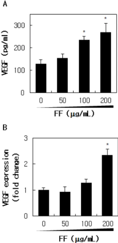
3.3. Effect of FF Extract on VEGF Expression To investigate in detail the effect of FF extract on VEGF production in keratinocytes, cells were treated with FF extract and the protein and mRNA expression levels of VEGF were analyzed. Released VEGF protein level was significantly increased by 2.1-fold in 200 µg/mL FF-treated medium compared to vehicle-treated control medium in a dose-dependent manner after 24 h (Figure 3A). As shown in Figure 3B, VEGF mRNA expression
level also was increased by FF extract treatment by up to 2.3-fold in a dose-dependent manner after 6 h.
Interestingly, keratinocyte-derived VEGF upregulation is regarded as a decreased microvessels’improver in aged human skin in vivo[15]. Thus, identification of potential agents regulating these deteriorated microvasculature might be a useful therapeutic strategy for the improve- ment of aged human skin.
3.4. FF-induced VEGF Production via p38 MAPK Activation
Chronic UV irradiation results in the decrease of skin vasculature; on the other hand, acute UV irradiation in- duces skin angiogenesis in human skin in vivo[21]. The Figure 2. Comparison of secretory protein profiles treated by
FF extract in HaCaT cells. (A) Cells were incubated in the absence or presence FF extract (200 µg/mL) for 24 h, and then the conditioned media were analyzed using protein array associated with an angiogenesis. 1: IGFBP-3, 2: IL-8, 3:
VEGF, and P: positive control. (B) Spots were quantified using a densitometry program Bio1D advanced, and presented as percentages of the non FF-treated control level. *p < 0.05 versus vehicle control.
Figure 3. Upregulation of VEGF by FF extract in HaCaT cells.
(A) VEGF protein levels were analyzed using sandwich ELISA specific for human VEGF in the supernatants of FF-treated or non FF-treated control cells after 24 h incubation. (B) VEGF mRNA expression (fold change) was analyzed by quantitative real-time RT-PCR after 6 h of FF treatment. The data represented mean ± S.E.M of 3 independent experiments. *p <
0.05 versus non FF-treated control.
mechanism of UV-induced angiogenic switch was ex- plained in previous studies. The ERK-dependent positive regulation of VEGF expression and the AKT-dependent negative regulation of TSP-1 (a potent endogenous in- hibitor of angiogenesis) expression are involved in the acute UV-induced angiogenic switch in human skin[22,23]. MAPKs are serine/threonine kinases that phosphorylate transcription factors and downstream ki- nases, mediating signal transduction from the cell surface to the nucleus[24]. They are responsible for many bio- logical activities and cellular processes, such as pro- liferation, differentiation, wound healing, and cancer for- mation[25].
To determine the mechanisms underlying VEGF regu- lation in response to FF extract, we attempted to identify the signaling pathway in MAPK activation. Cells were treated with 200 µg/mL of FF extract for 2, 5, 10, 30,
and 60 min, followed by extraction of the cellular protein.
The expressions of total and phosphorylated JNK, ERK1/2, and p38 MAPK were determined by western blot analysis. FF extract induced rapid increase in phos- pho-p38 MAPK, beginning as early as 2 min after the treatment of FF extract (Figure 4A). However, the phos- phorylation of SAPK/JNK was not detected at indicated time points and the phosphorylation of ERK1/2 was somewhat delayed compared with the phosphorylation of p38 MAPK after 30 min of FF extract treatment (data not shown). To confirm the ability of SB202190 to block p38 MAPK in HaCaT cells, cells were treated with a specific p38 MAPK inhibitor, SB202190 (10 µM) prior to FF ex- tract treatment. As shown in Figure 4B, this pretreatment completely inhibited the activation of p38 MAPK 5 min after FF extract treatment. To investigate the involvement of p38 MAPK activation in FF-induced VEGF pro- Figure 4. Involvement of p38 MAPK activation in FF-induced VEGF upregulation. (A) Kinetic activation of p38 MAPK was analyzed in HaCaT cells at indicated times after 200 µg/mL of FF extract treatment. (B) HaCaT cells were pretreated with a specific inhibitor of p38 MAPK, SB202190 (10 µM) and then detected by western blotting at 5 min after FF extract treatment. (C) The effects of SB202190 on FF-induced VEGF upregulation were examined using ELISA at 24 h and (D) quantitative real-time RT-PCR at 6 h post-FF extract treatment, respectively. The data represented mean ± S.E.M of 3 independent experiments. *p < 0.05 versus non FF-treated control. #p < 0.05 versus FF extract treated level.
duction, we examined VEGF expression in the presence and absence of SB202190. The FF-induced VEGF pro- duction was obviously inhibited by SB202190 pretreat- ment (Figure 4C). The FF-induced VEGF mRNA ex- pression was also significantly attenuated by a specific in- hibitor of p38 MAPK (Figure 4D). These findings demon- strate that the p38 MAPK pathway positively modulates VEGF production in FF extract-treated keratinocytes.
4. Conclusion
In this study, we aimed to investigate whether the FF extract regulates skin angiogenesis-associated responses and especially VEGF production in skin keratinocytes.
The blood vascular system forms a complicated network in the dermis of the skin, just up to the beneath of epidermis. The blood vessels play many crucial roles in- cluding maintenance of body temperature, supply of oxy- gen, nutrients and hormones, transport of inflammatory cells, organogenesis, as well as skin aging. We established here a novel regulatory effect of FF to induce VEGF pro- duction via p38 MAPK activation in human epidermal keratinocytes. These results suggest that the FF extract is useful as an ingredient with potent angiogenic activity and may help to improve age-dependent reduction of the microvasculature in aged human skin.
More than 100 compounds have been identified from FF, and the major constituents of FF include quercetin, rutaecarpine, forsythiaside A, betulinic acid, and for- sythialan A[16]. Recent pharmacological research in- dicates that forsythiaside, forsythin, and rutin are res- ponsible for the biological activities of FF[26-28].
Although the mechanism of active single compounds in FF extract was not elucidated in the present study, we first investigated the regulatory effect of FF on angio- genesis-associated responses, particularly FF-induced VEGF production in human keratinocytes. Further studies are needed on the involvement of specific active compo- nents to know the detailed mechanism of FF to elucidate the effective ingredient on angiogenic processes and skin anti-aging.
Reference
1. J. M. Waller and H. I. Maibach, Age and skin struc- ture and function, a quantitative approach (I): blood flow, pH, thickness, and ultrasound echogenicity, Skin Res. Technol., 11(4), 221 (2005).
2. I. Bentov and M. J. Reed, The effect of aging on the cutaneous microvasculature, Microvasc. Res., 100, 25 (2015).
3. A. G. Gunin, V. V. Petrov, N. N. Golubtzova, O. V.
Vasilieva, and N. K. Kornilova, Age-related changes in angiogenesis in human dermis, Exp. Gerontol., 55, 143 (2014).
4. J. H. Chung, J. Y. Seo, H. R. Choi, M. K. Lee, C.
S. Youn, G. Rhie, K. H. Cho, K. H. Kim, K. C. Park, and H. C. Eun, Modulation of skin collagen metabo- lism in aged and photoaged human skin in vivo, J.
Invest. Dermatol., 117(5), 1218 (2001).
5. J. H. Chung, K. Yano, M. K. Lee, C. S. Youn, J. Y.
Seo, K. H. Kim, K. H. Cho, H. C. Eun, and M.
Detmar, Differential effects of photoaging vs intrinsic aging on the vascularization of human skin, Arch.
Dermatol., 138(11), 1437 (2002).
6. D. Vybohova, Y. Mellova, K. Adamicova, M.
Adamkov, and G. Heškova, Quantitative changes of the capillary bed in aging human skin, Histol.
Histopathol., 27(7), 961 (2012).
7. K. Kajiya, Y. K. Kim, Y. Kinemura, J. Kishimoto, and J. H. Chung, Structural alterations of the cuta- neous vasculature in aged and in photoaged human skin in vivo, J. Dermatol. Sci., 61(3), 206 (2011).
8. Y. Tsuchida, The effect of aging and arteriosclerosis on human skin blood flow, J. Dermatol. Sci., 5(3), 175 (1993).
9. S. Vandekeere, M. Dewerchin, and P. Carmeliet, Angiogenesis revisited: an overlooked role of endo- thelial cell metabolism in vessel sprouting, Microcirculation, 22(7), 509 (2015).
10. T. D. Crafts, A. R. Jensen, E. C. Blocher-Smith, and T. A. Markel, Vascular endothelial growth factor:
therapeutic possibilities and challenges for the treat-
ment of ischemia, Cytokine, 71(2), 385 (2015).
11. S. Barrientos, O. Stojadinovic, M. S. Golinko, H.
Brem, and M. Tomic-Canic, Growth factors and cyto- kines in wound healing, Wound Repair Regen., 16(5), 585 (2008).
12. R. S. Ishak, S. A. Aad, A. Kyei, and F. S. Farhat, Cutaneous manifestations of anti-angiogenic therapy in oncology: review with focus on VEGF inhibitors, Crit. Rev. Oncol. Hematol., 90(2), 152 (2014).
13. K. E. Johnson and T. A. Wilgus, Vascular endothelial growth factor and angiogenesis in the regulation of cutaneous wound repair, Adv. Wound Care, 3(10), 647 (2014).
14. M. Sawane and K. Kajiya, Ultraviolet light-induced changes of lymphatic and blood vasculature in skin and their molecular mechanisms, Exp. Dermatol., 21(S1), 22 (2012).
15. J. H. Chung and H. C. Eun, Angiogenesis in skin ag- ing and photoaging, J. Dermatol., 34(9), 593 (2007).
16. Y. P. Guo, L. G. Lin, and Y. T. Wang, Chemistry and pharmacology of the herb pair Flos Lonicerae ja- ponicae-Forsythiae fructus, Chin. Med., 10, 16 (2015).
17. H. S. Kang, J. Y. Lee, and C. J. Kim, Anti-in- flammatory activity of arctigenin from Forsythiae fructus, J. Ethnopharmacol., 116(2), 305 (2008).
18. J. H. Lee, J. Y. Lee, T. D. Kim, and C. J. Kim, Antiasthmatic action of dibenzylbutyrolactone lignans from fruits of Forsythia viridissima on asthmatic re- sponses to ovalbumin challenge in conscious guin- ea-pigs, Phytother. Res., 25(3), 387 (2011).
19. P. C. Kuo, G. F. Chen, M. L. Yang, Y. H. Lin, and C. C. Peng, Chemical constituents from the fruits of Forsythia suspensa and their antimicrobial activity, BioMed Res. Int., 2014, 304830 (2014).
20. Y. Zhang, Y. Deng, T. Luther, M. Muller, R. Ziegler, R. Waldherr, D. M. Stern, and P. P. Nawroth, Tissue factor controls the balance of angiogenic and anti- angiogenic properties of tumor cells in mice, J. Clin.
Invest., 94(3), 1320 (1994).
21. K. Yano, K. Kadoya, K. Kajiya, Y. K. Hong, and M.
Detmar, Ultraviolet B irradiation of human skin in- duces an angiogenic switch that is mediated by up- regulation of vascular endothelial growth factor and by downregulation of thrombospondin-1, Brit. J.
Dermatol., 152(1), 115 (2005).
22. M. S. Kim, Y. K. Kim, H. C. Eun, K. H. Cho, and J. H. Chung, All-trans retinoic acid antagonizes UV-induced VEGF production and angiogenesis via the inhibition of ERK activation in human skin kera- tinocytes, J. Invest. Dermatol., 126(12), 2697 (2006).
23. M. S. Kim, Y. J. Oh, S. Lee, J. E. Kim, K. H. Kin, and J. H. Chung, Ultraviolet radiation attenuates thrombospondin 1 expression via PI3K-Akt activation in human keratinocytes, Photochem. Photobiol., 82(3), 645 (2006).
24. G. Pearson, F. Robinson, T. Beers Gibson, B. E. Xu, M. Karandikar, K. Berman, and M. H. Cobb, Mitogen-activated protein (MAP) kinase pathways:
regulation and physiological functions, Endocr. Rev., 22(2), 153 (2001).
25. M. J. Robinson and M. H. Cobb, Mitogen-activated protein kinase pathways, Curr. Opin. Cell Biol., 9(2), 180 (1997).
26. X. Fang, Y. Wang, J. Wang, J. Zhang, and X. Wang, Microwave-assisted extraction followed by RP-HPLC for the simultaneous extraction and determination of forsythiaside A, rutin, and phillyrin in the fruits of Forsythia suspensa, J. Sep. Sci., 36(16), 2672 (2013).
27. X. Pan, X. Cao, N. Li, Y. Xu, Q. Wu, J. Bai, Z. Yin, L. Luo, and L. Lan, Forsythin inhibits lip- opolysaccharide-induced inflammation by suppressing JAK-STAT and p38 MAPK signalings and ROS pro- duction, Inflamm. Res., 63(7), 597 (2014).
28. X. L. Piao, M. H. Jang, J. Cui, and X. Piao, Lignans from the fruits of Forsythia suspensa, Bioorg. Med.
Chem. Lett., 18(6), 1980 (2008).