11
두개결손부 모델에서 배양된 골막유래세포를 이용한 골이식 시 지지체로서 TCP의 효과
심경미*, 김세은**, 김종춘**, 배춘식**, 최석화***, 강성수**
남부대학교 방사선학과*, 전남대학교 수의과대학**, 충북대학교 수의과대학***
Effect of tricalcium phosphate (TCP) as a scaffold during bone grafting using cultured periosteum-derived cells in a rat calvarial defect model
Kyungmi Shim*, Seeun Kim**, Jongchoon Kim**, Chunsik Bae**, Seokhwa Choi*** and Seongsoo Kang**
Department of Radiology, Nambu University, Gwangju, Korea*; College of Veterinary Medicine, Chonnam National University, Gwangju, Korea**; College of Veterinary Medicine, Chungbuk National University, Cheongju, Korea***
요약
다능성 세포를 포함하는 골막은 골모세포와 연골세포로 분화될 수 있다. 그리고 배양된 골막유래세포는 골형성 능 력을 가지고 있다. 이 연구의 목적은 골막유래 세포들과 골이식재 간의 상호작용을 평가하는 것이다.
Sprague-Dawley 랫드의 두개골 골막에서 세포를 분리한 다음, 배양된 골막유래세포를 beta-tricalcium phosphate (β-TCP)와 함께 임계결손부 크기의 두개결손부에 이식하였다. 모든 랫드는 골이식 수술 후 8주째에 희생되었으며, 골이식부의 골형성 능력은 일반방사선, micro CT 및 조직검사를 통해 평가되었다. β-TCP와 함께 이식된 골막유래세 포는 골결손부에서 더욱 증가된 석회화작용을 나타내었으며, 골결손부 안쪽 및 가장자리에 골밀도 증가와 신생골이 형 성되었다. 특히 골막유래세포는 β-TCP만 단독으로 이식하였을때보다 함께 이식 시 효과적으로 신생골을 형성하였다.
이러한 결과는 배양된 골막유래세포가 골결손부에서 골형성을 증진시킬 수 있는 가능성을 보였다.
핵심어
:골재생
,골막유래세포
, β-TCP,랫드
Abstract
The periosteum contains multipotent cells that can differentiate into osteoblasts and chondrocytes. Cultured periosteum-derived cells (PDCs) have an osteogenic capacity. The purpose of this study was to evaluate the interaction of PDCs with bone graft biomaterial. After cell isolation from the calvarial periosteum of Sprague-Dawley rats, cultured PDCs were placed in critical-sized calvarial defects with beta-tricalcium phosphate (β-TCP). All rats were sacrificed 8 weeks after bone graft surgery, and the bone regenerative ability of bone grafting sides was evaluated by plain radiography, micro-computed tomography (CT), and histological examination. PDCs grafted with β-TCP displayed enhanced calcification in the defect site, density of regenerated bone and new bone formation within the defect and its boundaries. Furthermore, these PDCs more efficiently regenerated new bone as compared to grafted β-TCP only. The results suggest that cultured PDCs have the potential to promote osteogenesis in bone defects.
This study was financially supported by special research fund of Chonnam National University in 2007.
Corresponding Author: 강성수
주소:광주광역시 북구 용봉동 전남대학교 300 수의과대학. E-mail:[email protected], Tel: +82-62-530-2877
K ey Words: Bone regeneration, periosteum-derived cell, β-TCP, rat
I. Introduction
The goal of bone tissue engineering is to induce bone formation in the defect site and develop real bone tissue.
Three essential elements for bone formation by bone tissue engineering are osteoblast-induced bone formation
[1], the use of three-dimensional (3D) scaffolding (espe- cially polymers and bioceramics) to provide proper sites for the ingrowth of cells and vessels, and the use of oste- oinductive growth factors to facilitate induction and differentiation of bone tissue [2].
The importance of periosteum in bone development and fracture healing has been well demonstrated, and the osteogenic potential of cultured periosteal cells has been reported [3]. In the previous study, cultured human periosteum-derived cells showed osteogenic potential in a xenogeneic graft model using rat calvarial defects and showed the possibility of using cultured human periosteal cell/collagen complex grafts to form bone within in-vivo bone defects [3]. However, this new bone did not completely fill the defect by 35 days post grafting.
Therefore, some investigators have suggested the app- lication of bone growth factors [3] and scaffolds [4]are required to enhance osteogenic activity of periosteum- derived cells. Scaffolds to support cell-based tissue engineering are critical determinants of clinical efforts to regenerate and repair the body. Selection of a matrix carrier involves consideration of the role of the matrix as a scaffold for physical support and host tissue integration, as well as its ability to support synergistic osteoinduction of the implanted osteogenic cells. The aim of this study was to evaluate the effect of beta-tricalcium phosphate (β -TCP) to adherent cultured periosteum- derived cells (PDCs) as a scaffold on sinus augmentation with fibrin glue mixture in an allograft model using rat calvarial defects.
II. Materials and Methods
1. Animals.
Ten-week-old male Sprague-Dawley rats weighing 250–
280 g were used. They were kept under a standard light-dark schedule at a temperature of 20 ± 3oC and 50
± 10% humidity. Stock diet and tap water were available ad libitum. The care, maintenance and treatment of animals were carried out in accordance with the Guidelines for Animal Experiments of Chonnam National University.
2. Cells and cell culture.
Periosteum was harvested from the calvarium of9-month-old adult male rats. PDCs were isolated using a standard collagenase digestion protocol. Briefly, cells were washed three times with phosphate-buffered saline and then digested with 0.25% collagenase (Sigma-Aldrich, St.
Louis, MO, USA) for 2 h at 37°C with gentle shaking.
The sample was washed once in Dulbecco’s Modified Eagle’s Medium with 10% fetal bovine serum, and two more times with Hank’s Balanced Salt Solution. The washed cells were plated in T-75 flasks containing an osteogenic medium consisting of α-MEM (Gibco BRL, Grand Island, NY, USA) with 10% fetal bovine serum and 1% antibiotic solution (100 units/ml penicillin and 100 µg/ml streptomycin). The cultures were incubated under standard culture conditions (37°C with 5% carbon dioxide). Cells were checked daily for growth and fed fresh medium every other day. Cell cultures were serially passaged until the amplification number was attained. The cells were washed with Hank’s Balanced Salt Solution, detached from the flasks with trypsin/ethylenediamine- tetraacetic acid (0.05%/0.53 mM; Invitrogen, Carlsbad, CA, USA), and centrifuged. Cells passaged 2–4 times were used in the experiments described subsequently (1 x 10⁶ cells/β-TCP scaffold).
3. Surgical procedure.
Rats (n=24) were premedicated with a subcutaneous injection of atropine sulfate (Daihan Scientific, Seoul, Korea; 0.1 mg/kg) and enrofloxacine (Bytril® Bayer Korea, Seoul, Korea; 2.5 mg/kg SC), and then anes- thetized using an intraperitoneal injection of xylazine (Rompun®, Bayer Korea; 10 mg/kg) and ketamine-HCl (Huons, Seoul, Korea; 40 mg/kg). After a U-shaped skin incision and periosteal elevation, the calvarial defect was created with an 8 mm trephine bur and a Surgic XT Plus surgical motor (Shanghai NSK International Trading, Shanghai, China). The rats were divided into four groups (n=6 per group). Rats in the autobone group (autobone scaffold), β-TCP group (β-TCP scaffold) and β -TCP/PDCs group (β-TCP/PDCsscaffold) had the defects grafted with Greenplast fibrin glue mixture (Korea Green Cross, Seoul, Korea). Rats in the control group did not receive a graft. After grafting, the periosteum and skin were surgically closed.
4. Radiography.
The calvaria were radiographed using a Port-X dental X-ray instrument (Genoray, Seoul, Korea) and a Digi-X Sensor oral sensor (Hanjin Digi-X, Seoul, Korea) under conditions of 70 kVp and 0.15 mAs at 8 weeks after surgery.
5. Micro-computed tomography (CT).
At post-surgery week 8, all rats were sacrificed and calvaria were collected. Using a Skyscan 1172 Desktop X-ray Microtomograph micro-CT scanner (Skyscan, Aartselaar, Belgium), calvaria were scanned at 50 kV and 200 µA. After scanning, images were analyzed with NRecon Ver.1.4.4 software (Skyscan).
6. Histology.
The collected calvaria were fixed in 10% neutral formalin and decalcified with formic acid. All the
specimens were embedded in paraffin and 5 μm-thick axial sections were obtained. Sections were deparaffinized in xylene, rehydrated in graded alcohol and stained with hematoxylin and eosin.
III. Results
1. Radiological findings.
In the control group,little new bone was formed from the defect margin and showed only soft tissue-like radiopacities (Fig. 1A).The defect was almost completely repaired by the graft materials in the autograft group (Fig.
1B). In β-TCP and β-TCP/PDCs groups, β-TCP particles were radiopaque and round, and their density was variable.
Marginal new bone formation was evident at the defect margin in both groups (Fig. 1C, Fig. 1D). More pronounced bone formation of the scaffold and the margin of defect were evident in the β-TCP/PDCs group compared to the β-TCP group (Fig. 1C, Fig. 1D).
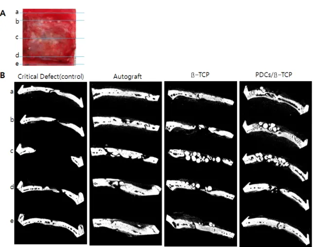
2. Micro-CT findings.
Micro-CT generated coronal tomograms of bone repair in 8 mm critical-size calvarial defects at 8 weeks. In the autograft group, the defect was almost completely repaired by graft materials (Fig. 2). In the β-TCP and β -TCP/PDCs groups, bone defect boundaries of host bone could be observed, with some remnant β-TCP, and a little new bone formation was observed at the margin of bone defect compared to the autograft group (Fig. 2). But the bottom margins of 8 mm critical defect were bridged either side in the β-TCP/PDCs group (Fig. 2).
3. Histological findings.
In the control group, the bone defect site was filled with soft tissue and blood vessels. A small amount of bone was formed only from existing bone (Fig. 3). In the autograft group, grafted bone particles were fused and new bone formation occurred with vascularization (Fig. 4). In the β-TCP grafted group, β-TCP particles absorbed and
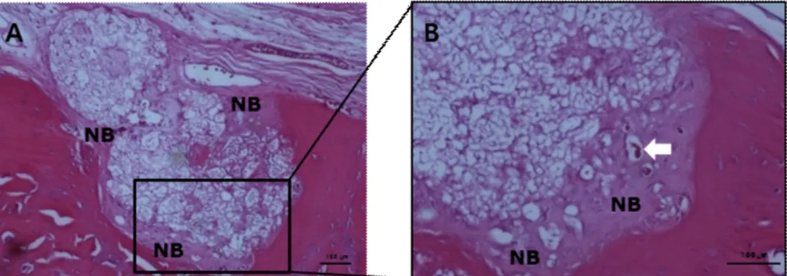
new bone formation occurred in and out of the β-TCP particles (Fig.5). Also, the fusion of β-TCP particles and normal bone was evident at the defect margin. In the β -TCP/PDCs group, β-TCP particles absorbed and new bone formation occurred in the β-TCP particles with neovascularization. Also, the fusion of β-TCP particles and normal bone was evident at the defect margin (Fig. 6).
IV. Discussion
β-TCP is used for bone tissue engineering due to its good biocompatibility and biodegradability [5]. This study combined cells capable of osteogenetic activity with an appropriate scaffolding material to stimulate bone regen- eration and repair. In particular, mesenchymal stem cells have a high proliferation capacity and multilineage potential. They can differentiate into osteoblasts [6], thus, they have been used to engineer bone tissue. The perio- steum is easier to obtain from patients than are bone marrow and periodontium, and the periosteum is a sufficient supply of osteogenic cells [7], chondrogenic cells
[8] and mesenchymal progenitor cells [9]. PDCs may serve as an optimal cell source for tissue engineering based on their accessibility, ability to proliferate rapidly and capa- bility to differentiate into multiple mesenchymal lineages
[10]. In our previous study, grafting of β-TCP particles proved to be an efficient means to regenerate new bone in a rat critical-sized calvarial defect model [11]. However, fusion with bone defect margins did not occur. Thus, this study was designed to test the practicality of placing PDCs and β-TCP particles into bone defects and to demonstrate the feasibility of calvarial defect repair using PDCs.
In our results, radiographic and histologic findings revealed near-complete repair of defects by the graft materials in the autograft group. However, in the β-TCP and β-TCP/PDCs groups, β-TCP particles were radiopaque and round, and their density was variable.
Marginal new bone formation of the β-TCP and β -TCP/PDCs groups was evident at the defect margin and around β-TCP particles. In the β-TCP/PDCs group,
micro-CT revealed that the bottom margins of 8 mm critical defect were bridged on either side compared the β -TCP group. Also, β-TCP particles had absorbed and new bone formation had occurred in the β-TCP particles, with neovascularization and the fusion of β-TCP particle and normal bone was seen at the defect margin. This indicated that β-TCP has been shown to be an appropriate scaffold for PDCs and to promote bone formation by PDCs.
Many different processing techniques have been developed for the design and fabrication of scaffolds that are required to seed osteoprogenitor cells for tissue re- generation. One study reported that bone formation was observed in the bone defect treated with periosteal cells from cultured medium within 35 days of grafting [3]. As in a previous study [12], while new bone induced from the grafted tissue was detected in the bone defect, thisnew bone did not completely fill the defect by 35 days post grafting. The scaffold for osteoprogenitor cells serves to promote progenitor cell migration, proliferation and differentiation, as well as vascularization [3] is needed. If more effective bone regeneration is to be achieved, the efficiency of cell introduction into β-TCP needs to be improved. Therefore, PDCs/β-TCP composites were implanted into critical defects using sinus augmentation with fibrin glue mixture. We successfully demonstrated that β-TCP had good biodegradability and osteoconductivity as a scaffold material for bone tissue engineering [5]. Similarly, Matsuno et al. [5] demonstrated that β-TCP bead/mes- enchymal stem cells are replaced by bone without any adv- erse reactions. However, some investigators have suggested the application of bone growth factors is required to enh- ance osteoblastic differentiation of periosteum-derived cells.
Hanada et al. [13] reported that chondrogenesis of rat periosteal cells is clearly enhanced by tumor growth factor- β and bone morphogenetic protien-2 treatment.
In conclusion, bone growth factors may provide useful enhancement of the osteogenic potential of graft materials such as cultured PDCs. Further studies about fusion technology with peptides and growth factors using of the combination of tissue engineering, cell engineering and
mechanical engineering are needed.
Acknowledgements
This study was financially supported by special research fund of Chonnam National University in 2007.
References
[1] P. S. Gomes, J. D. Santos and M. H. Fernandes, "Cell-induced response by tetracyclines on human bone marrow colonized hydroxyapatite and bonelike", Acta Biomater, Vol.4, No.3, pp.630-637, 2008.
[2] I. Drosse, E. Volkmer, R. Capanna, P. De Biase, W. Mutschler and M. Schieker, "Tissue engineering for bone defect healing:
an update on a multi-component approach", Injury, Suppl 2, pp.
S9-S20, 2008.
[3] Y. Sakata, T. Ueno, T. Kagawa, M. Kanou, T. Fujii, E.
Yamachika and T. Sugahara, "Osteogenic potential of cultured human periosteum-derived cells - a pilot study of human cell transplantation into a rat calvarial defect model" J
Craniomaxillofac Surg, Vol.34, No.8, pp.461-465, 2006.
[4] Q. Xu, H. Lu, J. Zhang, G. Lu, Z. Deng and A. Mo, "Tissue engineering scaffold material of
porousnanohydroxyapatite/polyamide 66", Int J Nanomedicine, Vol.5, pp.331-335, 2010.
[5] 5. T. Matsuno, Y. Hashimoto, S. Adachi, K. Omata, Y.
Yoshitaka, Y. Ozeki, Y. Umezu, Y. Tabata, M. Nakamura and T. Satoh, "Preparation of injectable 3D-formed beta-tricalcium phosphate bead/alginate composite for bone tissue engineering", Dent Mater J, Vol.27, No.6, pp.827-834, 2008.
[6] M. F. Pittenger, A. M. Mackay, S. C. Beck, R. K. Jaiswal, R.
Douglas, J. D. Mosca, M. A. Moorman, D. W. Simonetti, S.
Craig and D. R. Marshak, "Multilineage potential of adult human mesenchymal stem cells", Science, Vol.284, No.5411, pp.143-147, 1999.
[7] H. A. Declercq, R. M. Verbeeck, L. I. De Ridder, E. H.
Schacht and M. J. Cornelissen, "Calcification as an indicator of osteoinductive capacity of biomaterials in osteoblastic cell cultures", Biomaterials, Vol.26, No.24, pp.4964–4974, 2005.
[8] E. J. Jansen, P. J. Emans, N. A. Guldemond, L. W. Van Rhijn, T. J. Welting, S. K. Bulstra and R. Kuijer, "Human
periosteum-derived cells from elderly patients as a source for cartilage tissue engineering? " J Tissue Eng Regen, Vol.2, No.6, pp.331–339, 2008.
[9] C. De Bari, F. Dell’Accio, J. Vanlauwe, J. Eyckmans, I. M.
Khan, C. W. Archer, E. A. Jones, D. McGonagle, T. A.
Mitsiadis, C. Pitzalis and F. P. Luyten, "Mesenchymal multipotency of adult human periosteal cells demonstrated by single-cell lineage analysis", Arthritis Rheum, Vol.54, No.4, pp.1209–1221, 2006.
[10] H. Agata, I. Asahina, Y. Yamazaki, M. Uchida, Y. Shinohara, M. J. Honda, H. Kagami and M. Ueda, "Effective bone engineering with periosteum-derived cells", J DentRes Vol.86, No.1, pp.79-83, 2007.
[11] K. H. Yoo, S. E. Kim, K. M. Shim, H. J. Park, S. H. Choi and S. S. Kang, "Effect of porcine cancellous bones on regeneration in rats with calvarial defect", J Life Science, Vol.20, No.8, pp.1207-1213, 2010.
[12] M. Kanou, T. Ueno, T. Kagawa, T. Fujii, Y. Sakata, N. Ishida, J. Fukunaga and T. Sugahara, "Osteogenic potential of primed periosteum graft in the rat calvarial model", Ann Plast Surg, Vol.54, pp.71–78, 2005.
[13] K. Hanada, L. A. Solchaga, A. I. Caplan, T. M. Hering, V. M.
Goldberg, J. U. Yoo and B. Johnstone. "BMP-2 induction and TGF-beta 1 modulation of rat periosteal cell chondrogenesis", J Cell Biochem, Vol.81, No.2, pp.284-294, 2001.
Legend of figures
Figure 1. Radiographs of bone regeneration in 8 mm critical-size calvarial defects at 8 weeks postsurgery.
Figure 2. Micro-CT views of 8 mm critical-sized calvarial defects at 8 weeks postsurgery.
Figure 3. Histological sections of 8 mm critical-sized calvarial defect of the control group.
Figure 4. Histological sections of 8 mm critical-sized calvarial defect of the autograft group.
Figure 5. Histological sections of 8 mm critical-sized calvarial defect of the β-TCP grafted group.
Figure 6. Histological sections of 8 mm critical-sized calvarial defect of the β-TCP and the cultured PDCs groups.
Figure 1. Radiographs of bone regeneration in 8 mm critical-sized calvarial defects at 8 weeks postsurgery. In the untreated group (critical defect, A), little new bone was formed from the defect margin, and in the autograft group (B), the defect almost completely repaired by
the graft materials. In the
β-TCP (C) and cell/
β-TCP (D) groups,
β-TCP particles were evident as radiopaque and round, and their
density was variable. Marginal bone formation of
β-TCP and cell/
β-TCP groups was observed at the defect margin.
Figure 2. Micro-CT views of 8 mm critical-sized calvarial defects at 8 weeks postsurgery. A. Coronal lines of the calvarial defect as delineated for the CT-scan analysis. B.Micro-CT generated coronal tomograms of bone repair in 8 mm critical-size
calvarial defects at 8 weeks. In the autograft group, the defect was almost completely repaired by graft materials. In the
β-TCP and PDCs/
β-TCP groups, boundaries of bone defect of host bone could be observed, with some
β-TCP remnant, and
a little new bone formation was observed at the margin of bone defect.
Figure 3. Histological sections of 8 mm critical-sized calvarial defect of the control group (A and B). The bone defect site was filled with soft tissues and blood vessels. Arrow : blood vessels, arrow head : soft tissues in the bone defect (H&E
staining, scale bars = 100
μm).
Figure 4. Histological sections of 8 mm critical-sized calvarial defect of the autograft group (A and B). Grafted bone particles were fused and new bone formation occurred with vascularization. NB : newly formed bone,
arrow : blood vessels (H&E staining, scale bars = 100
μm).
Figure 5. Histological sections of 8 mm critical-sized calvarial defect of the β-TCP grafted group (A and B). β-TCP particles had been absorbing and new bone formation was occurred in and out of the β-TCP particles. Also, the fusion of β-TCP particle and normal bone was seen at the defect margin. NB : newly formed bone, arrow : blood vessels (H&E staining, scale bars = 100 μm).
Figure 6. Histological sections of 8 mm critical-sized calvarial defect of the β-TCP and the cultured PDCs groups (A and B). β-TCP particles absorbed and new bone formation occurred in the β-TCP particles with neovascularization. Also, the fusion of β-TCP particle and normal bone was seen at the defect margin. NB : newly formed bone, arrow : blood vessels(H&E staining, scale bars = 100 μm)