Identification and analysis of microRNAs in Candida albicans
Jin-Hyun Cho1 and Heon-Jin Lee2*
1Department of Prosthodontics, Kyungpook National University, School of Dentistry, Daegu 41940, Korea
2Department of Microbiology and Immunology, Kyungpook National University, School of Dentistry, Daegu 41940, Korea Received September 21, 2017 /Revised October 20, 2017 /Accepted November 15, 2017
Oral infection due to Candida albicans is a widely recognized and frequent cause of superficial in- fections of the oral mucosa (oral candidiasis). Although oral candidiasis is not a life-threatening funge- mia, it can cause severe problems in individuals under certain conditions. MicroRNAs (miRNAs) are noncoding, small RNA molecules, which regulate the expression of other genes by inhibiting the translation of target mRNAs. The present study was designed to identify miRNAs in C. albicans and determine their possible roles in this organism. miRNA-sized small RNAs (msRNAs) were cloned in C. albicans by deep sequencing, and their secondary structures were analyzed. All the cloned msRNAs satisfied conditions required to qualify them as miRNAs. Bioinformatics analysis revealed that two of the most highly expressed C. albicans msRNAs, Ca-363 and Ca-2019, were located in the 3' un- translated region of the corticosteroid-binding protein 1 (CBP1) gene in a reverse orientation. miRNA mimics were transformed into C. albicans to investigate their RNA-inhibitory functions. RNA oligonu- cleotide-transformed C. albicans was then observed by fluorescent microscopy. Quantitative PCR analy- sis showed that these msRNAs did not inhibit CBP1 gene expression 4 hr and 8 hr after ectopic miRNA transformation. These results suggest that msRNAs in C. albicans possess an miRNA-triggered RNA interference gene-silencing function, which is distinct from that exhibited by other eukaryotic systems.
Key words : Candida albicans, CBP1, deep sequencing, microRNA, msRNA, small RNA
*Corresponding author
*Tel : +82-53-660-6832, Fax : +82-53-425-6025
*E-mail : [email protected]
This is an Open-Access article distributed under the terms of the Creative Commons Attribution Non-Commercial License (http://creativecommons.org/licenses/by-nc/3.0) which permits unrestricted non-commercial use, distribution, and reproduction in any medium, provided the original work is properly cited.
Journal of Life Science 2017 Vol. 27. No. 12. 1494~1499 DOI : https://doi.org/10.5352/JLS.2017.27.12.1494
Introduction
MicroRNAs (miRNAs) are small, non-coding RNAs (20–
24 nt in length) that have regulatory functions. miRNAs, which are found in most eukaryotes and some viruses, are processed from precursor miRNAs (pre-miRNAs, approx- imately 60–80 nt) by RNase III (e.g., the enzyme Dicer), and are finally incorporated into the Ago-containing RNA-in- duced silencing complex (RISC). Although miRNA-sized small RNAs (msRNAs) from bacteria were recently identi- fied, miRNAs in fungi are still only vaguely understood.
Candida albicans is a fungus and type of budding yeast that causes candidiasis by infecting the oral mucosa. Oral candidiasis is not a life-threatening fungemia, but occurs of- ten and therefore causes severe problems in individuals whose immune systems are compromised by AIDS or the
use of immunosuppressant drugs [15]. Biofilms produced by C. albicans are also often observed on dental devices such as dentures and cause denture stomatitis [11]. This fungus also harbors Dicer (CaDcr1), an enzyme that is required for ribosomal and spliceosomal RNA maturation in addition to miRNA processing [2]. Therefore, the discovery of msRNAs in C. albicans [5] was not surprising, even though the func- tion of these small RNAs in this organism is unclear. In addi- tion, expression of the exogenous hairpin double-stranded RNA (dsRNA) does not result in downregulated expression of its target mRNA in C. albicans [16], suggesting that the RNA-silencing mechanism might differ from that in other eukaryotic systems.
In the present study, to examine the roles of these msRNAs in C. albicans, they were cloned and their secondary structures analyzed. All of the clones satisfied the conditions required to qualify them as miRNAs. We further trans- formed the miRNA mimics into C. albicans to investigate their potential RNA-inhibitory functions.
Martials and Methods
Culture conditions
The wild-type C. albicans strain SC5314 (ATCC MYA-2876) - Note -
was maintained on yeast peptone dextrose (YPD; 1% yeast extract, 2% peptone, and 2% glucose).
RNA extraction and small RNA cloning for sequencing Small RNA for the deep sequencing of C. albicans was isolated using the miRNeasy Mini Kit (Qiagen) according to the manufacturer’s protocols. The kit was further used to purify the small RNA-enriched RNA. Cloning of small RNAs was performed as described previously [7, 10]. The purified small-sized cDNA library was used for cluster gen- eration on Illumina’s cluster station (San Diego, CA, USA) and then sequenced on the Illumina GAIIx system according to the manufacturer instructions. Next-generation sequen- cingwas performed by LC Sciences (Houston, TX, USA).
Bioinformatics analysis of the sequence data Raw sequences were processed using Illumina’s pipeline software (details regarding the Illumina sequencing analysis software are provided in the Illumina Sequencing Analysis Software User Guide For Pipeline at http://support.illumina.
com) and then subjected to a series of data filtration steps to analyze the sequencing data using the ACGT101-miR soft- ware package (V3.5; LC Sciences, Houston, TX, USA). The C. albicans reference databases (Genome database: http://www.
candidagenome.org/download/sequence/C_albicans_SC5314/
Assembly21/current/C_albicans_SC5314_A21_chromosomes.
fasta.gz; mRNA database: http://www.candidagenome.org/
download/sequence/C_albicans_SC5314/Assembly21/cur- rent/C_albicans_SC5314_A21_orf_coding.fasta.gz) were used to map the small RNAs. Hairpin RNA structures were pre- dicted from adjacent 60-nt sequences in either direction, us- ing mfold software [18].
C. albicans transformation
C. albicans cells were grown overnight to the stationary phase. This culture was diluted in fresh YPD medium to an optical density of 0.3 at 600 nm and grown for an addi- tional 4 hr. Cells were collected by centrifugation and wash- ed twice in electroporation buffer (5 mM potassium phos- phate, 0.3 M sucrose, and 1 mM MgCl2). For electroporation, the cells were transferred to pre-chilled 2-mm electro- poration cuvettes (Bio-Rad Laboratories, Richmond, CA, USA). Five microliters of fluorescein isothiocyanate (FITC)- labeled RNA oligonucleotides (100 pM; synthesized by the Bioneer Corporation, Daejeon, Korea) was added to a 40-μL aliquot of electrocompetent cells. The same amount of
scrambled FITC-conjugated oligonucleotides was used as the negative control (NEG). Electroporation was performed us- ing a Bio-Rad gene pulser by applying a single pulse of 1.5 kV, with capacitance at 25 μF and resistance at 200 W.
Quantitative reverse transcription-polymerase chain reaction (qRT-PCR)
qRT-PCR was performed for amplification and quantifica- tion of the expression of CBP1 and ACT1 (as a reference).
Total RNA (1 μg) was reverse-transcribed using the Omni- script Reverse Transcription Kit (Qiagen, Germantown, MD, USA), and Power SYBR Green PCR Master Mix (Applied Biosystems) was used for the PCRs. The following primer sequences were used to assess CBP1 and ACT1 mRNA ex- pression: CBP1 forward 5'-ACTTGGATCATCGTTGGTGTA- 3' and CBP1 reverse 5'-TGTAAATTTTGGCCGTGTTCATAA- 3'; ACT1 forward 5'-TGGCAGAAGATTGAGAAGAAGTTT- 3' and ACT1 reverse 5'-AAGAATTGATTTGGCTGGTAGA GA-3'. All samples were measured in triplicate for each treatment. The data are presented as the ratio of expression of CBP1 to that of ACT1.
Results and Discussion
Factors such as stress, aberrant homeostasis, medical de- vices, or an imbalance in the host immune system can lead to excessive growth of organisms like C. albicans, resulting in severe infections such as oral candidiasis [11]. To prevent C. albicans-related diseases, the relation between the fungus and human immunity need to be closely investigated.
Recently, msRNAs from bacteria have gained attention as potential "communication molecules" between bacterial and human cells, and were suggested to modulate the immune signaling pathways of host cells [3, 4]. These RNAs in perio- dontal pathogens have been shown to decrease levels of cer- tain cytokines in host T cells, indicating that the bacterial msRNAs likely function as signaling molecules within other species as well.
In the current study, we found msRNAs in C. albicans, which were sequenced from the clones of a small RNA pool exhibiting a perfect hairpin fold-back structure. A total of 19,885,169 small-sized cDNA sequence reads were extracted and analyzed by next-generation sequencing for initial profiling. Raw sequences that did not meet the acceptance criteria of the filters (out of range and junk reads) were re- moved and the remaining sequences were considered
Fig. 1. Length distribution of the deep-sequenced miRNAs in C. albicans. The nucleotide (nt) lengths of the cloned miRNAs are shown on the x-axis and the number of total reads after deep sequencing are shown on the y-axis.
Table 1. Summary of the next-generation sequencing data
Characteristic Number of reads Percentage of
mappable reads
Number of unique miRNAs Raw reads1)
Total mappable reads Mapped to mRNA2)
Mapped to other RNAs (rRNA, tRNA, and other species) Total mapped RNAs of miRNA size (15-30 nt)
Genome within a hairpin3)
19,885,169 14,534,499 2,159,595 1,327,556 4,327,878 57,773
100 14.9 9.1 29.5 0.4
499
1)Total number of raw sequencing reads
2)Total number of reads mapped to mRNA
3)Total number of unique sequences
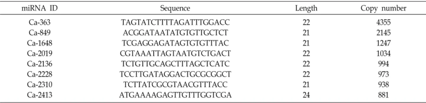
Table 2. List of miRNAs with high copy-number counts in C. albicans (see Additional file 1 for the full list)
miRNA ID Sequence Length Copy number
Ca-363 Ca-849 Ca-1648 Ca-2019 Ca-2136 Ca-2228 Ca-2310 Ca-2413
TAGTATCTTTTAGATTTGGACC ACGGATAATATGTGTTGCTCT TCGAGGAGATAGTGTGTTTAC CGTAAATTAGTAATGTCTGACT
TCTGTTGCAGCTTTAGCTCATC TCCTTGATAGGACTGCGCGGCT TCTTATCGCGTAACGTTTACC ATGAAAAGAGTTGTTTGGTCGA
22 21 21 22 22 22 21 24
4355 2145 1247 1034 994 973 938 881 mappable. The length distribution of the putative miRNAs
from the mappable reads is shown in Fig. 1. The majority of the mappable read sequences were between 20 and 24 nt in length. These small RNAs were considered putative miRNAs only if they formed hairpin folds within the ge- nome and were identified in 0.4% of the mappable reads
from the initial sequencing profile. The data from the next-generation sequencing are summarized in Table 1.
Overall, we profiled 499 miRNAs in C. albicans. A detailed list of the miRNAs showing relatively high expression levels is presented in Table 2.
Even though the functions of the miRNAs are not clear, the identified small RNAs satisfy the conventional rules for being considered miRNAs. Along with the previously dis- covered msRNAs in several bacteria, this small class of RNA exists in every domain of life and is evolutionarily well conserved.
Since their development requires the precise and efficient action of several enzymes on precursor RNAs, miRNAs are believed to be specific to eukaryotic cells or viruses that uti- lize their host systems [8]. Specifically, miRNAs need to meet the following criteria in order to be termed as such, according to the authors of the paper that coined the term and elucidated the nomenclature: consist of an approx- imately 22-nt-long sequence in a cDNA library synthesized from size-fractionated RNA and precursors, show a fold-back structure, and must include at least 16 bp within the first 22 nt of the miRNA and the other arm of the hairpin [1].
In general, miRNA biogenesis involves the preferential se-
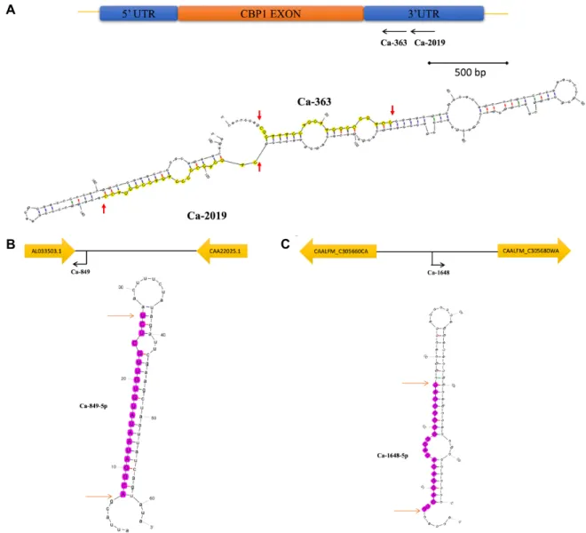
A
B C
Fig. 2. Genomic location and predicted secondary structures of the four most highly expressed miRNAs in C. albicans: Ca-365 and Ca-2019 (A), Ca-849 (B), and Ca-1648 (C). Schematic representations of the genomic locations of the miRNA candidates are also provided.
lection of a single strand of pre-miRNA for further action, while the other strand of the same pre-miRNA is degraded [13]. Many miRNAs are expressed from both arms of the same precursor, which distinguishes miRNAs as either -3p or -5p depending on the location of miRNA synthesis from the pre-miRNA [9]. In this study, we could not identify miRNA candidates that were located in the same precursor hairpin structure as often as detected in mammalian miRNAs, but we did find clustered miRNAs that are possi- bly regulated by the same promoter (Fig. 2A).
The predicted secondary structures and genome locations (from the National Center for Biotechnology Information da- tabase, NC_032089.1–NC_032096.1) of four highly expressed miRNA candidates are shown in Fig. 2. Interestingly, Ca-363
and Ca-2019 were located on the same precursors as some eukaryotic miRNAs and bacterial small RNAs [10, 12].
Among the highly expressed miRNAs, we chose Ca-363 and Ca-2019 to further investigate the potential RNA inter- ference (RNAi) role of the miRNAs in C. albicans. Since both of these miRNAs are located in a position complementary to the 3‘ untranslated region of the CBP1 gene, we specu- lated that they might inhibit the expression of the comple- mentary gene. To test this idea, we transformed synthetic FITC-labeled, single-stranded RNA oligonucleotides of Ca- 363 and Ca-2019 into C. albicans. However, we did not ob- serve a statistically significant change in CBP1 gene ex- pression levels when normalized to the level of the internal control gene (ACT1) 4 hr and 8 hr after transformation, even
A
B
Fig. 3. Transformation of ectopic miRNAs. (A) Microscopy im- ages of treated cells. Nuclei are stained blue (DAPI) and miRNAs appear green (FITC). Magnification is 1,000×.
(b) Relative expression levels measured by qRT-PCR af- ter Ca-363 and Ca-2019 transformation. The expression of CBP1 was not significantly affected by transformation.
The CBP1 expression level was normalized to that of ACT1 (internal control) after 4 hr and 8 hr of transfor- mation. NEG is the scrambled miRNA negative control.
Data show the means from three independent experi- ments (error bars indicate standard deviations).
though the presence of exogenous miRNAs was confirmed by fluoresce microscopy (Fig. 3).
C. albicans possesses important enzymes such as Drosha, Argonaute, and a non-canonical Dicer that are crucial for the RNAi machinery [5]. Moreover, exogenous hairpin dsRNA does not decrease the expression level of the target mRNA [16], indicating the existence of unknown functions of miRNAs in C. albicans. Moreover, the extracellular vesicles of C. albicans, similar to mammalian exosomes, can deliver macromolecules to other cells [6, 14, 17]. Therefore, the trans- portation of miRNAs in the extracellular vesicles of C. albi- cans requires further attention.
We also observed that transformation of the miRNA oli- gos did not inhibit transcription of the putative target genes, which suggests that the RNAi performed by the C. albicans miRNAs is different from that operating in other eukaryotic systems and probably has different functions. Recent evi-
dence of miRNAs produced via extracellular vesicles, which interact with the host, might provide clues regarding the functions of C. albicans miRNAs, and thus warrants further study.
Acknowledgement
This research was supported by the Kyungpook National University Research Fund, 2016.
References
1. Ambros, V., Bartel, B., Bartel D. P., Burge, C. B., Carrington, J. C., Chen, X., Dreyfuss, G., Eddy, S. R., Griffiths-Jones, S., Marchall, M., Matzke, M., Ruvkun, G. and Tuschl, T.
2003. A uniform system for microRNA annotation. RNA 9, 277-279.
2. Bernstein, D. A., Vyas, V. K., Weinberg, D. E., Drinnenberg, I. A., Bartel, D. P. and Fink, G. R. 2012. Candida albicans Dicer (CaDcr1) is required for efficient ribosomal and spli- ceosomal RNA maturation. Proc. Natl. Acad. Sci. USA 109, 523-528.
3. Celluzzi, A. and Masotti, A. 2016. How our other genome controls our epi-genome. Trends. Microbiol. 24, 1-11.
4. Choi, J. W., Um, J. H., Cho, J. H. and Lee, H. J. 2017. Tiny RNAs and their voyage via extracellular vesicles: Secretion of bacterial small RNA and eukaryotic microRNA. Exp. Biol.
Med. 242, 1475-1481.
5. Drinnenberg, I. A., Weinberg, D. E., Xie, K. T., Mower, J.
P., Wolfe, K. H., Fink, G. R. and Bartel, D. P. 2009. RNAi in budding yeast. Science 326, 544-550.
6. Gil-Bona, A., Llama-Palacios, A., Parra, C. M., Vivanco, F., Nombela, C., Monteoliva, L. and Gil, C. 2015. Proteomics unravels extracellular vesicles as carriers of classical cyto- plasmic proteins in Candida albicans. J. Proteome. Res. 14, 142-153.
7. Hafner, M., Landgraf, P., Ludwig, J., Rice, A., Ojo, T., Lin, C., Holoch, D., Lim, C. and Tuschl, T. 2008. Identification of microRNAs and other small regulatory RNAs using cDNA library sequencing. Methods 4, 3-12.
8. Lee, H. J. 2013. Exceptional stories of microRNAs. Exp. Biol.
Med. 238, 339-343.
9. Lee, H. J. 2014. Additional stories of microRNAs. Exp. Biol.
Med. 239, 1275-1279.
10. Lee, H. J. and Hong, S. H. 2012. Analysis of microRNA-size, small RNAs in Streptococcus mutans by deep sequencing.
FEMS Microbiol. Lett. 326, 131-136.
11. Nobile, C. J and Johnson, A. D. 2015. Candida albicans Biofilms and Human Disease. Annu. Rev. Microbiol. 69, 71- 92.
12. Okamura, K., Phillips, M. D., Tyler, D. M., Duan, H., Chou, Y. T. and Lai, E. C. 2008. The regulatory activity of microRNA* species has substantial influence on microRNA and 3' UTR evolution. Nat. Struct. Mol. Biol. 15, 354-363.
초록:
Candida albicans
의 마이크로RNA 동정과 분석조진현1․이헌진2*
(1경북대학교 치과대학 보철학교실, 2경북대학교 치과대학 구강미생물학교실)
Candida albicans에 의한 구강 감염(캔디다증)은 구강 점막에 빈번하게 발생하며 잘 알려진 질병이다. 구강 캔디 다증은 생명을 위협하는 정도의 곰팡이 감염증은 아니나, 특정상황에서 개인에게 심각한 위험을 초래할 수도 있 다. 마이크로 RNA는 세포 내에서 다른 타겟 유전자를 저해하는 작은 크기의 RNA 분자이며 단백질을 코딩하지는 않고 번역과정을 억제하는 조절자로서의 역할을 하고 있다. 본 연구는 C. albicans의 마이크로RNA를 처음으로 동 정하고 그러한 마이크로RNA가 지닌 기능을 조사하기 위함이다. 이를 위하여 C. albicans의 small RNA를 차세대 염기분석법을 통하여 분석하고 그러한 RNA들의 2차 구조를 생물정보학적 방법으로 조사하였다. 분석한 small RNA들은 마이크로 RNA라고 불리울 수 있는 특징들을 가지고 있었으며, 특별히 높게 발현되고 있는 두개의 마이 크로 RNA 정도 크기의 RNA가 CBP1 유전자의 3’ 말단 비번역구역(UTR)에서 반대방향으로 발현하는 것을 밝혀 내었다. 우리는 이러한 C. albicans의 RNA가 CBP1 유전자를 타겟으로 하여 조절하는지 알아보기 위해 RNA를 인위적으로 합성한 후 세포 내로 주입하고, 형광형미경으로 도입 사실을 확인하였다. 하지만 4시간과 8시간 후에 CBP1의 발현 변화는 관찰되지 않았다. 따라서, 이러한 결과는 C. albicans가 마이크로RNA에 의한 RNA 간섭 (RNAi) 작용이 다른 진핵세포와는 다르게 작용하는 것을 알 수 있다.
13. O'Toole, A. S., Miller, S., Haines, N., Zink, M. C. and Serra, M. J. 2006. Comprehensive thermodynamic analysis of 3' double-nucleotide overhangs neighboring Watson-Crick ter- minal base pairs. Nucleic Acids Res. 34, 3338-3344.
14. Rodrigues, M. L., Nimrichter, L., Oliveira, D. L., Nosanchuk, J. D. and Casadevall, A. 2008. Vesicular trans-cell wall trans- port in fungi: a mechanism for the delivery of viru- lence-associated macromolecules? Lipid Insights 2, 27-40.
15. Salvatori, O., Puri, S., Tati, S. and Edgerton, M. 2016. Innate immunity and saliva in Candida albicans-mediated oral diseases. J. Dent. Res. 95, 365-371.
16. Staab, J. F., White, T. C. and Marr, K. A. 2010. Hairpin dsRNA does not trigger RNA interference in Candida al
cans cells. Yeast 28, 1-8.
17. Vargas, G., Rocha, J. D. B., Oliveira, D. L., Albuquerque, P. C., Frases, S., Santos, S. S., Nosanchuk, J. D., Gomes, A.
M. O., Medeiros, L. C. A. S., Miranda, K., Sobreira, T. J.
P., Nakayasu, E. S., Arigi, E. A., Casadevall, A., Guimaraes, A. J., Rodrigues, M. L., Freire-de-Lima, C. G., Almeida, I.
C and Nimrichter, L. 2015. Compositional and immunobio- logical analyses of extracellular vesicles released by Candida albicans. Cell Microbiol. 17, 389-407.
18. Zuker, M. 2003. Mfold web server for nucleic acid folding and hybridization prediction. Nucleic Acids Res. 31, 3406- 3415.