• 검색 결과가 없습니다.

Identification of a High-yield Technique for Isolating Endometrial Epithelial Cells from the Mouse Uterus : A Comparison of Mechanical and Sedimentation-adherence Methods

N/A
N/A
Protected

Academic year: 2021

Share "Identification of a High-yield Technique for Isolating Endometrial Epithelial Cells from the Mouse Uterus : A Comparison of Mechanical and Sedimentation-adherence Methods"

Copied!
8
0
0

로드 중.... (전체 텍스트 보기)

전체 글

(1)

Identification of a High-yield Technique for Isolating Endometrial Epithelial Cells from the Mouse Uterus :

A Comparison of Mechanical and Sedimentation-adherence Methods

Jie Ohn Sohn

1,2

, Yoon Mi Jo

1

, Hye Jin Park

3

, Ji Yeon Ahn

2

, Hyun Jin Song

1

, Jeong Mook Lim

2,4,†

and Seung Tae Lee

3,5,†

1

Fertility Medical Center, Seoul Women’s Hospital, Bucheon 420-864, Korea

2

Dept. of Agricultural Biotechnology, Seoul National University, Seoul 151-921, Korea

3

Dept. of Animal Life Science, Kangwon National University, Chuncheon 200-701, Korea

4

Stem Cell and Bioevaluation, WCU Biomodulation Program, Seoul National University, Seoul 151-921, Korea

5

Division of Applied Animal Science, Kangwon National University, Chuncheon 200-701, Korea

ABSTRACT

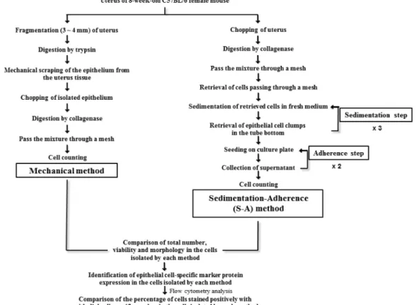
An in vitro assay following culture of endometrial epithelial cells is essential for understanding epithelial cell function in reproduction. Several diverse techniques have been developed for isolating endometrial epithelial cells, although an optimal technique has not been identified. In this study, we describe a sedimentation-adherence (S-A) isolation technique with a high-yield cell-separating ability to isolate endometrial epithelial cells from 8-week-old female C57BL/6 mice. We analyzed total cell number, viability, morphology, and expression of cytokeratin 18 as an endometrial epithelial cell-specific marker in cells isolated using a mechanical method compared to the S-A technique.

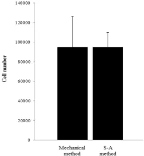
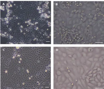
There were no significant differences in the total number, viability, or morphology of the putative endometrial epithelial cells with either method. In contrast, significantly more endometrial epithelial cells harvested using the S-A method were positively stained for cytokeratin 18 than those isolated using the mechanical method. These results confirm that the S-A method is more efficient for retrieving endometrial epithelial cells than a mechanical method.

(Key words : mouse, sedimentation-adherence method, isolation, endometrial epithelial cells, uterus)

This research was supported by a grant of the Korea Health Technology R&D Project (HI12C1404(A121515)), Ministry of Health and Welfare, Republic of Korea.

Correspondence : [email protected] and [email protected]

INTRODUCTION

The endometrium of the uterus consists of epithelial and stromal cells that undergo dynamic periodic changes in tissue differentiation, regeneration, and degeneration throughout the reproductive cycle (Deachapunva and O’Grady, 1998; Johnson et al., 1999; Arnold et al., 2001; Krikun et al., 2009). These hormone-dependent changes subsequently influence the recep- tivity of the endometrium to embryos, which is accompanied by histological and functional remodeling (Hewitt et al., 1979;

Morris and Potter, 1984; Lindenberg et al., 1988, 1989; Dea- chapunva and O’Grady, 1998; Srisuparp et al., 2003; Hantak et al., 2014), as well as abnormalities in endometrial thickness and endometriosis, which can cause pregnancy failure (Bongso et al., 1988; Cha et al., 2012; Kasius et al., 2014). Thus, the endometrium is a key regulator of the microenvironment favor- able for embryo implantation, and endometrial epithelial cell

culture is the basis for analyzing endometrium function. There have been numerous attempts to investigate endometrium func- tion during implantation (Tominaga, 1996; Thie et al., 1998;

Norwitz et al., 2001; Spencer et al., 2004; Cakmak and Taylor, 2011), the menstrual or estrus cycle (Bazer et al., 1986; Bell and Dore-Green, 1987; Boron and Boulpaep, 2005), and the endometrium-embryo interaction (Genbacev et al., 2003; Wang et al., 2004; Wollenhaupt et al., 2007; Margarit et al., 2010).

However, the reliability of experimental data has been hampered by the difficulty of successful isolation of endometrial cells.

Several techniques have been developed for the isolation of

epithelial cells from the endometrium. These include mechanical

procedures that scrape or squeeze endometrial tissue (Bigsby et

al., 1986; Lindenberg et al., 1988; Cheng et al., 2009; Eritja et

al., 2013; Janzen et al., 2013), and a sedimentation-adherence

(S-A) procedure that exploits differences in the weight and

attachment capacity of endometrial epithelial and stromal cells

(2)

Fig. 1. Experimental design.

(Riehl et al., 1983; Fujiwara et al., 2003; Azadbakht and Va- lojerdi, 2008). In addition, to improve the purity of endometrial cells, magnetic- (Chan et al., 2004; Gargett et al., 2004; Masuda et al., 2012; Messier et al., 2012) and fluorescence-activated (Chen et al., 2013; Janzen et al., 2013) cell sorting have been used after mechanical or enzymatic dissociation. However, to date, no studies have compared the purification yield among these isolation techniques, which have made it difficult to actively conduct endometrial epithelial cell research.

In this study, we investigated whether mechanical or S-A methods were more effective for the isolation of epithelial cells from endometrium by comparing the total yield, viability, morphology, phenotype, and cell-specific marker protein expre- ssion of epithelial cells.

MATERIALS AND METHODS

1. Animals

Uterine epithelial cells were obtained from 8-week-old female C57BL/6 mice (Japan SLC, Inc., Hamamatsu, Japan) and total sixty mice were equally allocated to each experiment group,

according to experimental design (Fig. 1). All animal housing, handling, and experimental procedures were approved by the Institutional Animal Care and Use Committee (IACUC) of Seoul National University (IACUC approval no. SNU-130225-1401) and conducted according to the Animal Care and Use Guide- lines of Seoul National University.

2. Mechanical Isolation of Endometrial Epithelial Cells from the Uterus Endometrial epithelial cells were isolated from the endome- trium using a previously described mechanical method with some modifications (Bigsby et al., 1986; Lindenberg et al., 1988). Briefly, uterine horns were rinsed with Hank’s balanced salt solution (HBSS; Gibco Invitrogen) supplemented with 2%

(v/v) penicillin-streptomycin (Gibco Invitrogen) and 1.25 μg/ml

Fungizone (Gibco Invitrogen) were dissected into 3 ∼4 mm

pieces and incubated in 0.25% trypsin (Sigma-Aldrich, St. Louis,

MO) at 4 ℃ for 1 h. After shaking for 30s, the epithelial tissue

was separated from the endometrial tissue by mechanical scra-

ping under a stereomicroscope (CK40M-32; Olympus, Tokyo,

Japan). Subsequently, the separated endometrial epithelial ti-

ssues were dissociated by incubating in HBSS supplemented

(3)

with 1.5 mg/ ml type I-S collagenase (Sigma-Aldrich) at 37 ℃ for 45 min and the dispersed cells were filtered through a 100 μm nylon mesh (Corning, Tewksbury, MA). Isolated endometrial epithelial cells were counted using a hemocytometer.

3. S-A Method for the Isolation of Endometrial Epithelial Cells from the Uterus

The isolation of endometrial epithelial cells from the endo- metrium using a S-A method was conducted as previously described (Azadbakht and Valojerdi, 2008) with some modifica- tions. Briefly, uterine horns were rinsed with HBSS containing 2% (v/v) penicillin-streptomycin and 1.25 μg/ml Fungizone, and split longitudinally to expose the luminal epithelium. The tissue was fragmented into fine pieces using surgical scissors and digested using 1.5 mg/ml type I-S collagenase in HBSS at 37 ℃ for 45 min, and the digested cells were filtered through 100 μm nylon mesh. A subsequent sedimentation step collected cell clumps in the tube bottom after separating the filtrated cells under unit gravity by incubating in a 15 ml tube at room temperature for 15 min. This procedure was repeated three times to remove stromal cells from cell clumps retrieved by sedimentation followed by an adherence step that retrieved the suspended cells after incubating cells clumps in a 100 mm cul- ture plate at 37 ℃ for 10 min that was repeated twice. Purified endometrial epithelial cells were counted using a hemocyto- meter.

4. Endometrial Cell Phenotype

The isolated endometrial epithelial cells were cultured in standard culture medium consisting of Dulbecco’s modified Eagle’s medium: nutrient mixture F-12 (DMEM/F12; Gibco Invitrogen) supplemented with 10% (v/v) heat-inactivated fetal bovine serum (FBS; Welgene, Daegu, Korea), 2% (v/v) peni- cillin-streptomycin, and 1.25 μg/ml Fungizone in a humidified atmosphere with 5% CO

2

at 37 ℃. The phenotype of the endo- metrial epithelial cells was observed at day 1 of culture under an inverted microscope (CKX41; Olympus) equipped with an EOS 600D digital camera (Canon, Tokyo, Japan).

5. Analysis of Cell Viability

Cell viability was assessed by incubating cells in 1 μg/ml propidium iodide (PI) staining solution (Sigma-Aldrich) dissolved in Dulbecco’s phosphate-buffered saline (DPBS; Welgene) at 4 ℃ for 1 min in the dark, and PI fluorescence was determined

using flow cytometry with the FL-2 channel of an FACS Cali- bur (Becton, Dickinson and Co., Franklin Lakes, NJ).

6. Immunocytochemistry

Cells fixed in 4% (v/v) formaldehyde (Sigma-Aldrich) for 20 min at room temperature were permeabilized in 0.1% (v/v) Triton X-100 (Sigma-Aldrich) solution for 10 min. After block- ing with DPBS supplemented with 2% (v/v) heat-inactivated FBS for 30 min at 4 ℃, the cells were incubated with uncon- jugated mouse anti-Cytokeratin 18 IgG primary antibody (1:100;

Santa Cruz Biotechnology, Dallas, TX) diluted in DPBS over- night at 4 ℃. Subsequently, primary antibody against cytokeratin 18 was detected using Texas Red-conjugated goat anti- mouse IgG secondary antibody (dilution rate = 1:100; Santa Cruz Bio- technology) for 1 h at 4 ℃. The stained cells were washed twice with DPBS, counterstained for 15 min with mounting medium for fluorescence with DAPI (Vector Laboratories, Inc., Burlin- game, CA), and observed under a fluorescence microscope (TE2000-U; Nikon, Tokyo, Japan) using NIS-Elements BR

TM

software (Nikon Instruments, New York, NY).

7. Flow Cytometry

Cells fixed with 0.01% (v/v) formaldehyde for 10 min at room temperature were washed with ice-cold HBSS and per- meabilized by incubating in HBSS containing 2% (v/v) heat- inactivated FBS and 0.1% (v/v) Triton X-100 for 15 min. Then the cells were stained for 30 min at 4 ℃ with unconjugated mouse anti-Cytokeratin 18 IgG primary antibody (1:100) and primary antibodies were detected using FITC-conjugated goat anti-mouse IgG secondary antibody (1:100) for 30 min at 4 ℃.

The stained cells were rinsed with DPBS and then sorted using a FACS Calibur (Becton, Dickinson and Co., Franklin Lakes, NJ). Analysis of data was performed using BD CellQuest Pro software (Becton, Dickinson and Co.).

8. Statistical Analysis

Statistical Analysis System software was used to analyze all numerical data. Comparisons among treatment groups were conducted using the Duncan’s method or the least-squares di- fference test, and the significance of main effects was evaluated by one-way analysis of variance (ANOVA). A value of p<0.05 was considered a statistically significant difference.

RESULTS

(4)

To elucidate a technique with high retrieval efficiency and low cytotoxicity, we compared mechanical and S-A methods by assessing total cell number and viability of putative endo- metrial epithelial cells. No significant differences in the total cell number (Fig. 2) and cell viability (Fig. 3) were found with these isolation techniques. However, cells isolated using the S-A method showed a 10% increase in viability compared to those isolated with the mechanical method (66.15±2.86 vs.

55.48±12.34, respectively).

We characterized the putative endometrial epithelial cells isolated with each method by assessing morphology and the expression of endometrial epithelial cell-specific marker protein.

Both isolation methods resulted in cells with typical epithelial cell-like morphology, described a polygonal in shape with more regular dimensions and growing attached to a substrate in discrete patches (Fig. 4) expressing cytoplasmic cytokeratin 18 (Fig. 5). The percentage of cells expressing cytokeratin 18 was significantly increased in cells isolated with the S-A method compared to the mechanical method (53.67±5.83% vs. 5.39±

2.69%, respectively). Moreover, the purity of cells harvested by the S-A method was significantly higher than those harvested by the mechanical method (Fig. 6). Thus, these results demon- strate that the S-A method can effectively isolate endometrial epithelial cells.

DISCUSSION

We investigated the most effective method for isolating endometrial epithelial cells from the mouse uterus from among those previously described (Bigsby et al., 1986; Lindenberg et al., 1988; Azadbakht and Valojerdi, 2008). Analyses revealed that 53.67% of the putative endometrial epithelial cells isolated using the S-A method expressed cytokeratin 18 as an endo- metrial epithelial cell-specific marker protein. Furthermore, the S-A method did not result in any significant reduction in total number or viability of the retrieved cells compared to the other methods. Thus, the S-A method produced the highest yield of purified cells.

As shown in Fig. 3 and 4, final viability and morphology of endometrial epithelial cells retrieved by each isolation method didn’t differ significantly and greatly, indicating that enzymatic and mechanical steps during entire isolation process don’t induce alteration of their characteristics. Accordingly, we can specu- late that endometrial epithelial cells have strong resistant and

Fig. 2. Comparison of total number of putative endometrial epithelial cells isolated from the mouse uterus with different isolation techniques. Putative cell populations were retrieved by me- chanical or S-A dissociation methods. Total cell number was determined using a hemocytometer. No significant difference in the total number of cells was detected. All data represent means±SE (standard error) of three independent experiments.

Fig. 3. Comparison of putative endometrial epithelial cell viability using different isolation techniques. Putative cell populations were retrieved by mechanical or S-A dissociation methods.

The viability of isolated cells was analyzed by flow cytometry

using propidium iodide. No significant difference in cell vi-

ability was observed. All data represent means±SE (standard

error) of three independent experiments.

(5)

Fig. 5. Translational expression of cytokeratin 18 as an endometrial epithelial cell-specific marker protein (red) in putative endometrial epithelial cell populations isolated with different isolation techniques, n=3. Scale bars=50 µm.

Fig. 4. Morphology of putative endometrial epithelial cells isolated with mechanical (A, B) and S-A methods (C, D). Retrieved cell populations were cultured for 1 day in standard epithelial cell culture medium. Cell morphology was observed by mi- croscopy. Most of the isolated cells populations were polygo- nal in shape with regular dimensions and had grown attached to a substrate in discrete patches, n=3. Scale bars =50 µm.

recovery capability against external stimulation, supported by the facts that they experience periodically dynamic alterations in structure and function during estrus cycle and preparation of blastocyst implantation in vivo (Valdez et al., 2015; Jeong et al, 2016).

During mechanical dissociation, a massive loss of endome- trial epithelial tissue or acquisition of stromal tissue adhering to endometrial epithelial tissue can occur when removing stromal fractions from the uterus after enzymatic digestion. This can reduce the number of putative endometrial epithelial cells deri- ved from small pieces of endometrial epithelial tissue. However, no significant decrease in the total number of the retrieved cells was observed, suggesting that there was a massive acquisition of stromal tissue adhering to endometrial epithelial tissue. This explains the lower proportion of purified endometrial epithelial cells in the cell suspension isolated using a mechanical method.

In contrast, we expected that there would be minimal loss

of endometrial epithelial tissues using the S-A method due to

enzymatic digestion of the entire uterus. Moreover, the increa-

sed density of cell clusters and slow adhesion of cells to the

bottom of the culture dish (Azadbakht and Valojerdi, 2008)

(6)

Fig. 6. The percentage of cytokeratin 18-expressing cells in the putative endometrial epithelial cell populations isolated using the mechanical or S-A method as analyzed by flow cytome- try. The yield of cytokeratin 18-positive cells in cells retrieved through the S-A method was significantly higher than that retrieved through the mechanical method. All data represent means±SE (standard error) of three independent experi- ments. * p<0.05.

may play a significant role in improving stromal cell removal from dissociated uterine cells, resulting in an increase in the proportion of real endometrial epithelial cells compared to pu- tative cells.

In conclusion, the S-A method was an efficient and conve- nient technique for the isolation of endometrial epithelial cells from the mouse uterus. It can contribute significantly to future endometrial epithelial cell researches related with implantation of blastocysts.

REFERENCES

Arnold JT, Kaufman DG, Seppälä M and Lessey BA. 2001.

Endometrial stromal cells regulate epithelial cell growth in vitro: A new co-culture model. Hum. Reprod. 16:836-845.

Azadbakht M and Valojerdi MR. 2008. Development of vitri- fied-warmed mouse embryos co-cultured with polarized or non-polarized uterine epithelial cells using sequential culture

media. J. Assist Reprod. Genet. 25:251-261.

Bazer FW, Vallet JL, Roberts RM, Sharp DC and Thatcher WW. 1986. Role of conceptus secretory products in establi- shment of pregnancy. J. Reprod. Fertil. 76:841-850.

Bell SC and Dore-Green F. 1987. Detection and characterization of human secretory “pregnancy-associated endometrial α 2- globulin” in uterine luminal fluid. J. Reprod. Immunol. 11:

13-29.

Bigsby RM, Cooke PS and Cunha GR. 1986. A simple effi- cient method for separating murine uterine epithelial and mesenchymal cells. Am. J. Physiol. 251:E630-636.

Bongso A, Gajra B, Lian NP, Wong PC, Soon-Chye N and Ratnam S. 1988. Establishment of human endometrial cell cultures. Hum. Reprod. 3:705-713.

Boron WF and Boulpaep EL. 2005. Medical Physiology: A Cellular and Molecular Approach. Elsevier Saunders. ISBN 1-4160-2328-3. OCLC 61527528.

Cakmak H and Taylor HS. 2011. Implantation failure: Mole- cular mechanisms and clinical treatment. Hum. Reprod. 17:

242-253.

Cha J, Sun X and Dey SK. 2012. Mechanisms of implantation:

strategies for successful pregnancy. Nat. Med. 18:1754-1767.

Chan RW, Schwab KE and Gargett CE. 2004. Clonogenicity of human endometrial epithelial and stromal cells. Biol.

Reprod. 70:1738-1750.

Chen JC, Erikson DW, Piltonen TT, Meyer MR, Barragan F, McIntire RH, Tamaresis JS, Vo KC, Giudice LC and Irwin JC. 2013. Coculturing human endometrial epithelial cells and stromal fibroblasts alters cell-specific gene expression and cytokine production. Fertil. Steril. 100:1132-1143.

Cheng SQ, He JL, Dong YL, Liu XQ, Ding YB, Gao RF, Tan Y, Ye Q, Tian ZL and Wang YX. 2009. Characterization of calreticulin expression in mouse endometrium during embryo implantation. Bio. Res. 42:505-516.

Deachapunya C and O'Grady SM. 1998. Regulation of chloride secretion across porcine endometrial epithelial cells by pro- staglandin E2. J. Physiol. 508:31-47.

Eritja N, Mirantes C, Llobet D, Yeramian A, Bergadà L, Dosil MA, Domingo M, Matias-Guiu X and Dolcet X. 2013. Long- term estradiol exposure is a direct mitogen for insulin/EGF- primed endometrial cells and drives PTEN loss-induced hyperplasic growth. Am. J. Pathol. 183:277-287.

Fujiwara H, Tatsumi K, Kosaka K, Sato Y, Higuchi T, Yo-

shioka S, Maeda M, Ueda M and Fujii S. 2003. Human

(7)

blastocysts and endometrial epithelial cells express activated leukocyte cell adhesion molecule (ALCAM/CD166). J. Clin.

Endocrinol. Metab. 88:3437-3443.

Gargett CE, Schwab KE, Zillwood RM, Nguyen HP and Wu D. 2004. Isolation and culture of epithelial progenitors and mesenchymal stem cells from human endometrium. Biol.

Reprod. 70:1738-1750.

Genbacev OD, Prakobphol A, Foulk RA, Krtolica AR, Ilic D, Singer MS, Yang ZQ, Kiessling LL, Rosen SD and Fisher SJ. 2003. Trophoblast L-selectin-mediated adhesion at the maternal-fetal interface. Science 299:405-408.

Hantak AM, Bagchi IC and Bagchi MK. 2014. Role of uterine stromal-epithelial crosstalk in embryo implantation. Int. J.

Dev. Biol. 58:139-146.

Hewitt K, Beer AE and Grinnell F. 1979. Disappearance of anionic sites from the surface of the rat endometrial epithe- lium at the time of blastocyst implantation. Biol. Reprod.

21:691-707.

Janzen DM, Cheng D, Schafenacker AM, Paik DY, Goldstein AS, Witte ON, Jaroszewicz A, Pellegrini M and Memarza- deh S. 2013. Estrogen and progesterone together expand murine endometrial epithelial progenitor cells. Stem Cells 31:808-822.

Jeong W, Jung S, Bazer FW, Song G and Kim J. 2016. Epi- dermal growth factor: Porcine uterine luminal epithelial cell migratory signal during the peri-implantation period of pre- gnancy. Mol. Cell Endocrinol. 420:66-74.

Johnson GA, Burghardt RC, Newton GR, Bazer FW and Spencer TE. 1999. Development and characterization of immortalized ovine endometrial cell lines. Biol Reprod. 61:1324-1330.

Kasius A, Smit JG, Torrance HL, Eijkemans MJ, Mol BW, Opmeer BC and Broekmans FJ. 2014. Endometrial thick- ness and pregnancy rates after IVF: A systematic review and meta-analysis. Hum. Reprod. 20:530-541.

Krikun G, Lockwood CJ and Paidas MJ. 2009. Tissue factor and the endometrium: From physiology to pathology. Th- romb Res. 124:393-396.

Lindenberg S, Hyttel P, Sjøgren A and Greve T. 1989. A com- parative study of attachment of human, bovine and mouse blastocysts to uterine epithelial monolayer. Hum. Reprod.

4:446-456.

Lindenberg S, Sundberg K, Kimber SJ and Lundblad A. 1988.

The milk oligosaccharide, lacto-N-fucopentaose I, inhibits attachment of mouse blastocysts on endometrial monolayers.

J. Reprod. Fertil. 83:149-158.

Margarit L, Taylor A, Roberts MH, Hopkins L, Davies C, Brenton AG, Conlan RS, Bunkheila A, Joels L, White JO and Gonzalez D. 2010. MUC1 as a discriminator between endometrium from fertile and infertile patients with PCOS and endometriosis. J. Clin. Endocrinol. Metab. 95:5320-5329.

Masuda H, Anwar SS, Bühring HJ, Rao JR and Gargett CE.

2012. A novel marker of human endometrial mesenchymal stem-like cells. Cell Transplant 21:2201-2214.

Messier EM, Mason RJ and Kosmider B. 2012. Efficient and rapid isolation and purification of mouse alveolar type II epithelial cells. Exp. Lung. Res. 38:363-373.

Morris JE and Potter SW. 1984. A comparison of developmen- tal changes in surface charge in mouse blastocysts and uterine epithelium using DEAE beads and dextran sulfate in vitro. Dev. Biol. 103:190-199.

Norwitz ER, Schust DJ and Fisher SJ. 2001. Implantation and the survival of early pregnancy. N. Engl. J. Med. 345:1400- 1408.

Riehl RM, Pathak RK and Harper MJ. 1983. A reliable method for isolating endometrial epithelial cells from rabbits, and preliminary studies of prostaglandin uptake. Biol. Reprod.

28:363-375.

Spencer TE, Johnson GA, Bazer FW and Burghardt RC. 2004.

Implantation mechanisms: Insights from the sheep. Repro- duction 128:657-668.

Srisuparp S, Strakova Z, Brudney A, Mukherjee S, Reierstad S, Hunzicker-Dunn M and Fazleabas AT. 2003. Signal trans- duction pathways activated by chorionic gonadotropin in the primate endometrial epithelial cells. Biol. Reprod. 68:

457-464.

Thie M, Röspel R, Dettmann W, Benoit M, Ludwig M, Gaub HE and Denker HW. 1998. Interactions between trophoblast and uterine epithelium: monitoring of adhesive forces. Hum.

Reprod. 13:3211-3219.

Tominaga T. 1996. Studies on the mechanism of embryo im- plantation. Nihon Sanka Fujinka Gakkai Zasshi 48:591-603.

Valdez-Morales FJ, Gamboa-Dominguez A, Vital-Reyes VS, Cruz JC, Chimal-Monroy J, Franco-Murillo Y and Cerbon M. 2015. Changes in receptivity epithelial cell markers of endometrium after ovarian stimulation treatments: Its role during implantation window. Reprod. Health. 12:45.

Wang X, Matsumoto H, Zhao X, Das SK and Paria BC. 2004.

Embryonic signals direct the formation of tight junctional

(8)

permeability barrier in the decidualizing stroma during em- bryo implantation. J. Cell Sci. 117:53-62.

Wollenhaupt K, Brüssow KP, Tiemann U and Tomek W. 2007.

The embryonic pregnancy signal oestradiol influences gene expression at the level of translational initiation in porcine

endometrial cells. Reprod. Domest. Anim. 42:167-175.

Received November 4, 2015, Revised December 24, 2015,

Accepted March 24, 2016

수치

Fig.  1.  Experimental  design.
Fig.  2.  Comparison  of  total  number  of  putative  endometrial  epithelial  cells  isolated  from  the  mouse  uterus  with  different  isolation  techniques
Fig.  5.  Translational  expression  of  cytokeratin  18  as  an  endometrial  epithelial  cell-specific  marker  protein  (red)  in  putative  endometrial  epithelial  cell  populations  isolated  with  different  isolation  techniques,  n=3
Fig.  6.  The  percentage  of  cytokeratin  18-expressing  cells  in  the  putative  endometrial  epithelial  cell  populations  isolated  using  the  mechanical  or  S-A  method  as  analyzed  by  flow  cytome-  try

참조

관련 문서