Ulmus macrocarpa Hance Reduces Cyclophosphamide-induced Toxicity in Mouse Liver
Deok Won Kim and Kyung Tae Chung*
Department of Clinical Laboratory Science Graduate School, Dong-Eui University, Busan 47340, Korea Received January 25, 2021 /Revised February 22, 2021 /Accepted February 23, 2021
Cyclophosphamide (CP) is widely used in cancer and lymphoma treatments and as an immunosup- pressant drug. CP is a DNA alkylating agent that metabolizes into 4-hydrocyclophosphamide (4H- CYP) and aldophosphamide in hepatocytes. However, its metabolites cause DNA synthesis disorder, leading to apoptosis and toxic side effects. The development of technology to minimize this side effect is essential to improve CP’s clinical application. Various bioactive compounds have been reported to have anti-cancer and antioxidant functions and preventive or therapeutic roles in metabolic diseases.
Many researchers have attempted to minimize the side effects and improve the efficacy of these drugs together with the use of bioactive compounds. Ulmus macrocarpa Hance has been used for the treat- ment of edema, mastitis, stomach pain, tumors, cystitis, and other inflammatory diseases. The aim of this study was to investigate at the histological level the protective function of U. macrocarpa Hance against CP’s side effects and any potential toxic effect of U. macrocarpa Hance in the liver and kidney.
Water extracts of U. macrocarpa Hance reduced CP-induced toxicity and did not induce any histo- logical damage in the liver and kidney. Therefore, U. macrocarpa Hance would be applicable in the pharmaceutical industry.
Key words :
Cyclophosphamide, histological damage, kidney, liver, Ulmus macrocarpa
*Corresponding author
*Tel : +82-51-890-2681, Fax : +82-51-890-2622
*E-mail : [email protected]
This is an Open-Access article distributed under the terms of the Creative Commons Attribution Non-Commercial License (http://creativecommons.org/licenses/by-nc/3.0) which permits unrestricted non-commercial use, distribution, and reproduction in any medium, provided the original work is properly cited.
Introduction
Cyclophosphamide (CP) is a DNA alkylating agent. When administered, CP is metabolized into 4-hydrocyclophos- phamide (4H-CYP) and aldophosphamide by the cyto- chrome P-450 enzyme present in the hepatocytes. Aldophos- phamide is further decomposed into phosphoramide mus- tard and acrolein, which are active metabolites. CP is widely used in cancer and lymphoma treatments as well as an im- munosuppressant drug [1, 11]. However, the clinical use of CP has been limited due to its ability to damage normal tissues which usually resulted in multiple organ toxicity mainly in the heart, testes, urinary bladder, and liver [2, 20, 26].
There are many reports on various bioactive compounds that have anti-cancer, antioxidant functions, and preventive and/or therapeutic roles in metabolic diseases [5, 24, 25].
In other words, simultaneously, bioactive compounds pro-
tect organs against harmful chemicals as well as other cell stress factors. Catechin is one of the most well-known bio- active compounds and has been reported for biological func- tion of anti-inflammation and anti-oxidation on cancer, car- diovascular disease, and metabolic diseases such as diabetes [9, 19]. Resveratrol is a strong anti-oxidant bioactive com- pound suppressing NADPH oxidase-mediated production of ROS [27]. Furthermore, resveratrol was reported for its anti-aging effects observed in lower organisms [3]. Quercetin is enriched in onion peel and can be ingested as a part of common diets and has anti-hypertensive actions mimicking verapamil, a Ca
2+ channel blocker that reduces blood pres- sure [16].
Recently, diverse biological functions of Ulmus macrocarpa Hance such as antioxidant, antihypertensive, anti-cancer, and anti-thrombotic activity have been studied [13, 18, 28].
Ulmus macrocarpa Hance is a deciduous tree (Ulmaceae) na-
tive to Korea [23]. In traditional medicine, it has been used
for the treatment of edema, mastitis, stomach pain, tumors,
cystitis as well as other inflammatory diseases [12]. Al-
though major bioactive compounds of U. macrocarpa Hance
have not been defined well, its reported biological function
must depend on bioactive compounds. For clinical use,
when administered orally, U. macrocarpa Hance should not
cause any adverse effects on any organ. All compounds are
- Note -
metabolized in the liver and excreted through the kidney.
In this study, we investigated any potential toxic effect at histological level of U. macrocarpa Hance in the liver and the kidney as well as protection function of Ulmus macro- carpa Hance against CP which causes side effects on many organs.
Materials and Methods
Experimental animals
12 weeks old male BALB/c mice (23±2 g) were purchased from Samtaco Bio Korea (Osan, Korea). These animals were kept under standard conditions with temperature maintain- ing 24±1℃, humidity 55±5%, and 12 hr dark-light cycle.
Food and water were freely accessible. All experimental procedures were followed by the guidelines of the Institu- tional Animal Care and Use Committee of Dong-Eui Uni- versity (R2014-017).
Preparation of Ulmus macrocarpa Hance water extract Cortex of Ulmus macrocarpa Hance was purchased from Dae-Han herbal medicine Inc. (Busan, Korea). Water ex- traction of the cortex of U. macrocarpa Hance was produced by heating at 95℃ for 6 hr and then filtered with an 80 mesh filter. The filtered extract was concentrated at 75℃
for 2 hr, and lyophilized at -45℃. The U. macrocarpa Hance water extract (UMWE) was dissolved in sterilized water be- fore the experiment [15].
Experimental design
The mice were divided into six groups (n=6); Normal control, CP administration, CP+UMWE 100 mg/kg, CP+
UMWE 200 mg/kg, UMWE 100 mg/kg, UMWE 200 mg/kg.
The mice of test groups were orally administered with UMWE for 14 days and the same amount of sterilized water was orally administered to the control group. Intraperito- neal injection of a dose of 120 mg/kg of cyclophosphamide (Sigma-Aldrich, USA) was administered to the test groups on the 13th day. All animals did not fast until the day of sacrifice.
Organ to body weight ratio
The liver and kidneys were dissected out from the sacri- ficed animals and the weights of each organ were measured.
The organ body weight ratio was calculated by the follow- ing formula [4].
Organ to body
weight ratio = Organ weight (mg) Body weight (g) ×100
Histological analysis
The liver and kidney were removed and fixed in 10%
neutral buffered formalin for 24 hr. The fixed tissues were dehydrated with a tissue processor (LEICA TP 1020, Leica Biosystems, Germany) and subjected to a clear, paraffin in- filtration process. Subsequently, the organs embedded in paraffin wax using a paraffin embedding station (Tissue- Tek® TEC
™5, Sakura, United States) and slides with a 3 μm section were cut using a microtome (LEICA RM 2235, Leica Biosystems, Germany) and stained with hematoxylin and eosin (H&E). For DAPI staining, tissue sections were deparaffinized and stained with DAPI. The slides were ex- amined under a fluorescence microscope (Axio Scope A1, Carl Zeiss, Germany). The number of hepatocyte nuclei ex- cluding kupffer cells were counted in four corner squares of 25×10
4pixel
2of a field of view.
Statistical analysis
Data were analyzed with GraphPad Prism 5 (GraphPad Software, United States). One-way ANOVA and Bonferroni post-test were used to compare multiple groups at sig- nificance level p<0.05. Results were expressed as mean ± standard deviation.
Results and Discussion
Body weight and organ weight are important indices in toxicity investigation of drugs and natural compounds [10].
In this case ratio of organ weight to body weight (organ weight index) is used to judge toxicity of any compound and especially for liver, organ index weight is required [17, 22]. According to the previous reports, decrease in body weight generally indicates toxicity of test materials [10]. To be consistent with previous reports, single administration of CP in this study showed decrease in body weight which is indicative of toxic effect. In body weight analysis, decrease of CP group’s body weight was statistically significant when compared with normal control group’s body weight (Table 1). Not only CP group but also body weight of CP+UMWE 100 and CP+UMWE200 groups was decreased. However, CP+UMWE200 group only showed statistical significance.
Similarly, the liver weight of CP+UMWE200 group was also
decreased. In order to justify decrease of liver weight of CP+
Fig. 1. Histological analysis of liver. Top panel: H&E-stained liver, bottom panel: DAPI-stained liver (x400). Open triangle indicates glycogen accumulation.
Table 1. Body weight and organ weight of each experimental group (n=6)
Parameter† Normal control CP UMWE100+CP UMWE200+CP UMWE100 UMWE200
Terminal body weight (g) Organ weight (g) Liver
Kidneys
Organ to body weight ratio (mg/g)
Liver Kidneys
25.90±1.45
1.270±0.239 0.358±0.038
48.800±7.101 13.800±0.816
24.08±1.06*
1.090±0.212 0.321±0.064
45.060±7.333 13.260±2.200
24.90±0.65
1.092±0.072 0.344±0.063
43.850±2.757 13.780±2.370
24.10±0.82*
1.012±0.152*
0.324±0.021
41.870±5.009 13.440±0.699
25.28±2.16
1.162±0.292 0.354±0.045
45.520±8.282 14.010±1.566
25.80±0.91
1.136±0.183 0.354±0.035
43.870±5.556 13.710±1.071
†Multiple groups of each parameter were analyzed by one-way ANOVA and Bonferroni post-test using GraphPad Prism 5.
*indicates statistically significant difference in groups.
Statistical significance means p<0.05.
UMWE200 group organ to body weight ratio was compared.
When organ indices were compared there was no statisti- cally significant difference among all groups, which indi- cates liver weight was proportionally decreased with body weight in CP+UMWE200 group due to CP, not to UMWE intake. Both UMWE100 and UMWE200 groups did not show any decrease in body weight as well as in organ index, sug- gesting that UMWE would not be toxic. Data of UMWE100 and UMWE200 groups were supporting this interpretation (Table 1). Body weight of UMWE200 group was 25.80±0.91 and liver weight was 1.136±0.0183, which is no statistically significant differences in body and liver weight of normal group.
Hematoxylin and Eosin (H&E) stain is the basic histo- logical staining for a general assessment of cell and tissue morphology. Cellular nucleus shows blue-purple and cyto- plasm and the cartilage matrix pinkish red by H&E staining, but neutrally charged molecules such as glycogen does not
stain leaving clear areas. When the mouse is fasting , the entire area of cytoplasm of hepatocyte is stained without clear areas by H&E staining because of no or weak accumu- lation of glycogen. However, when the mouse is fed nor- mally, the cytoplasm of hepatocyte shows clear areas by H&E staining because accumulated glycogen area in cyto- plasm is partially unstained.
All mice of each group had not fasted during the ex- perimental period. Hepatocytes of the normal control group showed a typical H&E stained pattern with obvious glyco- gen accumulation (Fig. 1). In contrast, H&E-stained hep- atocytes of the CP group did not show glycogen accumulat- ion. However, hepatocytes of both CP+UMWE100 and CP+
UMWE200 groups showed glycogen accumulation but not
as much as the amount of glycogen in normal group’s
hepatocytes. These data suggested that CP’s toxicity was
alleviated by UMWE intake. Hepatocytes of both UMWE100
and UMWE200 groups showed glycogen accumulation as
Fig. 2. Number of hepatocytes. DAPI-stained nuclei were count- ed in four corner squares of field of view. One square was 25x104 pixel2. All groups were analyzed by one-way ANOVA and Bonferroni post-test using GraphPad Prism 5. *: statistical difference compared with normal control,
#: statistical difference compared with CP. *** p<0.001,
**p<0.01, ##p<0.01
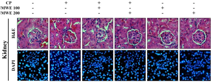
Fig. 3. Histological analysis of kidney. Top panel: H&E-stained kidney, bottom panel: DAPI-stained kidney (×400).
much as the amount of glycogen in normal group’s hepato- cytes . Based on these data CP seemed to cause a reduced intake of food which explains loss of body weight and no or less glycogen accumulation in CP-treated groups. On the other hand, UMWE seemed not to cause food take interfer- ence. We also analyzed nuclear change with DAPI staining for apoptosis caused by toxicity of CP or UMWE. No ob- vious apoptotic nucleus was identified (Fig. 1). One differ- ence was density of nucleus which varied among the groups.
Number of nuclei was counted to quantify cell numbers in the same size of area. Nuclei in four corner squares of 250,000 pixel
2were counted with three different slides of
each group and statistically analyzed. When compared with the normal group the number of cells of the CP group in- creased by 32.7% and the number of cells of CP+ UMWE100 also increased by 25.1%. However, the number of cells of CP+ UMWE200 was 27.5% less than that of CP group and remained similar to that of normal group (Fig. 2). These data indicate that volume of hepatocytes of CP group became smaller due to no glycogen accumulation caused by CP tox- icity, therefore, cell population increased within the area. It appears that UMWE reduced CP toxicity in both CP+UMWE 100 and CP+UMWE200 groups. Both UMWE100 and UMWE 200 groups without CP maintained as much as normal cell populations, which is again consistent with H&E stained pattern results.
The kidneys are responsible to maintain chemical compo- sition of cells by regulating the amount of water, electrolytes as well as many other molecules. Many drugs including anti- cancer drugs including cyclophosphamide have been re- ported to cause renal toxicity [21]. Susceptibility of the organ is due to receiving 20~25% of resting cardiac output, ex- posure to a higher concentration of drugs during filtration, increased intracellular concentrations of drugs via trans- porters, and high energy requirement of the tubules [8].
Recently, natural compounds are increasingly reported for treatment use of kidney diseases [14]. Natural compounds from medicinal plants have shown protective activity against nephrotoxicity. However, there is more likely no known study of U. macrocarpa effect on kidney under cyclophospha- mide administration. Our histological data of the kidney showed that structures of the glomerulus, glomerular capsu- le, and renal tubular cells were not altered in all groups (Fig.
3). One of the reasons would be a single administration in
this study. Kidney damage by cyclophosphamide has been reported by multiple administrations during a certain ex- perimental period. Ei-Shabrawy, et al, showed tubular and glomerular distortion in the kidney with 6 times admin- istrations during 3 weeks [7]. On the other hand, UMWE did neither cause any histological damage in the kidney by both 100 and 200 mg/kg concentrations, which suggests that UMWE does not have any toxic effect on the kidney after two weeks of a feeding period.
In summary, our data showed that single administration of CP caused histological change in the liver, and two weeks of UMWE feeding before CP administration reduced Cyclo- phosphamide-Induced toxicity and maintained histological structure close to the normal condition of the liver. Further- more, UMWE by itself did not show any histological struc- ture change of the liver. Two weeks of lab mouse is almost equivalent to one and half years of human lifespan [6].
Therefore, long-term intake of UMWE may not cause any adverse effect particularly in human liver and regular intake of UMWE would be applicable for nutraceutical tablets. No obvious histological change in kidney was identified by ei- ther CP or UMWE. However, this study did not present bio- chemical data of CT toxicity and UMWE effect which may link direct or indirect action against each other. Bichemical investigation with single and multiple administration of CP requires to better understand more precise beneficial roles of UMWE against CP.
The Conflict of Interest Statement
The authors declare that they have no conflicts of interest with the contents of this article.
References
1. Aladaileh, S. H., Abukhalil, M. H.,Saghir, S. A. M., Hanieh, H., Alfwuaires, M. A., Almaiman, A. A., Bin-Jumah, M. and Mahmoud, A. M. 2019. Galangin activates Nrf2 signaling and attenuates oxidative damage, inflammation, and apop- tosis in a rat model of cyclophosphamide-induced hepato- toxicity. Biomolecules 9, 346.
2. Almazor, M. E. S., Belseck, E., Shea, B., Wells, G. and Tugwell, P. 2000. Cyclophosphamide for rheumatoid arthritis.
Cochrane Database Syst. Rev. 4, CD001157.
3. Bhullar, K. S. and Hubbard, B. P. 2015. Lifespan and health- span extension by resveratrol. Biochim. Biophys. Acta 1852, 1209-1218.
4. Chan, P. C., Ramot, Y., Malarkey, D. E., Blackshear, P., Kis- sling, G. E., Travlos, G. and Nyska, A. 2010. Fourteen-week
toxicity study of green tea extract in rats and mice. Toxicol.
Pathol. 38, 1070-1084.
5. De la Iglesia, R., Loria-Kohen, V., Zulet, M. A., Martinez, J. A., Reglero, G. and de Molina, A. R. 2016. Dietary strat- egies implicated in the prevention and treatment of metabol- ic syndrome. Int. J. Mol. Sci. 17, 1877.
6. Dutta, S. and Sengupta, P. 2016. Men and mice: Relating their ages. Life Sci. 152, 244-248.
7. El-Shabrawy, M., Mishriki, A., Attia, H., Aboulhoda, B. E., Emam, M. and Wanas, H. 2020. Protective effect of tolvaptan against cyclophosphamide-induced nephrotoxicity in rat models. Pharmacol. Res. Perspect. 8, e00659.
8. Griffin, B. R., Faubel, S. and Edelstein, C. L. 2019. Biomark- ers of drug-induced kidney toxicity. Ther. Drug Monit. 41, 213-226.
9. He, H. F. 2017. Research progress on theaflavins: efficacy, formation, and preparation. Food Nutr. Res. 3, 1344521.
10. Hoffman, W. P., Ness, D. K. and Van Lier, R. B. 2002. Anal- ysis of rodent growth data in toxicology studies. Toxicol. Sci.
66, 313-319.
11. Iqubal, A., Iqubal, M. K., Sharma, S., Ansari, M. A., Najmi, A. K., Ali, S. M., Ali, J. and Haque, S. E. 2019. Molecular mechanism involved in cyclophosphamide-induced cardio- toxicity: Old drug with a new vision. Life Sci. 218, 112-131.
12. Kim, J. M., Choi, M. S., Cho, J. G., Jung, Y. M. and Park, T. W. 1994. Effect of Euonymus alatus and Ulmus clavidiana var japonica on the immune system. Kor. J. Vet. Res. 34, 307- 313.
13. Kim, T. M., Shin, S. K., Kim, T. W., Youm, S. Y., Kim, D.
J. and Ahn, B. 2012. Elm tree bark extract inhibits HepG2 hepatic cancer cell growth via pro-apoptotic activity. J. Vet.
Sci. 13, 7-13.
14. Khajavi, R. A., Mohebbati, R. and Hosseinian, S. 2017. Drug- induced nephrotoxicity and medicinal plants. Iran J. Kidney Dis. 11, 169-179.
15. Lee, S. D., Kim, D. W., Lee, I., Lee, J. H., Hyun, S. K., Kang, K. H., Hwang, H. J., Kim, C. M., Kim, B. W. and Chung, K. T. 2016. Ulmus macrocarpa Hance water extract improved splenocytes survival and NK cell cytotoxicity. J. Life Sci. 26, 109-116.
16. Marunaka, Y., Marunaka, R., Sun, H., Yamamoto, T., Kana- mura, N., Inui, T. and Taruno, A. 2017. Actions of quercetin, a polyphenol, on blood pressure. Molecules 22, 209.
17. Michael, B., Yano, B., Sellers, R. S., Perry, R., Morton, D., Roome, N., Johnson J. K., Schafer, K. and Pitsch, S. 2007.
Evaluation of organ weights for rodent and non-rodent tox- icity studies: a review of regulatory guidelines and a survey of current practices. Toxicol. Pathol. 35, 742-750.
18. Oh, K. S., Ryu, S. Y., Oh, B. K., Seo, H. W., Kim, Y. S. and Lee, B. H. 2008. Antihypertensive, vasorelaxant, and anti- oxidant effect of root bark of Ulmus macrocarpa. Biol. Pharm.
Bull. 31, 2090-2096.
19. Ohishi, T., Goto, S., Monira, P., Isemura, M. and Nakamura, Y. 2016. Anti-inflammatory action of green tea. Antiinflamm.
Antiallergy Agents Med. Chem. 15, 74-90.
20. Oyagbemi, A. A., Omobowale, O. T., Asenuga, E. R., Akin-
초록:Cyclophosphamide가 유발한 간 조직변화에 대한 느릅나무 열수추출물의 완화 효과
김덕원․정경태*
(동의대학교 대학원 임상병리학과)
Cyclophosphamide (CP)는 면역 억제제 뿐만 아니라 암 및 림프종 등의 치료에 널리 사용된다. CP는 DNA 알 킬화제로서, 간세포에서 대사되어 4-hydrocyclophosphamide (4H-CYP)와 aldophosphamide로 분리된다. Ulmus macrocarpa Hance는 부종, 유방염, 종양 및 기타 염증성 질환에 사용되어 왔다. 이 연구의 목적은 CP의 부작용에 대하여 U. macrocarpa Hance 열수 추출물이 조직학적 수준에서 CP의 부작용에 대한 간과 신장의 보호 기능과 U. macrocarpa Hance 자체의 잠재적 독성 영향을 조사하고자 하였다. 마우스 모델을 사용하여 헤마톡실린 및 에오 신(H&E) 염색과 DAPI 염색으로 간과 신장을 조직학적으로 분석하였다. CP 처리한 마우스에서 간세포의 형태는 글리코겐 축적을 나타내지 않았고, 세포 밀도도 감소하였다. 그러나 UMWE+CP 처리군에서는 간세포의 형태와 세포 밀도는 정상 간세포 패턴과 유사하였다. 또한, UMWE으로만 처리한 마우스에서도 간세포의 형태와 세포 밀도는 정상 간세포와 유사했다. 신장의 경우에는 정상 마우스와 비교했을 때 H&E 염색으로는 CP 또는 UMWE 처리된 마우스의 신장에서 명백한 차이를 나타내지 않았다. 즉, U. macrocarpa Hance의 열수추출물은 간과 신장에 아무런 영향을 유발하지 않으면서 CP가 유발한 독성을 감소시키는것으로 요약된다. 따라서 U. macrocarpa Hance 는 제약 산업에 사용될 수 있는 가능성을 나타내었다.
leye, A. S., Ogunsanwo, R. O. and Saba, A. B. 2016. Cyclo- phosphamide-induced hepatotoxicity in wistar rats: the modulatory role of gallic acid as a hepatoprotective and che- mopreventive phytochemical. Int. J. Prev. Med. 7, 51.
21. Santos, M. L. C., de Brito, B. B., da Silva, F. A. F., dos Santos Botelho, A. C. and de Melo, F. F. 2020. Nephrotoxicity in cancer treatment: An overview. World J. Clin. Oncol. 11, 190- 204.
22. Sellers, R. S., Mortan, D., Michael, B., Roome, N., Johnson, J. K., Yano, B. L., Perry, R. and Schafer, K. 2007. Society of toxicologic pathology position paper: organ weight rec- ommendations for toxicology studies. Toxicol. Pathol. 35, 751-755.
23. Seo, B. I., Ju, Y. S., Choi, H. Y., Park, J. H., Roh, S. S., Koo, J. S., Kim, J. J. and Kim, D. Y. 2011. Illustrated Book of Herbal Plants in Oriental Medicine. 1st ed., DaeWondang, Daegu.
24. Skrovankova, S., Sumczynski, D., Mlcek, J., Jurikova, T. and
Sochor, J. 2015. Bioactive compounds and antioxidant activ- ity in different types of berries. Int. J. Mol. Sci. 16, 24673- 24706.
25. Subramaniam, S., Selvaduray, K. R. and Radhakrishnan, A.
K. 2019. Bioactive compounds: natural defense against can- cer? Biomolecules 9, 758.
26. Üsküdar, C. D., Öztaş, E., Yilmaz, E. and Korkmaz, C. 2019.
Cyclophosphamide-induced severe acute hepatitis in a rheumatic disease: case-based review. Rheumatol. Int. 39, 377-385.
27. Xia, N., Daiber, A. and Förstermann, U. 2017. Antioxidant effects of resveratrol in the cardiovascular system. Br. J.
Pharmacol. 174, 1633-1646.
28. Yang, W. K., Lee, J. J., Sung, Y. Y., Kim, D. S., Myung, C.
S. and Kim, H. K. 2013. Extract of Ulmus macrocarpa Hance prevents thrombus formation through antiplatelet activity.
Mol. Med. Rep. 8, 726-730.