• 검색 결과가 없습니다.

A Case of Invasive Pulmonary Aspergillosis with Direct Invasion of the Mediastinum and the Left Atrium in an Immunocompetent Patient

N/A
N/A
Protected

Academic year: 2021

Share "A Case of Invasive Pulmonary Aspergillosis with Direct Invasion of the Mediastinum and the Left Atrium in an Immunocompetent Patient"

Copied!
6
0
0

로드 중.... (전체 텍스트 보기)

전체 글

(1)

A Case of Invasive Pulmonary Aspergillosis with Direct Invasion of the Mediastinum and the Left Atrium in an Immunocompetent Patient

Kyu-Hyun Han, M.D.

1

, Jung-Hyun Kim, M.D.

1,2

, Sun Young Shin, M.D.

1

, Hye Yun Jeong, M.D.

1

, Ji Min Chu, M.D.

1

, Hak Su Kim, M.D.

1

, Daejin Kim, M.D.

1

, Minjung Shim, M.D.

1

, Sang-Ho Cho, M.D.

3

and Eun Kyung Kim, M.D.

1,2

1

Department of Internal Medicine,

2

Division of Pulmonology,

3

Department of Pathology, CHA Bundang Medical Center, CHA University, Seongnam, Korea

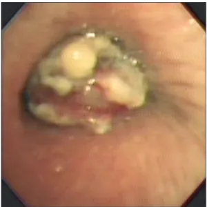
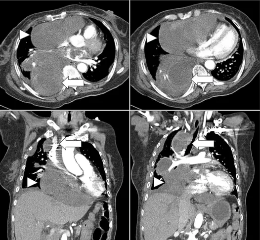
We report a case of invasive pulmonary aspergillosis invading the mediastinum and the left atrium. A 70-year-old woman was hospitalized for dyspnea. She had been well controlled for her diabetes mellitus and hypertension. The chest X-ray disclosed mediastinal widening, and the computed tomography scan of the chest showed that there was a large mediastinal mass and this lesion extended into the left atrium and right bronchus. The cardiac echocardiography showed that a huge mediastinal cystic mass compressed in the right atrium and a hyperechoic polypoid lesion in the left.

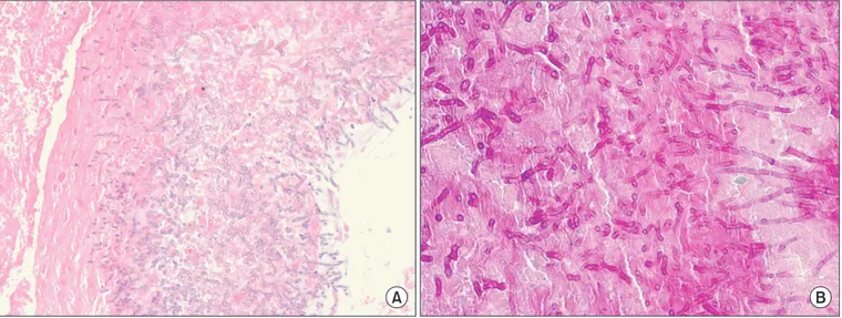
The pathology from the bronchoscopic biopsy observed abundant fungal hyphae which was stained with periodic acid- Schiff and Gomori's methenamine silver. Despite the treatment with antifungal agents, she died from cardiac tamponade after three months. Invasive pulmonary aspergillosis, which involves the mediastinum and the heart, is very rare in immunocompetent patients.

Keywords: Invasive Pulmonary Aspergillosis; Immunocompetence; Mediastinal Neoplasms; Heart Neoplasms

competent patients

1

. In addition, invasive aspergillosis, which involves mediastinum in an immunocompetent host, was reported only once in the literature

2

. To the best of our knowl- edge, this is the second case report of invasive pulmonary aspergillosis extending to the mediastium and the left atrium in an immunocompetent patient.

Case Report

A 70-year-old woman visited our hospital for mild dyspnea, cough, and sputum on March 6, 2012. She had been in a well- controlled state of her diabetes and hypertension since 2000.

The level of glycosylated hemoglobin A

1c

was 6.1% on admis- sion. She had no other medical history and did not administer medications such as corticosteroid or antibiotics. On admis- sion, body temperature was 37.4

o

C, blood pressure 100/70 mm Hg, and pulse rate 92 beats per minute. Breath sounds Copyright © 2014

The Korean Academy of Tuberculosis and Respiratory Diseases.

All rights reserved.

Introduction

Although invasive pulmonary aspergillosis is not rare in immunocompromised patients, it is very rare in immuno-

Address for correspondence: Eun Kyung Kim, M.D.

Division of Pulmonology, Department of Internal Medicine, CHA Bundang Medical Center, CHA University School of Medicine, 59 Yatap- ro, Bundang-gu, Seongnam 463-712, Korea

Phone: 82-31-780-6141, Fax: 82-31-780-6143 E-mail: imekkim@cha.ac.kr

Received: Mar. 20, 2014 Accepted: Apr. 9, 2014

cc

It is identical to the Creative Commons Attribution Non-Commercial

License (http://creativecommons.org/licenses/by-nc/3.0/).

(2)

Invasive pulmonary aspergillosis with mediastinal invasion

were diminished over the right lung. The laboratory test showed a white blood cell count of 7,760/mL and a C-reactive protein of 0.39 mg/dL. A plain chest radiograph showed prom-

inent mass opacity in the right hilum (Figure 1). A computed tomography (CT) scan of the chest revealed an ill-defined poorly enhanced mediastinal mass invading the subcarina,

Figure 1. The chest radiograph obtained on admission shows prominent opacities on the right hilum.

Figure 2. The chest computed tomogra- phy scan shows the mediastinal mass compressing the right atrium and the right ventricle (arrowheads). The medias- tinal mass is connected to the left atrium, the right hilum, and subcarina (arrows).

Figure 3. In bronchofiberscopic examination, the orifice of right bron-

chus intermedius is nearly completely obstructed by a bloody mass.

(3)

the right hilum, and the left atrium, a protruding lesion in the right bronchus intermedius, peribronchovascular interstitial thickening around the right bronchus, and a small amount of

pleural effusion in the right hemithorax. The mass in the me- diastinum compressed the right atrium and the right ventricle (Figure 2). The diagnostic thoracentesis revealed a lympho- Figure 4. (A) The bronchoscopic biopsy of the mass observed in abundant fungal hyphae (H&E stain, ×20). (B) Fungal hyphae were positive stained with periodic acid-Schiff stain (×40).

Figure 5. The polypoid mass in the left atrium was shown (arrows) in paraster- nal long axis view (A) and parasternal short axis view (B) of echocardiogram.

The mediastinal mass (arrowheads) compressed the right atrium and the right ventricle in subcostal view (C, D) of echocardiogram. Ao: aorta; AV: aortic valve; LA: left atrium; LV: left ventricle;

MV: mitral valve; PA: pulmonary artery;

RA: right atrium; RV: right ventricle.

(4)

Invasive pulmonary aspergillosis with mediastinal invasion

cyte dominant exudate, a white blood cell count of 1,630/mL (lymphocyte 43%), albumin of 2.2 g/dL, lactate dehydroge- nase of 883 U/L, glucose of 210 mg/dL, and adenosine deami- nase of 15I U/L. The cytologic examination observed no ma- lignant cells in the pleural effusion. A CT-guided percutaneous needle aspiration biopsy (PCNA) of the mediastinal mass was conducted. The pathology revealed necrotic tissues only with no evidence of malignancy. In the fiberoptic bronchoscopy, the orifice of the right bronchus intermedius was nearly totally obstructed by a bloody mass (Figure 3). The pathology from the bronchoscopic biopsy observed abundant fungal hyphae.

The result of stains with periodic acid-Schiff and Gomori’s me- thenamine silver for fungi was positive for fungal hyphae, and the acid-fasting staining showed a negative result for acid-fast bacilli (Figure 4). The serologic test for Aspergillus fumigatus was negative. An echocardiography showed an ejection frac- tion of 63% and normal global left ventricular systolic function with no regional wall motion abnormality. However, there was a huge mediastinal mass compressing the heart and a hyperechoic polypoid lesion in the left atrium (Figure 5). She was diagnosed with invasive pulmonary aspergillosis involv- ing the heart and thus, amphotericin B (0.25 mg/kg/day) was prescribed intravenously.

Fourteen days after hospitalization, dyspnea got more

worsened. A follow-up chest radiograph showed aggravated pleural effusion in the right hemithorax and worsened cardio- megaly. The effusion was surgically drained by video-assisted thoracic surgical drainage. The antifungal agent was voricon- azole, initially, 6 mg/kg intravenously every 12 hours, and then changed to 4 mg/kg intravenously every 12 hours.

Two weeks later, the chest tube was removed, but found a cardiac tamponade. This was why the sono-guided pig-tail catheter was inserted into the pericardium. Seven days later, the catheter was removed and the endobronchial aspergil- loma was removed using fiberoptic bronchoscope for pallia- tion. Two months later, a small amount of pleural effusion still remained. The mediastinal mass decreased and her dyspnea improved. She was discharged from a hospital.

One week later, her dyspnea got worsened. The laboratory test showed thrombocytopenia of the platelet cell count of 26,000/mL. The body temperature was 36.5

o

C, blood pressure 90/60 mm Hg, and pulse rate 130 beats per minute. A CT scan showed that the amount of pleural effusion and pericardial fluid increased more. The mediastinal mass increased in size and still compressed the heart, and the heart was deviated to the left side (Figure 6). The echocardiography observed increased polypoid lesion in the left atrium, and the lesion shifted to mitral valve during the diastolic phase (Figure 7).

Figure 6. The chest computed tomogra-

phy scan observed increased right hilar

and subcarinal mass (arrows) compress-

ing the superior vena cava, increased

right mediastinal mass compressing the

right atrium and right ventricle (arrow-

heads), and the heart deviated to the left.

(5)

A pericardiocentesis with drainage was performed to relieve cardiac tamponade. Despite the medical treatment, she wasn’t improved. When the patient and her family refused to receive any more intensive cares, she died seven days later. An autopsy was also refused.

Discussion

Aspergillus fumigatus is a common pathogen in human airways and causes various diseases ranging from mild hy- persensitivity reaction to more severe invasive diseases. The major form of pulmonary aspergillosis ranges from benign aspergilloma to invasive aspergillosis, which is rapidly pro- gressive, and its mortality rate is as high as at 50%, despite the treatment with proper antifungal agents including amphoteri- cin B and voriconazole

3,4

. In this case, her disease progressed rapidly and the antifungal treatment was not effective any lon- ger. The time to take from the initial manifestation to her death was only three months.

Invasive pulmonary aspergillosis in an immunocompetent

host is very rare. In this case, she had been treated for diabetes mellitus, but she was in a well-controlled state. There was no past medical history of immunosuppression or underlying malignancy. There was no leukocytopenia in the laboratory findings, and the test for human immunodeficiency virus was negative. This is why we concluded that she was an immuno- competent host.

On admission, we suspected that the right hilar mass would be a lung cancer. However, the specimens obtained from PCNA showed necrotic tissues only and no evidence of malig- nancy. The pathology from the bronchoscopic biopsy showed abundant fungal hyphae. Because the endobrochial mass was connected to the mediastium in chest CT, we diagnosed this mediastinal mass as invasive aspergillosis. But there was a possibility of contamination by fungi in the air on broncho- scopic biopsy.

Initially, we thought that the intracardiac lesion would be thrombus with invasive aspergillosis. But there was no re- sponse of anticoagulation, and the CT scan revealed that the mediastinal mass was directly bridged to the endobroncheal lesion and the left atrial intracardiac mass. So, we thought that Figure 7. The polypoid mass in the left atrium is shown (arrow). The mass shifted to the mitral valve during the dia- stolic phase in two apical chamber views (A: diastolic phase, B: systolic phase) of echocardiogram. The mediastinal mass (arrowheads) increased more and com- pressed the right atrium and the right ventricle in the parasternal short asix view (C) and the two apical chamber views (D) of echocardiogram. LA: left atrium; LV: left ventricle; MV: mitral valve;

RA: right atrium; RV: right ventricle.

(6)

Invasive pulmonary aspergillosis with mediastinal invasion

the endocardium in the left atrium was also directly invaded by the Aspergillus. However, in our case, we had only an imag- ing evidence of cardiac involvement of aspergillosis and no histological evidence, for her family refused to do autopsy.

This was the limitation for this case.

In our case, the pathology from the bronchoscopic biopsy showed invasive aspergillosis. But the result from galacto- mannan assay (GMA) was negative. The galactomannan, a principal ingredient of Aspergillus hypae cell membrane, was released into the blood when Aspergillus invaded the blood vessel

5

. So, the GMA using enzyme immunoassay was used to diagnose Aspergillus infection. The galactomanan assay has a low sensitivity and high specificity. In one study, sensitivity, specificity, positive predictive value, or negative predictive value of the GMA was at 50% (95% confidence interval [CI], 26−74%), 88% (95% CI, 81−93%), 38% (95% CI, 19−59%), or 93% (95% CI, 86−97%), respectively

6

. The GMA was also af- fected by the activity of Aspergillus invasion. The sensitivity of GMA is very low in immunocompetent patients or in non- invasive Aspergillus infection. However, the sensitivity was high in immunocompromised patients or in the fast progres- sion of disease

7-9

. When the host immunity is normal and the aspergillus invasion of blood vessel is less than the standard, the result from GMA can be normal in invasive aspergillosis patients. In our case, although aspergillus invaded the blood vessel and the disease was progressive fast, we thought that the negative result of the GMA resulted from the normal host immunity and low sensitivity of the GMA.

Invasive pulmonary aspergillosis is responsible for a high mortality rate ranging from 30% to 50%, despite the proper treatment

3,4

. And the prognosis of aspergillosis with cardiac involvement is very poor, usually due to the delayed diagno- sis

10,11

. As we think that the causes of death are heart failure and cardiac tamponade, the progression of disease from ad- mission to death is very rapid and aggressive. To improve the prognosis of invasive aspergillosis, it is important to recognize the clinical features of extra-pulmonary aspergillosis includ- ing cardiac invasion and to institute the aggressive anti-fungal treatment

12

.

In conclusion, Invasive pulmonary aspergillosis with car- diac involvement occurs even in immunocompetent patients.

The prognosis of aspergillosis with cardiac invasion is very poor, even though it is occurred in immunocompetent pa- tients. Thus, it is important to recognize the clinical features of extra-pulmonary aspergillosis as quickly as possible and to institute the aggressive proper treatment as early as possible.

New approaches and new therapies would be also needed to improve the outcome of invasive aspergillosis in the patient groups of high mortality.

Conflicts of Interest

No potential conflict of interest relevant to this article was reported.

References

1. Allam MF, Del Castillo AS, Diaz-Molina C, Navajas RF. Inva- sive pulmonary aspergillosis: identification of risk factors.

Scand J Infect Dis 2002;34:819-22.

2. Shakoor MT, Ayub S, Ayub Z, Mahmood F. Fulminant invasive aspergillosis of the mediastinum in an immunocompetent host: a case report. J Med Case Rep 2012;6:311.

3. Herbrecht R, Denning DW, Patterson TF, Bennett JE, Greene RE, Oestmann JW, et al. Voriconazole versus amphotericin B for primary therapy of invasive aspergillosis. N Engl J Med 2002;347:408-15.

4. Patterson TF, Kirkpatrick WR, White M, Hiemenz JW, Wing- ard JR, Dupont B, et al. Invasive aspergillosis. Disease spec- trum, treatment practices, and outcomes. I3 Aspergillus Study Group. Medicine (Baltimore) 2000;79:250-60.

5. Mennink-Kersten MA, Donnelly JP, Verweij PE. Detection of circulating galactomannan for the diagnosis and manage- ment of invasive aspergillosis. Lancet Infect Dis 2004;4:349- 57.

6. Song KH, Lee S, Jang HC, Jeon JH, Park WB, Park KU, et al.

Diagnostic usefulness of galactomannan assay for invasive aspergillosis. Infect Chemother 2009;41:82-9.

7. Aquino VR, Goldani LZ, Pasqualotto AC. Update on the contribution of galactomannan for the diagnosis of invasive aspergillosis. Mycopathologia 2007;163:191-202.

8. Balloy V, Huerre M, Latge JP, Chignard M. Differences in pat- terns of infection and inflammation for corticosteroid treat- ment and chemotherapy in experimental invasive pulmonary aspergillosis. Infect Immun 2005;73:494-503.

9. Pasqualotto AC, Denning DW. Post-operative aspergillosis.

Clin Microbiol Infect 2006;12:1060-76.

10. Woods GL, Wood RP, Shaw BW Jr. Aspergillus endocarditis in patients without prior cardiovascular surgery: report of a case in a liver transplant recipient and review. Rev Infect Dis 1989;

11:263-72.

11. Mullen P, Jude C, Borkon M, Porterfield J, Walsh TJ. Aspergil- lus mural endocarditis. Clinical and echocardiographic diag- nosis. Chest 1986;90:451-2.

12. Hori A, Kami M, Kishi Y, Machida U, Matsumura T, Kashima T.

Clinical significance of extra-pulmonary involvement of inva-

sive aspergillosis: a retrospective autopsy-based study of 107

patients. J Hosp Infect 2002;50:175-82.

수치

Figure 2. The chest computed tomogra- tomogra-phy scan shows the mediastinal mass  compressing the right atrium and the  right ventricle (arrowheads)
Figure 5. The polypoid mass in the left  atrium was shown (arrows) in  paraster-nal long axis view (A) and parasterparaster-nal  short axis view (B) of echocardiogram
Figure 6. The chest computed tomogra- tomogra-phy scan observed increased right hilar  and subcarinal mass (arrows)  compress-ing the superior vena cava, increased  right mediastinal mass compressing the  right atrium and right ventricle  (arrow-heads), an

참조

관련 문서

A source of light waves moving to the right with velocity 0.7c. The frequency is higher on the right, and lower

However, all greetings are usually used to welcome people and wish them

– Head rotation to the left causes extension of left arm and leg and flexion of right arm and leg; head rotation to the right causes extension of right arm and leg and

웹 표준을 지원하는 플랫폼에서 큰 수정없이 실행 가능함 패키징을 통해 다양한 기기를 위한 앱을 작성할 수 있음 네이티브 앱과

• The supply and demand curves cross at the equilibrium price and quantity.. • You can read off approximate equilibrium values

_____ culture appears to be attractive (도시의) to the

systemic circulation, in the right ventricle and oxygenated blood from the lungs, or pulmonary circulation, in the left ventricle, as in birds and mammals.. Two vessels,

The index is calculated with the latest 5-year auction data of 400 selected Classic, Modern, and Contemporary Chinese painting artists from major auction houses..