─ 304 ─ elSSN 2287-1683
plSSN 1738-8767
Journal of Trauma and Injury Vol. 26, No. 4, December, 2013
� Case Report � [ J Trauma Inj 2013;26:304-307 ]
� Address for Correspondence : Seong Yup Kim, M.D.
Department of Trauma Surgery, National Medical Center, Euljiro 6-Ga, Jung-Gu, Seoul, 100-799, Korea
Tel : 82-2-2260-7540, Fax : 82-2-2269-0750, E-mail : [email protected]
Submitted : October 30, 2013 Revised : November 16, 2013 Accepted : December 20, 2013
This case report was presented at the poster session, 1st Pan-Pacific Trauma Congress, 2013.
고의로 섭취한 커터칼날의 내시경 및 보존적 치료 증례 보고
국립중앙의료원 외상외과
박종민, 김성엽, 정일용, 김우식, 신용철, 김영철, 박세혁
- Abstract -
A Case of Successful Endoscopic and Conservative Treatment for Intentional Ingestion of Sharp Foreign Bodies
in the Alimentary Tract
Jong-Min Park, M.D., Seong Yup Kim, M.D., Il Yong Chung, M.D., Woo-Shik Kim, M.D., Yong-Chul Shin, M.D., Yeong Cheol Kim, M.D., Sei Hyeog Park, M.D.
Department of Trauma Surgery, National Medical Center, Seoul, Korea
Food bolus impaction is the most common cause of esophageal foreign body obstruction in adults. Other causes include intentional ingestion in psychiatric patients or prison inmates. We experienced successful treat- ment of a patient with intentional ingestion of multiple sharp foreign bodies(25 cutter and razor fragments). A 47-year-old male patient who was suffering from chronic alcoholism was admitted, via the emergency room, with dysphagia and neck pain. He was suffering from alcoholic liver cirrhosis and psychiatric problems, such as chronic alcoholism, anxiety disorder and insomnia. The patient had intended to leave the hospital after having swallowed the sharp objects. Plain radiographs and computed tomography (CT) scan showed multiple, scat- tered metal fragments in the esophagus, stomach, and small bowel. We performed emergent endoscopy and successfully removed one impacted blade in the upper esophagus using by a snare with an overtube. The rest of the fragments had already passed through the pylorus, so we could not find them with endoscopy. We checked the patient with simple abdominal radiographs and careful physical examinations every day. All remaining frag- ments were uneventfully excreted through stool during the patient’s 6 day hospital stay. Finally, we were able to confirm the presence of the objects in the stool, and radiographs were negative. The patient was discharged without complications after 14 days hospital stay and then was followed by the Department of Psychiatry .
Key Words: Foreign bodies, Gastrointestinal tract, Endoscopy, Observation
─ 305 ─
Jong-Min Park, et al.: Sharp Foreign Bodies in the Alimentary Tract
I. Introduction
Accidental foreign body or large food bolus inges- tion occurs primarily in children and in edentulous or mentally impaired elderly patients. Food bolus impaction is the most common cause of esophageal foreign body obstruction in adults. Other causes include intentional ingestion in psychiatric disor- ders, mental retardation, or impairment caused by alcohol, and those seeking some secondary gain from access to a medical facility.(1-4) Endoscopic intervention within 24 hours from the time of ingestion should be considered early in adults, because delaying intervention may produce more symptomatic esophageal ulcerations with odynopha- gia.(5) More than 80 percent of ingested foreign bodies pass without the need for intervention.
However, in the setting of intentional ingestions, endoscopic intervention is required in up to 76 per- cent of patients, and surgical intervention is required up to 16 percent.(6,7) We experienced suc-
cessful endoscopic and conservative treatment of a patient with intentional ingestion of multiple sharp metal foreign bodies(25 fragments of cutter knife blade).
II. Case
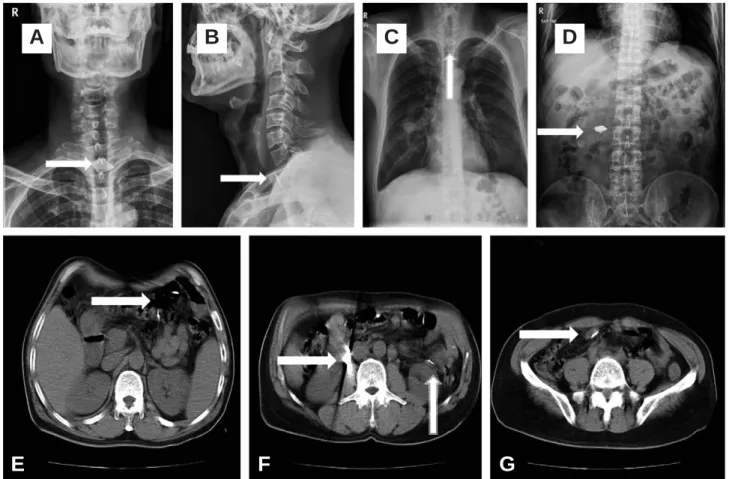
A 47-year-old male patient who has chronic alco- holism was admitted via emergency room with dys- phagia and neck pain. At that time of the transfer, he was a long-stay patient in another hospital for treating his chronic alcoholism. He was suffering from alcoholic liver cirrhosis and psychiatric prob- lem, such as chronic alcoholism, anxiety disorder and insomnia. He swallowed sharp fragments of cutter knife blade with intent to leaving the hospital the previous day. Plain radiographs (Fig. 1A-D) and computed tomography (CT) scan (Fig. 1E-G) showed multiple, scattered metal fragments in esophagus, small bowel, and colon. We performed emergent endoscopy and successfully removed one impacted
Fig. 1. Plain radiographs (A, B, C, D) show multiple fragments of cutter knife (white arrow) and also, computed tomography scans (E, F, G) show them in alimentary tract at admission.
A B C D
E F G
blade in upper esophagus using snare with overtube (Fig. 2A-C). After extraction of the impacted blade, complete esophagogastroduodenostomy was per- formed immediately to detect any underlying disor- der or complications. It showed multiple linear mucosal laceration and ulcer without active bleeding in esophagus and stomach. (Fig. 2D, E) The rest of fragments already passed through the pylorus and we could not find them with endoscopy. We admin- istered antiulcer drugs and checked the patient with simple abdominal radiographs and careful physical examinations every day. All remainders were uneventfully excreted through anus on 6 days of hospital stay. Finally, we were able to confirm objects in stool and negative radiographs. The patient was discharged without complications on 14 days of hospital stay and followed by department of psychiatry.
III. Discussion
Most foreign bodies that enter the stomach will pass in four to six days, and conservative manage- ment is appropriate for most blunt objects in asymptomatic patients. Even small sharp objects can pass alimentary tract without serious complications exceptions include disk batteries, magnets, objects longer than 6cm, and objects with a diameter >2.5 cm. Patients with negative radiographs who are asymptomatic can be followed expectantly. Other patients will likely require endoscopic or surgical intervention.(7,8) Even though these guidelines are consistent with our experience, the approach to management depends upon the type of object ingested, the location of the object, and the patient’
s clinical status. We should be well aware of appro- priate selection of therapeutic modality and the tim- ing of endoscopy at the moment.
─ 306 ─
- Journal of Trauma and Injury Vol. 26, No. 4 -
Fig. 2. A impacted fragment of cutter knife was noted on upper esophagus (A). This fragment was removed by snare with over tube (B, C). D and E view showed multiple mucosal lacerations in stomach and esophagus. The rest of fragments already passed through the pylorus.
A
B C
D E
─ 307 ─
Jong-Min Park, et al.: Sharp Foreign Bodies in the Alimentary Tract
REFERENCES
01) Schunk JE, Harrison AM, Corneli HM, Nixon GW.
Fluoroscopic foley catheter removal of esophageal foreign bodies in children: experience with 415 episodes. Pediatrics 1994; 94: 709-14.
02) Webb WA. Management of foreign bodies of the upper gas- trointestinal tract: update. Gastrointest Endosc 1995; 41: 39-51.
03) Singh B, Kantu M, Har-El G, Lucente FE. Complications associated with 327 foreign bodies of the pharynx, larynx, and esophagus. Ann Otol Rhinol Laryngol 1997; 106: 301-4.
04) Kamal I, Thompson J, Paquette D M. The hazards of vinyl glove ingestion in the mentally retarded patient with pica: new implications for surgical management. Can J Surg 1999; 42:
201-4.
05) Wu WT, Chiu CT, Kuo CJ, Lin CJ, Chu YY, Tsou YK, et al.
Endoscopic management of suspected esophageal foreign body in adults. Dis Esophagus 2011; 24: 131-7.
06) Palta R, Sahota A, Bemarki A, Salama P, Simpson N, Laine L. Foreign-body ingestion: characteristics and outcomes in a lower socioeconomic population with predominantly inten- tional ingestion. Gastrointest Endosc 2009; 69: 426-33.
07) ASGE Standards of Practice Committee, Ikenberry SO, Jue TL, Anderson MA, Appalaneni V, Banerjee S, et al.
Management of ingested foreign bodies and food impactions.
Gastrointest Endosc 2011; 73: 1085-91.
08) Weiland ST, Schurr MJ. Conservative management of ingest- ed foreign bodies. J Gastrointest Surg 2002; 6: 496-500.
09) Eisen GM, Baron TH, Dominitz JA, Faigel DO, Goldstein JL, Johanson JF, et al. Guideline for the management of ingested foreign bodies. Gastrointest Endosc 2002; 55: 802-6.