eISSN 2287-1683 pISSN 1738-8767 Journal of Trauma and Injury Vol. 30, No. 2, June, 2017 http://dx.doi.org/10.20408/jti.2017.30.2.47
Case Report
Address for Correspondence : Maru Kim, M.D.
Department of Trauma Surgery, Uijeongbu St. Mary’s Hospital, 271, Cheonbo-ro, Uijeongbu-si, Gyeonggi-do, Korea
Tel : 82-31-820-5266, Fax : 82-31-847-2717, E-mail : [email protected]
Submitted : January 5, 2017 Revised : January 12, 2017 Accepted : January 12, 2017
Hydronephrosis during Conservative Treatment for a Renal Injury Patient
Maru Kim, M.D., Joongsuck Kim, M.D.1, Sung Jeep Kim, M.D., Hang Joo Cho, M.D.
Department of Trauma Surgery, Uijeongbu St. Mary’s Hospital, College of Medicine, The Catholic University of Korea
1Department of Trauma Surgery, Cheju Halla General Hospital, Jeju, Korea
A 21-year-old male visited our emergency room. He could not remember the mechanism of injury. He was found beside a motorcycle. Initial vital sign was stable. Observation and conservative treatment were planned at the intensive care unit (ICU). On the third day at ICU, he complained sudden flank pain. It was colicky and hard to control. Without the pain, he had no specific symptom, sign, or laboratory findings. On computed tomography, renal pelvis was filled with hematoma which induced hydronephrosis. Double-J catheter and percutaneous nephrostomy was implemented by an intervention radiologist. Hematome in the renal pelvis was aspirated during the procedure. Symptom of the patient was subsided after the procedure. He was discharged without specific complications. [ J Trauma Inj 2017; 30: 47-50 ]
Key Words: Hydronephrosis, Hematoma, Wounds and injuries
- Journal of Trauma and Injury Vol. 30, No. 2 -
Table 1. Result of laboratory examinations
Laboratory marker Normal range Initial *HD #2 HD #3 HD #4
Hemoglobin 13.0-18.0 15.2 12.7 13.0 12.5
Hematocrit 40.0-54.0 44.5 37.8 37.7 35.9
Platelet 150-450 206 144 140 161
ESR 00-10 003 033 056
CRP 00.-0.3 000.03 001.66 007.76 008.37
BUN 08.0-20.0 11.0 07.9 06.3 07.1
Cr 0.5-1.3 001.04 000.93 000.87 000.77
* HD: hospital day
ESR: erythrosite sedimentation rate CRP: C-reactive protein
BUN: blood urea nitrogen Cr: creatine
Normal range was made by department of laboratory medicine in author’s institution.
Fig. 1. Initial CT showing left renal injury (grade 3). Fig. 2. Follow-up CT showing dilated pelvis filled with hematoma.
´ ´
Maru Kim, et al. Hydronephrosis during Conservative Treatment for a Renal Injury Patient
Fig. 3. Segmental filling defects that imply hematoma were identified on nephrogram.
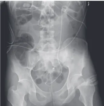
Fig. 4. Double-J-catheter and percutaneous nephrostomy catheter were inserted.
REFERENCES
01) Serafetinides E, Kitrey ND, Djakovic N, Kuehhas FE, Lumen N, Sharma DM, et al. Review of the current management of upper urinary tract injuries by the EAU Trauma Guidelines Panel. Eur Urol 2015; 67: 930-6.
02) Bryk DJ, Zhao LC. Guideline of guidelines: a review of uro- logical trauma guidelines. BJU Int 2016; 117: 226-34.
03) Morey AF, Brandes S, Dugi DD, 3rd, Armstrong JH, Breyer 2014; 192: 327-35.
04) Santucci RA, Bartley JM. Urologic trauma guidelines: a 21st century update. Nat Rev Urol 2010; 7: 510-9.
05) Bent C, Iyngkaran T, Power N, Matson M, Hajdinjak T, Buchholz N, et al. Urological injuries following trauma. Clin Radiol 2008; 63: 1361-71.
06) Stein DM, Santucci RA. An update on urotrauma. Curr Opin Urol 2015; 25: 323-30.
07) Santucci RA, Wessells H, Bartsch G, Descotes J, Heyns CF, McAninch JW, et al. Evaluation and management of renal injuries: consensus statement of the renal trauma subcommit- tee. BJU Int 2004; 93: 937-54.
08) Setia S, Jackson J, Herndon CD, Corbett S. Delayed Partial Nephrectomy for Hydronephrosis after Renal Trauma. Urology 2016.
09) Burks FN, Santucci RA. Management of iatrogenic ureteral injury. Ther Adv Urol 2014; 6: 115-24.
10) Esparaz AM, Pearl JA, Herts BR, LeBlanc J, Kapoor B.
Iatrogenic urinary tract injuries: etiology, diagnosis, and man- agement. Semin Intervent Radiol 2015; 32: 195-208.
11) Malik T, Khan S. An unusual cause of hydronephroureter. J Coll Physicians Surg Pak 2014; 24: 766-7.
12) Tseng PC, Liu TY, Pan SJ, Sung DS. Spontaneous perirenal urinoma associated with ureteropelvic junction obstruction in a child: a case report. Pediatr Neonatol 2009; 50: 121-4.
13) Lee WJ, Lin HJ, Cheng TC. Ureteral injury due to blunt abdominal trauma. Eur J Emerg Med 2006; 13: 244-6.
14) Bauab T, Jr. Renal pelvic hematoma in hydronephrotic kid- ney. Urology 1985; 25: 547.
- Journal of Trauma and Injury Vol. 30, No. 2 -