Received: December 1, 2019 Accepted: December 13, 2019 Trauma and InJury
Correspondence to Seong Beom Oh, M.D.
Department of Emergency Medicine, Dankook University Hospital, College of Medicine, Dankook University, 201 Manghyang-ro Dongnamgu, Cheonan 31116, Korea
Tel: +82-41-550-6840 Fax: +82-41-556-0524 E-mail: [email protected]
a Carbon monoxide Poisoning Case in Which Hyperbaric oxygen Thera- py Was not Possible due to Iatrogen- ic Pneumothorax after unnecessary Central Catheterization
Hyung Il Kim, M.D., Seong Beom Oh, M.D.
Department of Emergency Medicine, Dankook University Hospital, College of Medicine, Dankook University, Cheonan, Korea
Hyperbaric oxygen therapy (HBOT) is used to treat carbon monoxide (CO) poisoning.
However, untreated pneumothorax is an absolute contraindication for HBOT. More caution is needed with regard to monoplace hyperbaric chambers, as patient monitor- ing and life-saving procedures are impossible inside these chambers. Central catheter- ization is frequently used for various conditions, but unnecessary catheterization must be avoided because of the risk of infection and mechanical complications. Herein, we describe a case of CO poisoning in which iatrogenic pneumothorax developed after unnecessary subclavian central catheterization. The patient did not need to be cathe- terized, and HBOT could not be performed because of the pneumothorax. Hence, this case reminds us of basic—but nonetheless important—principles of catheterization.
Keywords: Hyperbaric oxygenation; Complications; Pneumothorax; Catheterization, Central venous
INTRODUCTION
The basic principle of hyperbaric oxygen therapy (HBOT) is to provide patients with
pure oxygen at a pressure that exceeds atmospheric pressure. HBOT has various clin-
ical applications, including carbon monoxide (CO) poisoning, air embolism, wound
healing, decompression sickness, and radiation injury. Traditionally, the most com-
mon therapeutic use of HBOT in Korea has been to treat CO poisoning. However,
a Carbon monoxide Poisoning Case in Which Hyperbaric oxygen Thera- py Was not Possible due to Iatrogen- ic Pneumothorax after unnecessary Central Catheterization
Hyung Il Kim, M.D., Seong Beom Oh, M.D.
Department of Emergency Medicine, Dankook University Hospital, College of Medicine, Dankook University, Cheonan, Korea
Hyperbaric oxygen therapy (HBOT) is used to treat carbon monoxide (CO) poisoning.
However, untreated pneumothorax is an absolute contraindication for HBOT. More caution is needed with regard to monoplace hyperbaric chambers, as patient monitor- ing and life-saving procedures are impossible inside these chambers. Central catheter- ization is frequently used for various conditions, but unnecessary catheterization must be avoided because of the risk of infection and mechanical complications. Herein, we describe a case of CO poisoning in which iatrogenic pneumothorax developed after unnecessary subclavian central catheterization. The patient did not need to be cathe- terized, and HBOT could not be performed because of the pneumothorax. Hence, this case reminds us of basic—but nonetheless important—principles of catheterization.
Keywords: Hyperbaric oxygenation; Complications; Pneumothorax; Catheterization, Central venous
untreated pneumothorax is a contraindication for HBOT.
Particular caution is needed with regard to monoplace hyperbaric chambers because it is not possible to monitor patients inside these chambers, which medical personnel cannot enter.
Central catheterization is frequently used when caring for critically ill patients as a way to infuse vasopressors, monitor central venous pressure, and obtain emergency vascular access. However, unnecessary catheterization must be avoided because catheter-induced infections and mechanical complications can develop.
We experienced a case in which iatrogenic pneumotho- rax after unnecessary subclavian central catheterization prohibited a patient from being treated with HBOT.
Therefore, this case reminds us of the basic principles of catheterization.
CASE REPORT
A 28-year-old woman was transferred to the emergency department (ED) of a local hospital with depressed mental status. She had no previous medical history and had lived by herself in a small efficiency apartment. She was dis- covered in her bathroom with a burnt charcoal briquette.
Non-contrast brain computed tomography (CT) and brain diffusion magnetic resonance imaging (MRI) were performed at the local ED. She was transferred to our ED for HBOT. She was comatose, with a Glasgow coma scale (GCS) score of 8 (E4V2M2). Her blood pressure was 110/67 mmHg, her heart rate was 100/min, her respirato- ry rate was 16 breaths/min, and her body temperature was 36.9℃. Her pupils were 5 mm in size and symmetrical. A grade 3 pressure sore measuring 5×8 cm was noted on her right buttock (Fig. 1). The initial PaO
2was 104 mmHg,
A b
Fig. 1. (A) A grade 3 pressure sore measuring 5×8 cm was initially observed on the patient’s right buttock. (B) After surgical debridement.
Fig. 2. Brain magnetic resonance images. Acute hypoxic encephalopathy was noted.
and the initial PaCO
2was 33 mmHg. Her carboxyhemo- globin level was 48% (reference range: <0.8%). The white blood cell count was 12,310/μL, and the platelet count was 115,000/μL. The levels of other blood test parameters were as follows: aspartate transaminase, 157 U/L; alanine transaminase, 78 U/L; amylase, 423 U/L (reference range:
28-100 U/L); lipase, 888 U/L (reference range: 13-60 U/L);
creatine phosphokinase, 3,691 U/L (reference range: 26- 174 U/L), and troponin T, 0.117 ng/mL (reference range:
<0.1 ng/mL). Other laboratory findings were unremark- able. There was no definite lesion on brain CT, but acute ischemic encephalopathy was observed on brain MRI (Fig. 2). HBOT was needed to minimize the possibility of delayed neurological sequelae and to relieve systemic CO toxicity. However, central catheterization with a 7-Fr catheter had been established in the right subclavian area at the local ED. Iatrogenic pneumothorax at the right upper apex was detected on the chest X-ray and chest
CT images obtained at the local hospital (Figs. 3, 4). The hyperbaric chamber in Dankook University Hospital was a monoplace chamber. Although the pneumothorax was not large, patient monitoring was not possible in the chamber and other medical personnel could not enter the chamber with the patient; therefore, HBOT could not be performed. The patient was admitted to the intensive care unit and surgical debridement of the pressure sore was performed. The pneumothorax resolved spontaneously after several days. She did not regain consciousness, and had a GCS score of 9 on the discharge day.
DISCUSSION
HBOT is used to provide patients with pure oxygen at higher than atmospheric pressure (2–3 atmospheres abso- lute), enabling more oxygen to be delivered to the tissue in
A b C
Fig. 3. Chest X-ray images. (A) The initial X-ray before central catheterization. No pneumothorax was noted. (B) An X-ray after central catheterization
at the right subclavian area. Pneumothorax was noted. The pleural margin of the lung (arrows). (C) An X-ray obtained on the second hospital day. The amount of the pneumothorax had increased. The pleural margin of the lung (arrows).
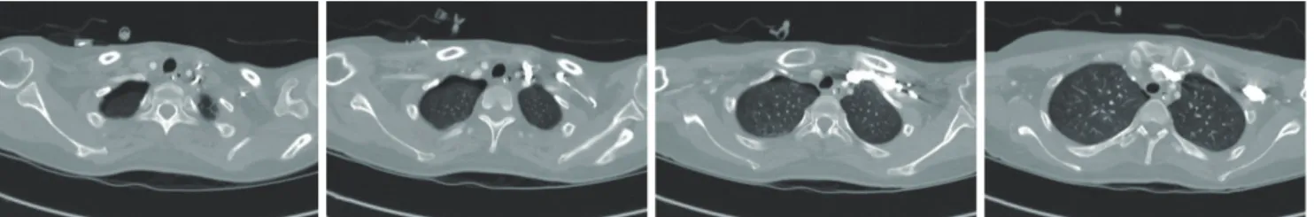
Fig. 4. Chest computed tomography images. Pneumothorax was noted at the right lung apex.