Original Article
Received May 9, 2014, Revised July 11, 2014, Accepted July 19, 2014 Correspondence to: Hunho Song
Department of Internal Medicine, Kandong Sacred Heart Hospital, University of Hallym College of Medicine, 445, Gil-dong, Gangdong-gu, Seoul 134-701, Korea
Tel: +82-2-2224-2691, Fax: +82-2-478-6925, E-mail: [email protected]
This is an Open Access article distributed under the terms of the Creative Commons Attribution Non-Commercial License (http://creativecommons.org/licenses/by-nc/3.0) which permits unrestricted non-commercial use, distribu- tion, and reproduction in any medium, provided the original work is properly cited.
Safety and Efficacy of Peripherally Inserted Central Catheters in Terminally Ill Cancer Patients: Single
Institute Experience
Kwonoh Park, M.D., Hyoung Gun Lim, M.D.*, Ji Yeon Hong, M.D. and Hunho Song, M.D., Ph.D.
†Department of Internal Medicine, *Radiology, KEPCO Medical Center,
†Department of Internal Medicine, Kandong Sacred Heart Hospital, University of Hallym College of Medicine, Seoul, Korea
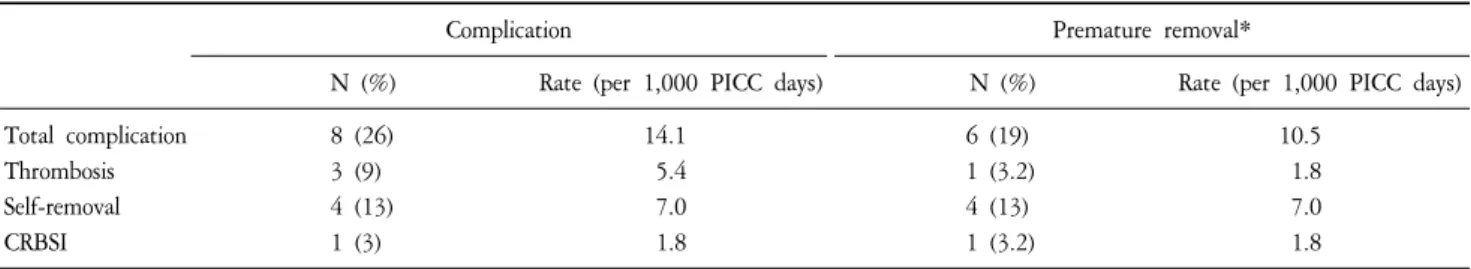
Purpose: We investigated the safety and efficacy of peripherally inserted central catheters (PICCs) in terminally ill cancer patients. Methods: A retrospective review was conducted on patients who underwent PICC at the hospice-palliative division of KEPCO (Korea Electric Power Corporation) Medical Center between January 2013 and December 2013. All PICCs were inserted by an interventional radiologist. Results: A total of 30 terminally ill cancer patients received the PICC procedure during the study period. Including one patient who had had two PICC insertions during the period, we analyzed a total of 31 episodes of catheterization and 571 PICC days. The median catheter life span was 14.0 days (range, 1∼90 days). In 25 cases, catheters were maintained until the intended time (discharge, transfer, or death), while they were removed prematurely in six other cases (19%;
10.5/1000 PICC days). Thus, the catheter maintenance success rate was 81%. Of those six premature PICC removal cases, self-removal due to delirium occurred in four cases (13%; 7.0/1000 PICC days), and catheter-related blood stream infection and thrombosis were reported in one case, each (3%; 1.8/1000 PICC days). Complication cases totaled eight (26%; 14.1/1000 PICC days). The time to complication development ranged from two to 14 days and the median was seven days. There was no PICC complication-related death. Conclusion: Considering characteristics of terminally ill cancer patients, such as a poor general condition, vulnerability to trivial damage, and a limited period of survival, PICC could be a safe intravenous procedure.
Key Words: Peripheral venous catheterization, Central venous catheterization, Hospice care, Terminal care, Palliative care
INTRODUCTION
Oral administration of medicine and nutrition is often difficult in terminally ill cancer patients because of progressive difficulties in swallowing, nausea and vomiting, intestinal obstruction, and consciousness disturbance (1). So, reliable intravenous (IV) access is an important issue in terminally ill cancer patients (2,3). However, these terminally ill cancer patients had limited or no peripheral venous access due to
edema or thrombophlebitis caused by long-term IV therapy including chemotherapy and blood transfusions. Thus, intravenous access has been provided by central venous catheter (CVC).
There are some options for applying CVC in cancer patients;
subclavian venous catheter (SVC), chemo-port (CP), and the
peripherally inserted central catheter (PICC). The PICC offers
certain advantages over other forms of CVC. PICC shows mi-
nimal procedure-related catastrophic risk (e.g., pneumothorax, or
hemothrorax) at the time of insertion, because it is inserted via
the peripheral vein (usually upper limb) without requiring surgical procedure (4). Compared with SVC, PICC has a longer dwelling time (5), and less chance of catheter related blood stream in- fection (CRBSI) (6-8). Compared with CP, PICC has a shorter durability of dwelling (9), but has many advantages, including cost-effectiveness (5,7,8), well-tolerated insertion (5), no issues with wound dehiscence in the insertion site, and ease of removal (4).
Terminally ill cancer patients are vulnerable to minor trauma due to poor performance and general conditions and may have behavior problems due to mental changes or delirium (10). In addition, most of these patients have a limited survival duration of 1∼2 months. Hence, when considering such aspects of terminally ill cancer patients, safe insertion without procedure- related complications, comfortable insertion and an intermediate term of IV access maintenance are needed for CVC. So, the PICC might be an attractive alternative methods of CVC.
However, only limited data exist regarding the safety and efficacy of PICC in terminally ill cancer patient with only 1 published study worldwide (11). In Korean, thus far, only two reports have described PICC in a general oncology setting (9,12), and no study has reported in a homogeneous cohort of terminally ill cancer patients. Thus, we conducted the present retrospective cohort study to investigate the safety and efficacy of PICC to provide the guidance for PICC utilization in terminally ill cancer patients.
METHODS 1. Patients and study design
A retrospective review was conducted on all terminally ill cancer patients who underwent PICC at hospice-palliative part of KEPCO (Korea Electric Power Corporation) Medical Center between January and December 2013. The hospice-palliative part of KEPCO Medical Center is composed of 1 attending physician, 1 social worker and 20 volunteers, and a mean of 6∼7 inpatients are maintained. Inpatients are usually referred from home and institutions of the same city for end-of-life care, intensive symptom control, and/or to pro- vide a family respite. Terminally ill cancer patient represents patient without additional anti-cancer treatment who have an estimated survival of 1∼2 months or less. Of these patients, those who needed an administration of artificial hydration, total parenteral nutrition (TPN), and medication but with
limited peripheral line access were recommended for PICC insertion. If the prior PICC was removed by unexpected events or planned discharge, another catheter was placed at a site remote from the original site. For the analysis, we counted each PICC placement as a new event. This study was appro- ved by the institutional review board of KEPCO Medical Center, which waived the requirement for informed consent due to the retrospective design of this study (HIRB-2014-005).
2. PICC insertion procedure and management
All PICCs were inserted by an interventional radiologist in the angiography room using ultrasound guidance and fluoro- scopic imaging. All operators wore aseptic gowns, masks, and gloves, and all of the patients received a dressing with aseptic drapes. Seldinger’s technique was used routinely. The PICC lines contained double lumens and were made of second-/
third-generation polyurethane. The location of the catheter tip was confirmed by chest radiography. None of the PICCs was sutured; they were held in place with StatLock
ⓇCatheter Stabilization Device.
No patient was adminstered prophylactic antibiotics or anticoagulation drugs for infection or thrombosis. Catheter replacement over a guidewire was strictly prohibited at our hospital. All of the patients received a closed dressing dampened with betadine on the catheter insertion site every 3 days.
3. Catheter monitoring and data collection
We obtained data from the patient’s medical records, temperature-pulse-respiratory chart, and daily nurse’s checklist to identify patients with an indwelling PICC and reviewed the clinical complications, such as pain, edema, and local systemic catheter-related infections. Microbiology reports were reviewed to identify systemic catheter-related bloodstream infections (CRBSIs). CRBSIs were defined by catheter tip culture and at least one positive peripheral blood culture of the same orga- nism. On suspicion of a catheter-related infection, the line was removed, and the tip was sent for analysis in the microbiology laboratory, whereas routine tip-culture was not performed.
Catheter-related thrombosis was suspected when the catheter
flow rate was impossible to back flush or when patients
complained of arm edema or pain. However, all of the patients
who had thrombosis symptoms, with or without a confirmatory
test such as Doppler ultrasonography or venography, were in-
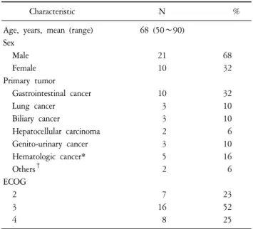
Table 1. General Characteristics (N=31).
Characteristic N %
Age, years, mean (range) 68 (50∼90) Sex
Male 21 68
Female 10 32
Primary tumor
Gastrointestinal cancer 10 32
Lung cancer 3 10
Biliary cancer 3 10
Hepatocellular carcinoma 2 6
Genito-urinary cancer 3 10
Hematologic cancer* 5 16
Others
†2 6
ECOG
2 7 23
3 16 52
4 8 25
*Myelodysplastic syndrome 2, Lymphoma 2, Multiple myeloma 1,
†