Open Access
Comparison of Breast Conserving Surgery Followed by Radiation Therapy with Mastectomy Alone for Pathologic N1 Breast Cancer Patients in the Era of Anthracycline Plus Taxane-Based Chemotherapy: A Multicenter Retrospective Study (KROG 1418)
Original Article
Purpose
We compared the oncologic outcomes of breast-conserving surgery plus radiation therapy (BCS+RT) and modified radical mastectomy (MRM) under anthracycline plus taxane-based (AT) regimens and investigated the role of adjuvant radiation therapy (RT) in patients with pathologic N1 (pN1) breast cancer treated by mastectomy.
Materials and Methods
We retrospectively reviewed the medical records of 2,011 patients with pN1 breast cancer who underwent BCS+RT or MRM alone at 12 institutions between January 2006 and December 2010. Two-to-one propensity score matching was performed for balances in vari- ables between the groups.
Results
The median follow-up duration for the total cohort was 69 months (range, 1 to 114 months).
After propensity score matching, 1,074 patients (676 in the BCS+RT group and 398 in the MRM-alone group) were analyzed finally. The overall survival, disease-free survival, locore- gional failure-free survival, and regional failure-free survival (RFFS) curves of the BCS+RT group vs. MRM-alone group were not significantly different. The subgroup analysis revealed that in the group with both lymphovascular invasion (LVI) and histologic grade (HG) III, the BCS+RT showed significantly superior RFFS (p=0.008). Lymphedema (p=0.007) and radi- ation pneumonitis (p=0.031) occurred more frequently in the BCS+RT group than in the MRM-alone group, significantly.
Conclusion
There are no differences in oncologic outcomes between BCS+RT and MRM-alone groups under the AT chemotherapy regimens for pN1 breast cancer. However, BCS+RT group showed superior RFFS to MRM-alone group in the patients with LVI and HG III. Adjuvant RT might be considerable for pN1 breast cancer patients with LVI and HG III.
Key words
Breast neoplasms, Pathologic N1, Breast conserving surgery, Radiotherapy, Mastectomy, Anthracyclines, Taxane, Survival
Gyu Sang Yoo, MD
1Won Park, MD, PhD
1Jeong Il Yu, MD, PhD
1Doo Ho Choi, MD, PhD
1Yeon-Joo Kim, MD
2Kyung Hwan Shin, MD, PhD
3Chan Woo Wee, MD
3Kyubo Kim, MD, PhD
4Kyung Ran Park, MD, PhD
5Yong Bae Kim, MD, PhD
6Sung Ja Ahn, MD, PhD
7Jong Hoon Lee, MD, PhD
8Jin Hee Kim, MD, PhD
9Mison Chun, MD, PhD
10Hyung-Sik Lee, MD, PhD
11Jung Soo Kim, MD, PhD
12Jihye Cha, MD, PhD
13+ + + + + + + + + + + + + + + + + + + + + + + + + + + + + + + + + + + + + + + + + + + + + + + + + + + + + + + + + + + + + + + + + + + + + + + + + + + + + + + + + + + + + + + + + + + + + + + + + + + + + + + + + + + + + + + + + + + + + + + + + + + + + + + + + + + + + + + + + + + + + + + + + + + + + + + + + + + + + + + + + + + + + + + + + + + + + + + + + + + + + + + + + + + + + + + + + + + + + + + + + + + + + + + + + + + + + + + + + + + + + + + + + + + + + + + + + + + + + + + + + + + + + + + + + + + + + + + + + + + + + + + + + + + + + + + + + + + + + + + + + + + + + + + + + + + + + + + + + + + + + + + + + + + + + + + + + + + + + + + + + + + + + + + + + + + + + + + + + + + + + + + + + + + + + + + + + + + + + + + + + + + + + + + + + + + + + + + + + + + + + + + + + + + + + + + + + + + + + + + + + + + + + + + + + + + + + + + + + + + + + + + + + + + + + + + + + + + + + + + + + + + + + + + + + + + + + + + + + + + +
Correspondence: Won Park, MD, PhD Department of Radiation Oncology, Samsung Medical Center, Sungkyunkwan University School of Medicine, 81 Irwon-ro, Gangnam-gu, Seoul 06351, Korea Tel: 82-2-3410-2438
Fax: 82-2-3410-2619
E-mail: [email protected]
Co-correspondence: Kyung Hwan Shin, MD, PhD Department of Radiation Oncology,
Seoul National University College of Medicine, 101 Daehak-ro, Jongno-gu, Seoul 03080, Korea Tel: 82-2-2072-2524
Fax: 82-2-765-3317 E-mail: [email protected]
Received July 26, 2018 Accepted October 22, 2018 Published Online November 1, 2018
*A list author’s a!liations appears at the end
of the paper.
Introduction
Randomized trials comparing breast-conservation surgery (BCS) plus adjuvant radiation therapy (RT) with mastectomy alone in early-stage breast cancer have shown no differences in oncologic outcomes [1-6]. On the other hand, several recent population-based studies reported that BCS plus RT (BCS+RT) resulted in better oncologic outcomes than mas- tectomy according to T or N category [7-11]. The role of adju- vant RT after mastectomy for pN1 breast cancer is also a controversial issue. Key randomized trials showed better oncologic outcomes in the post-mastectomy RT (PMRT) group compared with the non-PMRT group for breast cancer with nodal metastasis [12,13]. Subgroup analysis by the Dan- ish Breast Cancer Cooperative Group (DBCG) 82 B&C ran- domized trial [13] and meta-analysis from the Early Breast Cancer Trialists' Collaborative Group (EBCTCG) [14] repor- ted that PMRT had the benefit of local control for subgroups with 1-3 axillary nodal metastases. The National Comprehen- sive Cancer Network (NCCN) clinical practice guideline rec- ommends strong consideration of PMRT for N1 breast cancer [15]. However, the chemotherapeutic regimens of these stud- ies are not the modern one, but mostly the CMF (cyclophos- phamide, methotrexate, and 5-fluorouracil) regimen, which is less effective [16,17].
Anthracycline plus taxane-based (AT) chemotherapy is the currently recommended adjuvant treatment in the breast cancer, especially with nodal metastasis [15]. This regimen showed benefits in local control as well as overall survival (OS) comparing with previous chemotherapy regimens [18,19]. In the era of the AT regimen, the role of PMRT for pN1 disease is still unclear [20,21]. Therefore, we performed this study to compare oncologic outcomes between BCS+RT and modified radical mastectomy (MRM) alone under the AT regimen and to investigate the role of adjuvant RT in pati- ents with pN1 breast cancer treated by mastectomy.
Materials and Methods
1. Patients
We retrospectively reviewed the medical records of 2,011 patients with pathologic N1 breast cancer who underwent BCS+RT or MRM alone at 12 institutions in South Korea bet- ween Jan 2006 and Dec 2010.
Information was obtained from medical records regarding pathologic tumor features, including molecular subtype, tumor size, resection margin, lymphovascular invasion (LVI),
nuclear grade (NG), histologic grade (HG), number of lymph nodes (LN) with metastasis, and extracapsular extension (ECE). Patients were excluded because of non-AT chemother- apy (n=47), BCS without adjuvant RT (n=37) and PMRT (n=157), and insufficient medical records (n=296). Finally, 1,474 patients were included in the analysis.
2. Treatment
BCS+RT was performed for 1,047 patients (71.0%). The median dose of adjuvant RT on the whole breast was 50 Gy (range, 45 to 50.4 Gy). Tumor bed boost with median dose of 10 Gy (range, 5 to 16 Gy) was applied to 1,026 patients. RT to the supraclavicular fossa with a median dose of 50 Gy (range, 45 to 50.4 Gy) was performed in 320 patients (30.6%), and 37 (3.5%) of them received RT to the internal mammary area with a median dose of 50.4 Gy (range, 45 to 50.4 Gy). RT plans followed the general principles of RT to the whole breast. Median number of axillary LN dissections performed was 16 (range, 1 to 47). Most patients (99.8%) were treated with an AC (adriamycin and cyclophosphamide) plus T (tax- ane) regimen, while others were administrated the FAC (flu- orouracil, adriamycin, and cytoxan) plus T regimen (0.1%) or EC (epirubicin and cyclophosphamide) plus T regimen (0.1%).
MRM alone was performed for 427 patients (29.0%). All patients in the MRM-alone group received a median of 19 axillary LN dissections (range, 2 to 43). AC plus T was admi- nistered to most patients (99.8%) in the MRM group. Other patients received the FEC (fluorouracil, epirubicin, and cytoxan) plus T regimen (0.2%). None of this group under- went adjuvant RT.
3. Statistical analysis
OS, disease-free survival (DFS), locoregional failure-free survival (LRFFS), and regional failure-free survival (RFFS) were defined as the interval from surgery to death, cancer recurrence, locoregional recurrence, and regional recurrence, respectively. The chi-square test or Fisher exact test was used to compare patient characteristics and patterns of failure bet- ween the BCS+RT and MRM-alone group. The Kaplan-Meier method was used to estimate survival curves. Log-rank tests were performed to compare survival between groups for various variables. Cox regression analysis was chosen for multivariate analysis to determine the independent prognos- tic factors for outcomes. A two-sided p-value of < 0.05 was considered statistically significant. Statistical analyses were performed with SPSS ver. 22.0 (IBM Corp., Armonk, NY).
Analyses were also performed for subgroups defined accord-
ing to the number of risk factors identified as significant in
multivariate analyses for DFS.
Two-to-one propensity score matching was performed to eliminate imbalances in variables between the two treatment groups. Matching variables were age, menopause status, site, pathology, pathologic T staging, number of LNs with metas-
tasis, LVI, NG, HG, molecular subtype, and ECE. R Statistical Software ver. 3.2.3 (The R foundation for Statistical Analyses, Vienna, Austria) was utilized in propensity score matching.
Before matching After matching
Characteristic BCS+RT MRM alone
p-value BCS+RT MRM alone
p-value
(n=1,047) (n=427) (n=676) (n=398)
Age (yr) 47.4±8.6 48.9±9.4 0.004 48.1±8.9 48.7±9.3 0.319
Menopausal status
Premenopause 676 (64.6) 251 (58.8) 0.043 415 (61.4) 238 (59.8) 0.604
Postmenopause 371 (35.4) 176 (41.2) 261 (38.6) 160 (40.2)
Site
Left 504 (48.1) 217 (50.8) 0.380 330 (48.8) 199 (50.0) 0.732
Right 543 (51.9) 210 (49.2) 346 (51.2) 199 (50.0)
Pathology
IDC 984 (94.0) 413 (96.7) 0.044 648 (95.9) 384 (96.5) 0.601
Non-IDC 63 (6.0) 14 (3.3) 28 (4.1) 14 (3.5)
Pathologic T category
1 507 (48.4) 160 (37.5) 0.000 280 (41.4) 160 (40.2) 0.897
2 532 (50.8) 265 (62.1) 393 (58.1) 236 (59.3)
3 8 (0.8) 2 (0.5) 3 (0.4) 2 (0.5)
No. of LN metastases
1 603 (57.6) 231 (54.1) 0.070 381 (56.4) 218 (54.8) 0.722
2-3 444 (42.4) 196 (45.9) 295 (43.6) 180 (45.2)
LN management
SLNB only 78 (7.4) 24 (5.6) 0.258 46 (6.8) 22 (5.5) 0.438
ALND 969 (92.6) 403 (94.4) 630 (93.2) 376 (94.5)
Positive LN ratio
! 0.1 506 (48.3) 233 (54.6) 0.034 338 (50.0) 219 (55.0) 0.101
> 0.1 541 (51.7) 194 (45.4) 338 (50.0) 179 (45.0)
LVI
No 370 (35.3) 273 (63.9) 0.000 397 (58.7) 244 (61.3) 0.068
Yes 677 (64.7) 154 (36.1) 279 (41.3) 154 (38.7)
Nuclear grade
1-2 599 (57.2) 200 (46.8) 0.000 324 (47.9) 200 (50.3) 0.412
3 448 (42.8) 227 (53.2) 352 (52.1) 198 (49.7)
Histologic grade
I-II 642 (61.3) 216 (50.6) 0.000 301 (44.5) 185 (46.5) 0.482
III 405 (38.7) 211 (49.4) 375 (55.5) 213 (53.5)
Molecular subtype
Luminal A 459 (43.8) 192 (45.0) 0.736 375 (55.4) 216 (54.3) 0.736
Non-luminal A 588 (56.2) 235 (55.0) 301 (44.6) 182 (45.7)
ECE
No 578 (55.2) 267 (62.5) 0.012 382 (56.5) 239 (60.1) 0.247
Yes 469 (44.8) 160 (37.5) 294 (43.5) 159 (39.9)
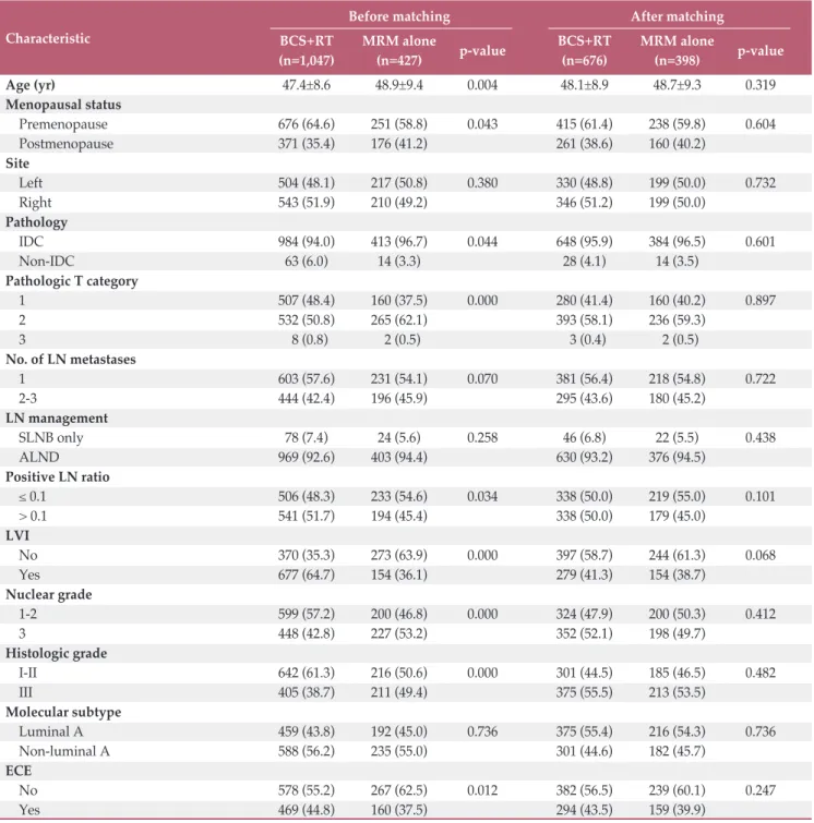
Table 1. Baseline characteristics of study patients
Values are presented as mean±standard deviation or number (%). BCS, breast conserving surgery; RT, radiation therapy;
MRM, modified radical mastectomy; IDC, invasive ductal carcinoma; LN, lymph node; SLNB, sentinel lymph node biopsy;
ALND, axillary lymph node dissection; LVI, lymphovascular invasion; ECE, extracapsular extension.
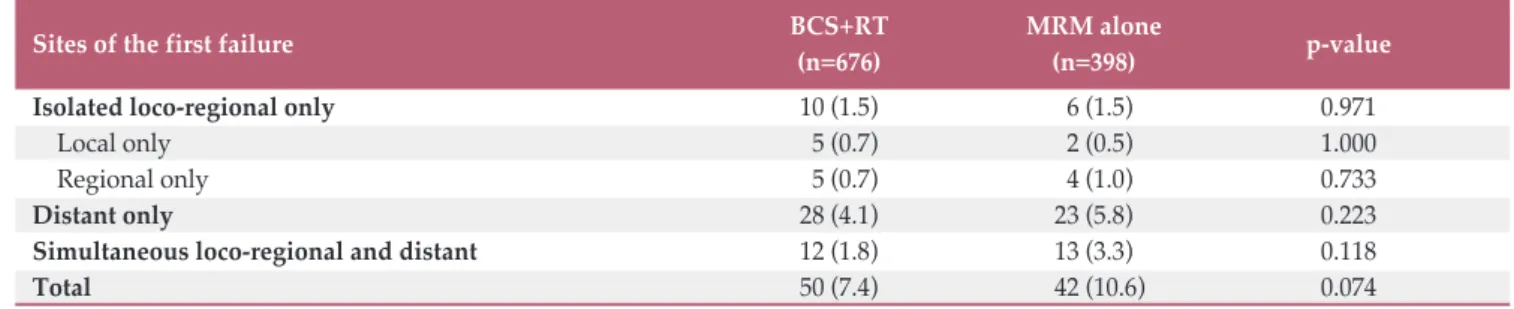
Table 2. Patterns of the first failure according to field of radiotherapy
Sites of the first failure BCS+RT MRM alone
p-value
(n=676) (n=398)
Isolated loco-regional only 10 (1.5) 6 (1.5) 0.971
Local only 5 (0.7) 2 (0.5) 1.000
Regional only 5 (0.7) 4 (1.0) 0.733
Distant only 28 (4.1) 23 (5.8) 0.223
Simultaneous loco-regional and distant 12 (1.8) 13 (3.3) 0.118
Total 50 (7.4) 42 (10.6) 0.074
Values are presented as number (%). BCS, breast conserving surgery; RT, radiation therapy; MRM, modified radical mas- tectomy.
Fig. 1. Survival curves according to treatment group. Overall survival (OS) (A), disease-free survival (DFS) (B), locoregional failure-free survival (LFFS) (C), and regional failure-free survival (RFFS) (D). BCS+RT, breast conserving surgery plus radi- ation therapy; MRM, modified radical mastectomy.
A
BCS+RT MRM alone p=0.088
Su rv iv al ra te (% )
100
0 20 40 60
0
Time (mo) OS
100 80
40 60
20 120
80
676 398
667 394
654 384
192 132 458 291
51 41
1 1 No. at risk
BCS+RT MRM alone
B
BCS+RT MRM alone p=0.107
Su rv iv al ra te (% )
100
0 20 40 60
0
Time (mo) DFS
100 80
40 60
20 120
80
676 398
663 385
626 366
182 132 430 270
48 39
1 1 No. at risk
BCS+RT MRM alone
C
BCS+RT MRM alone p=0.254
Su rv iv al ra te (% )
100
0 20 40 60
0
Time (mo) LFFS
100 80
40 60
20 120
80
676 398
665 391
643 376
190 135 449 281
50 41
1 1 No. at risk
BCS+RT MRM alone
D
BCS+RT MRM alone p=0.102
Su rv iv al ra te (% )
100
0 20 40 60
0
Time (mo) RFFS
100 80
40 60
20 120
80
676 398
666 391
647 377
189 135 451 282
50 41
1 1 No. at risk
BCS+RT
MRM alone
4. Ethical statement
This study was approved by the Institutional Review Board (IRB) of each hospital and performed in accordance with the principles of the Declaration of Helsinki. Each IRB approved a waiver of informed consent.
Results
1. Patient characteristics
The median follow-up duration for the total cohort was 69 months (range, 1 to 114 months). The 5-year rates of OS, DFS, LRFFS, and RFFS of the total cohort were 98.0%, 92.4%,
Characteristic Univariate analysis Multivariate analysis
5-Year OS rate (%) p-value HR (95% CI) p-value Age (yr)
! 55 97.7 0.828 - -
> 55 97.5 -
Menopause status
Premenopause 98.6 0.072 0.592 (0.295-1.190) 0.141
Postmenopause 96.0 -
Site
Left 97.3 0.737 - -
Right 97.9 -
Pathology
IDC 97.6 0.725 - -
Non-IDC 97.7 -
Pathologic T category
1 97.9 0.731 - -
2-3 97.4 -
No. of LN metastases
1 97.8 0.443 - -
2 97.4 -
LVI
No 98.0 0.342 - -
Yes 97.2 -
Nuclear grade
1-2 98.9 0.002 0.544 (0.174-1.704) 0.296
3 96.2 -
Histologic grade
I-II 99.0 0.004 0.773 (0.269-2.220) 0.632
III 95.9 -
Molecular subtype
Luminal A 99.1 0.001 0.327 (0.129-0.825) 0.018
Non-luminal A 96.3 -
ECE
No 97.6 0.771 - -
Yes 97.7 -
Modality
BCS+RT 98.6 0.088 0.597 (0.297-1.200) 0.147
MRM alone 96.1 -
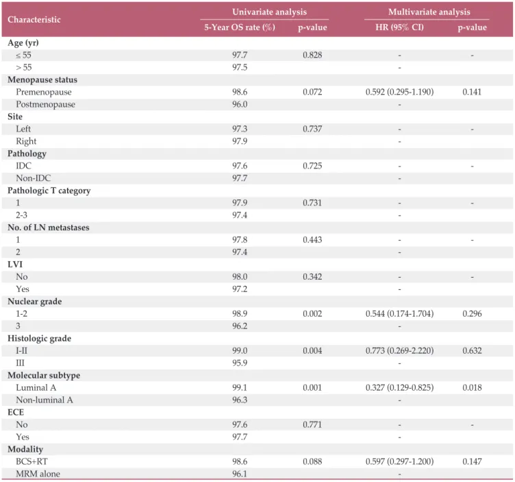
Table 3. Univariate and multivariate analysis (OS)
OS, overall survival; HR, hazard ratio; CI, confidence interval; IDC, invasive ductal carcinoma; LN, lymph node; LVI, lym-
phovascular invasion; ECE, extracapsular extension; BCS, breast conserving surgery; RT, radiation therapy; MRM; modified
radical mastectomy.
97.0%, and 97.6%, respectively. Among the total cohort, there were significant differences in age, menopausal status, pathology, pathologic T category, LVI, NG, HG, and ECE between the BCS+RT and MRM-alone groups. After propen- sity score matching, a total of 1,074 patients (676 in the BCS+
RT group and 398 in the MRM-alone group) were included for analysis. The patient characteristics are summarized in
Table 1. The number of dissected LNs were 16 (range, 1 to 42) in BCS+RT group and 18 (range, 2 to 42) in MRM-alone group.
Characteristic Univariate analysis Multivariate analysis
5-Year DFS rate (%) p-value HR (95% CI) p-value Age (yr)
! 55 91.9 0.987 - -
> 55 92.3 -
Menopause status
Premenopause 92.5 0.248 - -
Postmenopause 91.0 -
Site
Left 90.7 0.171 1.239 (0.818-1.875) 0.311
Right 93.2 -
Pathology
IDC 91.9 0.598 - -
Non-IDC 93.2 -
Pathologic T category
1 95.0 0.001 0.665 (0.415-1.064) 0.089
2-3 89.6 -
No. of LN metastases
1 93.2 0.126 0.823 (0.545-1.244) 0.356
2-3 90.4 -
LVI
No 94.6 < 0.001 0.472 (0.304-0.731) 0.001
Yes 88.7 -
Nuclear grade
1-2 95.4 < 0.001 0.715 (0.389-1.314) 0.280
3 88.0 -
Histologic grade
I-II 96.4 < 0.001 0.254 (0.134-0.481) 0.001
III 86.1 -
Molecular subtype
Luminal A 94.4 0.024 0.849 (0.544-1.326) 0.472
Non-luminal A 90.0 -
ECE
No 91.8 0.771 - -
Yes 92.0 -
Modality
BCS+RT 93.3 0.107 0.702 (0.463-1.065) 0.096
MRM alone 89.7 -
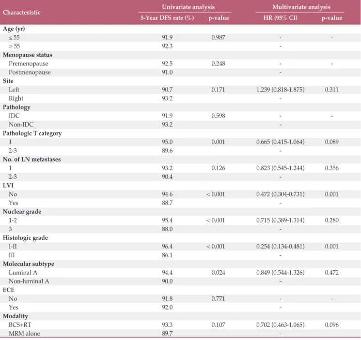
Table 4. Univariate and multivariate analysis (DFS)
DFS, disease-free survival; HR, hazard ratio; CI, confidence interval; IDC, invasive ductal carcinoma; LN, lymph node; LVI,
lymphovascular invasion; ECE, extracapsular extension; BCS, breast conserving surgery; RT, radiation therapy; MRM; mod-
ified radical mastectomy.
2. Treatment outcome
Among 1,074 patients, 92 (8.6%) experienced disease recu- rrence. The patterns of first failure were not significantly dif- ferent between the groups (Table 2). The OS, DFS, LRFFS, and RFFS rates of the BCS+RT group vs. MRM-alone group at 5 years were 98.6% vs. 96.1% (p=0.088), 93.3% vs. 89.7%
(p=0.107), 97.6% vs. 95.2% (p=0.254), and 98.3% vs. 95.7%
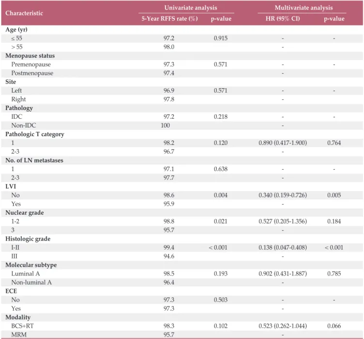
(p=0.102), respectively (Fig. 1). On multivariate analysis, luminal A type was identified as an independent prognostic factor associated with better OS (Table 3). The multivariate analyses revealed that LVI and HG were independent prog- nosticators associated with DFS and RFFS (Tables 4 and 5).
Three subgroups were determined according to the num-
Characteristic Univariate analysis Multivariate analysis
5-Year RFFS rate (%) p-value HR (95% CI) p-value Age (yr)
! 55 97.2 0.915 - -
> 55 98.0 -
Menopause status
Premenopause 97.3 0.571 - -
Postmenopause 97.4 -
Site
Left 96.9 0.571 - -
Right 97.8 -
Pathology
IDC 97.2 0.218 - -
Non-IDC 100 -
Pathologic T category
1 98.2 0.120 0.890 (0.417-1.900) 0.764
2-3 96.7 -
No. of LN metastases
1 97.1 0.638 - -
2-3 97.7 -
LVI
No 98.6 0.004 0.340 (0.159-0.726) 0.005
Yes 95.9 -
Nuclear grade
1-2 98.8 0.021 0.527 (0.205-1.356) 0.184
3 95.7 -
Histologic grade
I-II 99.4 < 0.001 0.138 (0.047-0.408) < 0.001
III 94.6 -
Molecular subtype
Luminal A 98.5 0.193 0.902 (0.431-1.887) 0.785
Non-luminal A 96.4 -
ECE
No 97.3 0.503 - -
Yes 97.3 -
Modality
BCS+RT 98.3 0.102 0.523 (0.262-1.044) 0.066
MRM 95.7 -
Table 5. Univariate and multivariate analysis (RFFS)
RFFS, regional failure-free survival; HR, hazard ratio; CI, confidence interval; IDC, invasive ductal carcinoma; LN, lymph node; LVI, lymphovascular invasion; ECE, extracapsular extension; BCS, breast conserving; RT, radiation therapy; MRM;
modified radical mastectomy.
Morbidity BCS+RT (n=676) MRM alone (n=398)
p-value Grade 1 Grade ! 2 Total Grade 1 Grade ! 2 Total
Lymphedema 69 (10.2) 22 (3.3) 91 (13.5) 26 (6.5) 6 (1.5) 32 (8.0) 0.007
Pneumonitis 8 (1.2) 1 (0.1) 9 (1.3) 0 ( 0 ( 0 ( 0.031
Table 6. Treatment-related toxicities
Values are presented as number (%). BCS, breast conserving surgery; RT, radiation therapy; MRM, modified radical mas- tectomy.
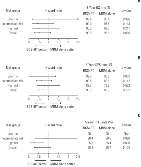
Fig. 2. Subgroup analyses according to risk group for overall survival (OS) (A), disease-free survival (DFS) (B), and regional failure-free survival (RFFS) (C). BCS+RT, breast conserving surgery plus radiation therapy; MRM, modified radical mastec- tomy; NA, not applicable,
a)Hazard ratio was not calculated for RFFS in the low risk group because there was no regional failure in that subgroup.
Risk group Low risk Intermediate risk
High risk Overall
Hazard ratio 5-Year OS rate (%)
BCS+RT MRM alone p-value 0.970 0.113 0.211 0.088
99.4 99.0 96.3 98.6
98.4 95.8 92.7 96.1
0 0.5 1 1.5 2 2.5
A
MRM alone better BCS+RT better
Risk group Low risk Intermediate risk
High risk Overall
Hazard ratio 5-Year DFS rate (%)
BCS+RT MRM alone p-value 0.052 0.142 0.522 0.107
99.5 93.6 83.7 93.3
96.8 88.6 79.6 89.7
0 0.5 1 1.5 2 2.5
B
MRM alone better BCS+RT better
Risk group Low risk Intermediate risk
High risk Overall
Hazard ratio 5-Year RFFS rate (%)
BCS+RT MRM alone p-value NA
a)0.890 0.008 0.102
100 98.4 95.8 98.3
100 96.6 85.4 95.7
0 0.5 1 1.5 2 2.5
C
MRM alone better
BCS+RT better
ber of risk factors (positive LVI and HG of III) identified in multivariate analysis for DFS and RFFS: the low risk group (n=346) had no risk factors, the intermediate risk group (n=512) had one risk factor, and the high risk group (n=216) had two risk factors. For the high risk group, BCS+RT showed significantly superior RFFS (99.5% vs. 96.8%, p=0.008) (Fig. 2). Although there was also a tendency for BCS+RT to have better DFS for all risk groups, there were no statistically significant differences (Fig. 2).
3. Toxicity
Lymphedema and radiation pneumonitis occurred more frequently in the BCS+RT group than in the MRM-alone group (Table 6). A total of 8.0% of patients in the MRM-alone group showed lymphedema, while 13.5% of patients pre- sented with lymphedema after BCS+RT (p=0.007). Radiation pneumonitis was present in 1.3% of patients in the BCS+RT group, whereas no patient in the MRM group showed radi- ation pneumonitis (p=0.031).
Discussion
Randomized trials comparing BCS+RT with mastectomy in early-stage breast cancer have shown comparable onco- logic outcomes by long-term follow-up data [1-6]. However, several recent population-based studies reported that BCS+RT resulted in superior oncologic outcomes compared to mastectomy [7-11]. There are several reasons for this inconsistency in results between historic randomized trials and population-based studies. Those randomized trials were conducted in the 1980s with outdated diagnostic and thera- peutic procedures; therefore, local recurrence was much higher [11,22]. Differences in study designs and patient pop- ulations may be additional reasons for inconsistent results between randomized trials and population-based studies [23]. The evolution of chemotherapy for early breast cancer may also deviate from the results of historic randomized tri- als [18,19]. Therefore, special caution is required in interpret- ing those randomized trials in current practice. Those popu- lation-based studies also have some limitations. The chemo- therapy regimens were not standard. In addition, compar- isons between treatment modalities were stratified only according to pathologic T or N category, not according to any other histologic or molecular subtype-based characteristics.
To our knowledge, the current research is the first study to compare treatment outcomes between BCS+RT and MRM alone under a modern and homogeneous chemotherapy reg- imen. In addition, the treatment outcomes were compared
according to subgroups stratified with various pathological variables for pN1 breast cancer. The inhomogeneity of the demographic and histologic properties was amended by propensity score matching.
The oncologic outcomes were not significantly different between treatment modalities. Among population-based studies, two reported the outcome of the pN1 subgroup.
Chen et al. [8] used the National Cancer Database and reported the adjusted hazard ratio of mastectomy alone as 1.44 (range, 2.31 to 1.53; p < 0.001) over BCS+RT in patients with pN1 breast cancer. van Maaren et al. [11] analyzed pati- ents selected from the Netherlands Cancer Registry and reported a higher mortality risk rate in the mastectomy com- pared with the BCS+RT. However, in those studies, mastec- tomy groups contained higher proportions of patients with large and multi-centric tumors, which are known prognostic factors associated with inferior LRFFS and OS. In addition, information regarding detailed chemotherapy regimen was not clarified, and a considerable proportion of patients treated with chemotherapy were included in the study pop- ulation. In the modern chemotherapy era, the outcome of mastectomy alone has improved [18,19]. In particular, some studies have specifically reported that patients with 1-3 pos- itive LNs showed improvement in oncologic outcomes [20,24,25]. Therefore, the results are inconsistent, with the current study showing better outcomes for the mastectomy group than those studies.
The role of PMRT is also a controversial issue for pN1 non- metastatic breast cancer. The DBCG 82 B&C trial and EBC- TCG revealed that PMRT reduced mortality rates regardless of the number of LN metastases [6,13,14]. On the other hand, recent retrospective studies showed that addition of PMRT in patients with pN1 breast cancer seems to have no signifi- cant impact on oncologic outcomes in the modern chemo- therapy era [20,24,25]. In the current study, the benefit of PMRT was not evaluated because patients who received PMRT were excluded from analyses. However, in the high- risk subgroup with LVI and HG 3, which were identified as independent factors associated with poor DFS, the BCS+RT group showed significantly better RFFS than the MRM-alone group. This implied that there might be a role of adjuvant RT in reducing regional recurrence for patients with LVI and HG 3, which are well-known risk factors related to poor locore- gional control and LN metastasis [20,26-28]. Therefore, PMRT is a consideration for patients with LVI and HG III even in the modern chemotherapy era. Further large scale randomized trials with stratification according to detailed risk groups are necessary to identify the role of PMRT in pN1 non-metastatic breast cancer.
There are several limitations in the current study. First,
there is inevitable selection bias due to its retrospective
nature. Although the propensity score matching method was
utilized to balance treatment groups, some variables that were excluded from score matching might have been unevenly distributed. The pathologic data were collected from several different institutions, so bias regarding patho- logic information also could not be excluded. Finally, the rel- atively short follow-up duration of the current study is also a limitation. The median follow-up duration of the total cohort was 69 months, which is not enough to detect late recurrence and treatment-related sequelae [29]. Therefore, further follow-up is required to compare long-term oncologic outcomes and adverse effects between treatment groups.
In conclusion, there are no differences in OS, DFS, LRFFS, and RFFS between BCS+RT and MRM alone under the AT chemotherapy regimen for patients with pN1 non-breast can- cer. However, BCS+RT showed superior RFFS in patients with LVI and HG III, implying that adjuvant RT potentially has a role in reducing regional recurrence. Therefore, PMRT might be considerable for patients with LVI and HG 3.
Conflicts of Interest
Conflict of interest relevant to this article was not reported.
Acknowledgments
This study was supported by a grant from the National R&D Pro- gram for Cancer Control, Ministry of Health & Welfare, Republic of Korea (1720170).
Author Details
1
Department of Radiation Oncology, Samsung Medical Center, Sungkyunkwan University School of Medicine, Seoul,
2Center for Breast Cancer, Research Institute and Hospital, National Cancer Center, Goyang,
3Department of Radiation Oncology, Seoul Natio- nal University College of Medicine, Seoul,
4Department of Radiation Oncology, Ewha Womans University Mokdong Hospital, Ewha Womans University School of Medicine, Seoul,
5Department of Radiation Oncology, Kosin University Gospel Hospital, Busan,
6
Department of Radiation Oncology, Yonsei Cancer Center, Yonsei University College of Medicine, Seoul,
7Department of Radiation Oncology, Chonnam National University Medical School, Gwangju,
8