Korean Circulation Journal
Introduction
Conditions leading to sinus node dysfunction have considerable clinical importance. Sick sinus syndrome (SSS) manifesting as sinus bradycardia, sinus arrest, and/or sinoatrial block is the most frequent worldwide clinical indication for pacemaker implantation.
1)Benson et al.
2)screened SCN5A as a candidate gene in ten patients from families diagnosed with congenital SSS on the basis of disorders of cardiac rhythm and conduction during the first decade of life.
Moreover, several investigations have linked variations in SCN5A, the gene encoding the pore-forming α-subunit of the cardiac-type sodium ion (Na
+) channel, Nav1.5, with familial SSS.
3-5)Furthermore, the SCN5A gene is known to be associated with different forms of arrhythmias such as long QT syndrome, Brugada syndrome,
Print ISSN 1738-5520 • On-line ISSN 1738-5555
Genetic Variation of SCN5A in Korean Patients with Sick Sinus Syndrome
Young Soo Lee, MD 1 *, Michael A Olaopa, PhD 2 * , Byung Chun Jung, MD 3 , Sang Hee Lee, MD 4 , Dong Gu Shin, MD 4 , Hyoung Seob Park, MD 5 , Yongkeun Cho, MD 6 , Sang Mi Han, MS 7 , Myung Hoon Lee, PhD 7,8 , and Yoon Nyun Kim, MD 5
1
Division of Cardiology, Catholic University of Daegu, Daegu, Korea,
2Krannert Institute of Cardiology, Indiana University School of Medicine, Indianapolis, IN, USA,
3
Daegu Fatima Hospital, Daegu,
4Yeungnam University, Daegu,
5Department of Internal Medicine, Keimyung University, Daegu,
6Kyungpook National University, Daegu,
7
D&P Biotech, Daegu,
8Department of Biochemistry and Cell Biology, Kyungpook National University, Daegu, Korea
Background and Objectives: Due to recent studies that have shown an association between the genetic variation of SCN5A and sick sinus syndrome (SSS), we sought to determine if a similar correlation existed in Korean patients with SSS.
Subjects and Methods: We enrolled 30 patients with SSS who showed a sinus pause (longer than 3.0 s) in Holter monitoring, in addition to 80 controls. All exons including the putative splicing sites of the SCN5A gene were amplified by polymerase chain reaction and sequenced either directly or following subcloning. Wild-type and single nucleotide polymorphisms were expressed in human embryonic kidney cells, and the peak sodium current (I
Na) was analyzed using the whole-cell patch-clamp technique.
Results: A total of 9 genetic variations were identified: 7 variations (G87A-A29A, IVS9-3C>A, A1673G-H558R, G3823A-D1275N, T5457C- D1819D, T5963G-L1988R, and C5129T-S1710L) had been previously reported, and 2 variants (A3075T-E1025D and T4847A-F1616Y) were novel; the potential structural effects of F1616Y were analyzed in a three-dimensional model of the SCN5A domain. Patch-clamp studies at room temperature demonstrated that the peak I
Nawas significantly increased by 140% in HEK cells transfected with F1616Y compared with wild-type (-335.13 pA/pF±24.04, n=8 vs. -139.95 pA/pF±23.76, n=7, respectively). Furthermore, the voltage dependency of the activation and steady-state inactivation of F1616Y were leftward-shifted compared with wild-type (V
hactivation=-55.36 mv±0.22, n=8 vs. V
hactivation=-44.21 mV±0.17, n=7; respectively; V
hinactivation=-104.47 mV±0.21, n=7 vs. V
hinactivation=-84.89 mV±0.09, n=12, respectively).
Conclusion: F1616Y may be associated with SSS. (Korean Circ J 2016;46(1):63-71) KEY WORDS: SCN5A protein, human; Polymorphism, single nucleotide; Sick sinus syndrome.
Received: February 20, 2015 Revision Received: May 18, 2015 Accepted: July 7, 2015
Correspondence: Yoon Nyun Kim, MD, Department of Internal Medicine, Keimyung University, 56 Dalseong-ro, Jung-gu, Daegu 41931, Korea Tel: 82-53-250-7432, Fax: 82-53-250-7034
E-mail: ynkim@dsmc.or.kr
* These authors contributed equally to this work.
• The authors have no financial conflicts of interest.
This is an Open Access article distributed under the terms of the Creative Commons Attribution Non-Commercial License (http://creativecommons.
org/licenses/by-nc/3.0) which permits unrestricted non-commercial use,
distribution, and reproduction in any medium, provided the original work
is properly cited.
progressive cardiac conduction defect, atrial fibrillation, dilated cardiomyopathy, and overlapping syndromes.
6)7)Voltage-gated Na
+channels are transmembrane proteins that produce the fast inward Na
+current responsible for the depolarization phase of the cardiac action potential. Inherited variations in SCN5A result in a spectrum of disease entities termed as Na
+channelopathies.
To date, although many variations of the SCN5A gene have been documented in various cardiac diseases, SCN5A variations in Korean SSS patients have not yet been studied in detail. Therefore, we carried out complete sequencing of the coding regions of the SCN5A gene, excluding untranslated regions, in Korean SSS patients in order to identify any potential variations associated with SSS.
Subjects and Methods
Patient population
This study was approved by the Ethics Committee at each hospital;
consent was obtained from all individuals before enrollment into the study. We enrolled 30 Korean patients with SSS and 80 controls with no cardiac symptoms. The diagnostic criterion for SSS was the presence of a pause longer than 3 seconds in the day-time during Holter monitoring or surface electrocardiography. Normal sinus function was confirmed in controls through a cardiac electrophysiological study after ablation for supraventricular tachycardia. We excluded the patients with atrial and ventricular arrhythmia such as atrial and ventricular tachycardia, atrial fibrillation and atrial flutter, atrioventricular block, Brugada syndrome, known malignancy, and a structurally abnormal heart.
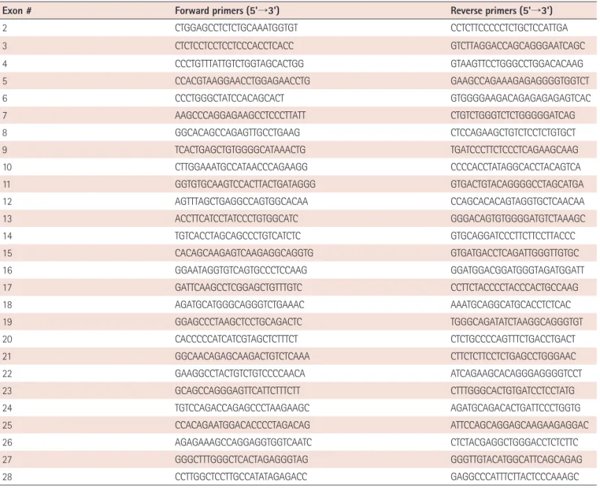
Table 1. Multiplex polymerase chain reaction primers for amplification of the coding region of SCN5A
Exon # Forward primers (5'→3') Reverse primers (5'→3')
2 CTGGAGCCTCTCTGCAAATGGTGT CCTCTTCCCCCTCTGCTCCATTGA
3 CTCTCCTCCTCCTCCCACCTCACC GTCTTAGGACCAGCAGGGAATCAGC
4 CCCTGTTTATTGTCTGGTAGCACTGG GTAAGTTCCTGGGCCTGGACACAAG
5 CCACGTAAGGAACCTGGAGAACCTG GAAGCCAGAAAGAGAGGGGTGGTCT
6 CCCTGGGCTATCCACAGCACT GTGGGGAAGACAGAGAGAGAGTCAC
7 AAGCCCAGGAGAAGCCTCCCTTATT CTGTCTGGGTCTCTGGGGGATCAG
8 GGCACAGCCAGAGTTGCCTGAAG CTCCAGAAGCTGTCTCCTCTGTGCT
9 TCACTGAGCTGTGGGGCATAAACTG TGATCCCTTCTCCCTCAGAAGCAAG
10 CTTGGAAATGCCATAACCCAGAAGG CCCCACCTATAGGCACCTACAGTCA
11 GGTGTGCAAGTCCACTTACTGATAGGG GTGACTGTACAGGGGCCTAGCATGA
12 AGTTTAGCTGAGGCCAGTGGCACAA CCAGCACACAGTAGGTGCTCAACAA
13 ACCTTCATCCTATCCCTGTGGCATC GGGACAGTGTGGGGATGTCTAAAGC
14 TGTCACCTAGCAGCCCTGTCATCTC GTGCAGGATCCCTTCTTCCTTACCC
15 CACAGCAAGAGTCAAGAGGCAGGTG GTGATGACCTCAGATTGGGTTGTGC
16 GGAATAGGTGTCAGTGCCCTCCAAG GGATGGACGGATGGGTAGATGGATT
17 GATTCAAGCCTCGGAGCTGTTTGTC CCTTCTACCCCTACCCACTGCCAAG
18 AGATGCATGGGCAGGGTCTGAAAC AAATGCAGGCATGCACCTCTCAC
19 GGAGCCCTAAGCTCCTGCAGACTC TGGGCAGATATCTAAGGCAGGGTGT
20 CACCCCCATCATCGTAGCTCTTTCT CTCTGCCCCAGTTTCTGACCTGACT
21 GGCAACAGAGCAAGACTGTCTCAAA CTTCTCTTCCTCTGAGCCTGGGAAC
22 GAAGGCCTACTGTCTGTCCCCAACA ATCAGAAGCACAGGGAGGGGTCCT
23 GCAGCCAGGGAGTTCATTCTTTCTT CTTTGGGCACTGTGATCCTCCTATG
24 TGTCCAGACCAGAGCCCTAAGAAGC AGATGCAGACACTGATTCCCTGGTG
25 CCACAGAATGGACACCCCTAGACAG ATTCCAGCAGGAGCAAGAAGAGGAC
26 AGAGAAAGCCAGGAGGTGGTCAATC CTCTACGAGGCTGGGACCTCTCTTC
27 GGGCTTTGGGCTCACTAGAGGGTAG GGGTTGTACATGGCATTCAGCAGAG
28 CCTTGGCTCCTTGCCATATAGAGACC GAGGCCCATTTCTTACTCCCAAAGC
Sampling and DNA extraction
All patient and control samples were recruited from the four participating medical centers: Keimyung University, Yeungnam University, Catholic University of Daegu, and Daegu Fatima Hospital.
Peripheral bloods were collected in ethylenediamine tetraacetic acid (EDTA) containing tubes, and DNA was extracted from whole blood samples using the QIAamp DNA blood mini kit (Qiagen, Hilden, Germany). DNA concentration was determined using a NanoDrop ND1000 spectrophotometer, and the purity of the DNA was assessed
based on the 260/280 nm absorbance ratio.
Multiplex polymerase chain reaction and sequence analysis The coding region (exon 2-exon 28) of SCN5A was amplified by multiplex polymerase chain reaction (PCR) using newly designed primers (Table 1). PCR conditions were as follows: after an initial denaturation at 95℃ for 15 min, denaturation at 94℃ for 30 sec, annealing at 68-70℃ for 30-60 sec, and extension at 72℃ for 60- 90 sec were repeated for 30-35 cycles. Following multiplex PCR, Table 2. Baseline demographics of patients
No Age Gender Height (cm) Weight
(kg) DM HTN Symptom LVEF (%) Family history
of SSS Sinus pause (sec)
1 60 Male 164 88 Yes Yes Dizziness 58 No 3.3
2 57 Female 154 55 No Yes Dizziness 68 No 3.2
3 42 Male 172 73 No Yes Dizziness 64 No 3.4
4 47 Female 161 51 No No Syncope 57 No 6.1
5 60 Male 153 65 No Yes Syncope 68 No 6.0
6 44 Female 160 59 No No Dizziness 64 No 3.4
7 65 Female 153 59 No No Dizziness 68 No 3.7
8 58 Male 173 68 Yes No Dizziness 66 No 4.0
9 58 Female 162 57 No No Dizziness 71 No 3.7
10 65 Male 171 80 No No Syncope 57 No 8.4
11 57 Female 153 59 No Yes Syncope 67 No 6.3
12 58 Female 157 72 No No Dizziness 71 Yes 3.9
13 61 Female 171 77 Yes No Dizziness 64 No 3.4
14 58 Female 158 62 Yes Yes Syncope 76 No 6.2
15 65 Female 155 65 Yes Yes Dyspnea 77 No 4.3
16 27 Male 168 70 No No Dizziness 46 No 3.6
17 63 Female 154 64 No Yes Dyspnea 67 No 3.6
18 62 Female 146 63 Yes Yes Syncope 68 No 6.5
19 61 Female 148 59 Yes No Dizziness 57 No 4.8
20 39 Female 158 63 No No Dizziness 70 No 4.7
21 60 Female 147 58 No No Dizziness 75 No 4.2
22 59 Female 160 88 Yes No Dizziness 63 No 4.6
23 62 Male 167 74 No No Syncope 53 No 10.0
24 55 Female 157 61 No No Dizziness 71 No 3.1
25 52 Female 152 52 No No Syncope 71 No 5.8
26 59 Female 146 55 Yes No Dyspnea 59 No 3.2
27 45 Male 168 60 No No Dyspnea 60 No 4.2
28 59 Female 155 52 Yes Yes Syncope 52 No 5.6
29 64 Male 166 62 No No Dizziness 72 No 4.0
30 47 Female 155 51 No No Dizziness 64 No 4.6
DM: diabetes mellitus, HTN: hypertension, LVEF: left ventricular ejection fraction, SSS: sick sinus syndrome
the reaction mixture was electrophoresed in a 2% agarose gel and stained with ethidium bromide (EtBr). Amplified PCR products were purified using the QIAquick PCR purification kit (Qiagen, Hilden, Germany), and directly sequenced using the BigDye Terminator Ver 3.1 cycle sequencing kit (Applied Biosystems, Foster city, CA, USA) and an ABI PRISM 3100 Genetic Analyzer (Applied Biosystems, Foster City, CA, USA). Sequencing results were compared with reference sequences (SCN5A/NM_198056.2/ENSG00000183873/ENST00000333535) using the alignment program BLAST 2.0 of the national center of biotechnology information (NCBI; Bethesda, MD, USA), and the portion of variation that occurred was determined.
Three-dimensional modeling of SCN5A
The structural model of the human SCN5A domain was obtained from the Automated SWISS-MODEL version 8.05 (Swiss Institute of Bioinformatics Biozentrum, Basel, Switzerland).
8-10)Superimposition, model building, construction of insertion regions, structure validation, and calculation of structural properties were carried out
using the subprograms ProMod, SPDBV, Loop, LoopDB, Parameters, and Topologies, which are available in the Automated Swiss-Model Package Program (http://swissmodel.expasy.org). Pymol v0.99 and Cn3D (www. ncbl.nlm.nih.gov/Structure/CN3D/cn3d.html) were used to display the three-dimensional model structures.
Patch-clamp recording
Whole-cell configuration of the voltage-clamp technique was used as described elsewhere.
11)Briefly, whole-cell configuration was made in Tyrode’s solution. Pipette resistances were 1.5–3 MΩ. After achieving a gigaseal, the test-pulse current was nulled by adjusting the pipette capacitance compensator with both fast and slow components. After break-in, the whole-cell charging transient was nulled by adjusting whole-cell capacitance and series resistance. Voltage control protocols were generated with an Axopatch 200B amplifier/Digidata 1440A acquisition system using the pCLAMP-10 software (Molecular Devices/
Axon, Sunnyvale, CA, USA). Whole-cell recording was analyzed using Clampfit 10.2 (Axon, Sunnyvale, CA, USA). For measuring I
Na, we Table 3. Genetic variations in the SCN5A gene and their frequency in Korean patients with sick sinus syndrome (N=30) and normal controls (N=80)
dbSNP ID Variation N Exon Genotype frequency (N/%)
rs6599230 G87A (A29A)
Case Control 30
80 2 GG GA AA
11 (36.7) 15 (50.0) 4 (13.3) 36 (45.0) 36 (45.0) 8 (10.0)
rs41312433 IVS9-3C>A
Case
Control 30
80 intron 9 CC CA AA
22 (73.3) 8 (26.7) 0 (0.0) 61 (76.3) 19 (23.8) 0 (0.0)
rs1805124 A1673G (H558R)
Case
Control 30
80 12 AA AG GG
22 (73.3) 8 (26.7) 0 (0.0) 59 (73.8) 21 (26.3) 0 (0.0) A3075T (E1025D)
Case Control
30 80 17
AA AT TT 29 (96.7) 1 (3.3) 0 (0.0) 80 (100.0) 0 (0.0) 0 (0.0) G3823A (D1275N)
Case Control
30
80 21 GG GA AA
28 (93.3) 2 (6.7) 0 (0.0) 80 (100.0) 0 (0.0) 0 (0.0) T4847A (F1616Y)
Case Control
30
80 28 TT TA AA
29 (96.7) 1 (3.3) 0 (0.0) 80 (100.0) 0 (0.0) 0 (0.0) C5129T (S1710L)
Case Control
30
80 28 CC CT TT
29 (96.7) 1 (3.3) 0 (0.0) 80 (100.0) 0 (0.0) 0 (0.0)
rs1805126 T5457C (D1819D)
Case Control 30
80 28
TT TC CC
11 (36.7) 8 (26.7) 11 (36.7) 8 (10.0) 52 (65.0) 20 (25.0) T5963G (L1988R)
Case
Control 30
80 28 TT TG GG
27 (90.0) 3 (10.0) 0 (0.0)
78 (97.5) 2 (2.5) 0 (0.0)
dbSNP ID: database single nucleotide polyporphysm
used Tyrode’s solution (see above) as the bath solution. The pipette solution contained (in mM) NaF 10, CsF 110, CsCl 20, EGTA 10, and HEPES 10 (pH 7.35 adjusted with CsOH). The HEK293 cells used in our experiments did not show significant persistent I
Nain the absence or presence of tetrodotoxin as previously described.
11)12)Results
Patient demographics
Table 2 shows patient demographics. The mean age is 56 years old, and the mean left ventricular ejection fraction was 65%. 9 patients were male. The mean sinus pause detected by surface ECG or Holter monitoring was 4.7 sec.
Gene sequencing
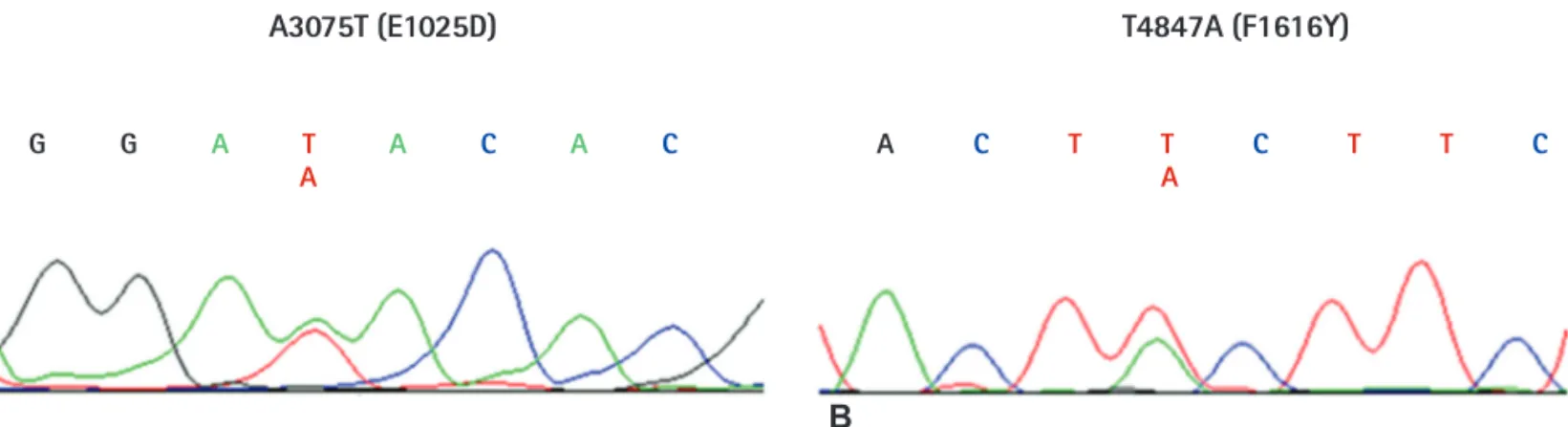
We sequenced all exons (exon 2-exon 28) of the SCN5A gene except for untranslated regions. Although we analyzed only 30 samples from Korean SSS patients, we identified 9 genetic variations, consisting of 2 synonymous, 6 non-synonymous, and 1 at the splicing donor site of the intron9-exon10 boundary (Table 3). Among these, 7 variations (G87A-A29A, IVS9-3C>A, A1673G-H558R, G3823A-D1275N, T5457C- D1819D, T5963G-L1988R, and C5129T-S1710L) have previously been reported in the American, Japanese, and Han Chinese populations, and their allele frequencies were similar to that in the Japanese or Chinese series. In addition, 2 novel variations (A3075T-E1025D and T4847A- F1616Y) were also found in two patients (Fig. 1). To compare with the control population, we analyzed normal samples using the same methods in order to determine whether the same variations existed in
A3075T (E1025D) T4847A (F1616Y)
G G A T A C A C
A A C T T C T T C
A
Fig. 1. DNA sequencing analysis results of the newly discovered SCN5A gene variants in this study. (A) DNA sequencing results of SCN5A exon 17. A3075T is a heterozygous nucleotide change, and causes an amino acid change from glutamine to aspartate. (B) DNA sequencing results of SCN5A exon 28.
T4847A is a heterozygous nucleotide change, and causes an amino acid change from phenylalanine to tyrosine. All DNA sequence electropherograms show non-synonymous nucleotide changes in the sick sinus syndrome patients.
A B
Fig. 2. Two variation sites (F1616Y and S1710L) within the 3D model of the SCN5A domain. F1616Y is located in the center of the second loop, which is well exposed, while the S1710L variation is located immediately following the 6th alpha helix.
S1710L S1710L 90°
F1616Y
F1616Y
their genes. Five variations (G87A-A29A, IVS9-3C>A, A1673G-H558R, T5963G-L1988R, and T5457C-D1819D) were also found in the control group, however the novel genetic variations (A3075T-E1025D and T4847A-F1616Y) were not present in any of the 80 normal controls.
Three-dimensional model of sinlge nucleotide polyporphysm We successfully generated a three-dimensional model of the SCN5A domain (amino acid residues 1525-1750), which revealed 8 alpha-helices and 7 loops or kinks. We were able to use this model to analyze one of the novel variations (F1616Y) found in this study, and one of the known variations (S1710L) (Fig. 2). The location of the S1710L variation was immediately after the 6
thalpha helix. The long 6
thloop started from this area. In addition, the serine to leucine change likely generated an extended 6
thalpha-helical structure.
F1616Y was located in the center of the second loop, which is well exposed. Three-dimensional modeling of the other variations was not successful due to a lack of available crystal structures for the homologous templates.
Patch-clamp recording
Figs. 3 and 4 show the representative tracings and time course of I
Naat a frequency of 20/min. The I
Nawas induced by a repetitive depolarization pulse (to –10 mV for 300 ms) from a holding potential of –140 mV. All experiments were carried out at room temperature.
The peak I
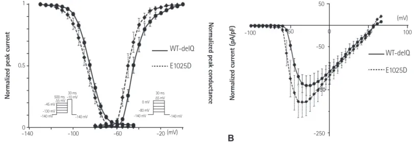
Naand voltage-dependency in cells transfected with mutant E1025D were not significantly changed compared with wild- type (Fig. 3). However, the voltage-dependency of the Na
+channel in mutant F1616Y-expressing HEK cells was significantly leftward- shifted compared with wild-type (V
hinactivation of F1616Y- delQ=-104.47 mV±0.21, n=7 vs. V
hinactivation of WT-delQ=-84.89 mV±0.09, n=12, p<0.005; V
hactivation of F1616Y-delQ=-55.36 mv±0.22, n=8 vs. V
hactivation of WT-delQ=-44.21 mV±0.17, n=7, p<0.005) (Fig. 4A). Furthermore, the peak I
Nain cells transfected with F1616Y demonstrated a 140% increase compared with wild-type (-335.13 pA/pF±24.04, n=8 vs -139.95 pA/pF±23.76, n=7, respectively, p<0.005) (Fig. 4B).
WT-deIQ E1025D 1
0.5
0
N ormaliz ed p eak c urr en t Normaliz ed c urr en t (pA /p F)
N ormaliz ed p eak c ondu cta nc e
-140 -100 -60 -20 (mV)
WT-deIQ E1025D
WT-deIQ
5 ms
1 nA
1 nA 5 ms
E1025D
(mV) 50
50 0
-100 100
-50
-250
-45 mV -130 mV -140 mV
∆5 mV ∆5 mV
0 mV -80 mV
-140 mV -140 mV
500 ms -10 mV
-140 mV
30 ms 30 ms
Fig. 3. Gating kinetics of WT and mutant E1025D sodium channels at 25°C in HEK293 cells. (A) Voltage-dependent kinetics of the steady-state fast inactivation and peak conductance at 25°C.
Steady-state inactivation was estimated by 500-ms pre-pulse protocols from a holding potential of
−140 mV. Normalized peak currents were plotted against the pre-pulse membrane potentials.
Conductance G (V) was calculated by the equation: G (V)=I
peak/(V
m−E
rev), where I
peakis the peak current, E
revis the measured reversal potential, and V
mis the membrane potential. The normalized peak conductance was plotted against the membrane potentials. Inactivation data were fitted with the Boltzmann equation: y={1+exp ([V
m−V
h]/k)}
−1; conductance data were fitted with the Boltzmann equation: y={1+ exp ([V
h−V
m]/k)}
−1, where y represents variables; V
h, midpoint; k, slope factor; and V
m, membrane potential. Note: V
h(E1025D-delQ)=-90.29 mv±0.31 (n=12) is slightly leftward-shifted compared with V
h(WT-delQ)=-84.89 mV±0.09 (n=10), p<0.1; k (E1025D- delQ)=7.73±0.27, k (WT-delQ)=6.33±0.08. V
hactivation (E1025D-delQ)=-52.14 mv±0.43 (n=6) is significantly leftward-shifted compared with V
hactivation (WT-delQ)=-44.21 mV±0.17 (n=7) (p<0.05). Data are presented as the mean±SE. (B) I–V relationships at 25°C. I
peak(E1025D- delQ)=-178.61 pA/pF±34.75 (n=6), I
peak(WT-delQ)=-139.95 pA/pF±23.76 (n=7) (p<0.1). (C) Superimposed macroscopic sodium current traces obtained from the cells expressing WT-delQ (upper panel) and E1025D-delQ (lower panel). The currents were induced by step-pulse protocols from a holding potential of −140 mV. WT: wild type, SE: standard error.
A
C
B
Discussion
In the present study, the genetic nucleotide sequence of SCN5A was determined in Korean SSS patients and compared with that of the controls. We found two novel genetic variations (A3075T-E1025D and T4847A-F1616Y). In addition, the normalized I
Nain mutant F1616Y-expressing HEK cells showed a leftward-shift in inactivation and activation, and the peak I
Nawas increased.
The SCN5A gene consists of 28 exons and encodes a protein of 2016 amino acids, with a molecular mass of 227 kDa.
13)In our patients with SSS, there were 9 sites of nucleotide change from exon 2 to exon 28 of the SCN5A gene. Among these, 2 sites (G87A- A29A and IVS9-3C>A) have been reported as genetic variations in a Western study,
14)and T5457C-D1819D has already been reported as a variation or polymorphism without functional effects to the channel in Asian studies.
15-17)Recently, Gui et al.
18)reported that A1673G-H558R has variation-specific effects on SCN5A-related SSS.
In addition, G3823A-D1275N, which is reported as a heterozygous loss-of-function variation, is related to reduced whole-cell currents due to impaired cell surface localization.
19)Since a synonymous nucleotide change in the SCN5A gene is unlikely to induce a functional change in the Na
+channel, a non- synonymous substitution was considered as a candidate for the variation associated with this disorder. The two new variations found in this study are non-synonymous and heterozygous changes.
The variations may not affect the pathogenesis of SSS due to the opposite normal allele. Therefore, it is reasonable to suggest that the functional consequences of these changes should be assessed by measuring their effect on Na
+channel activity, allowing assessment of a direct relationship between the variations and disease.
We can deduce the functional effect of the variations by structural simulations, which provide direction for subsequent studies. In the present study, we obtained some information from a structural model of SCN5A on the functional effect of the variations S1710L
N ormaliz ed p eak c urr en t N ormaliz ed curr ent (p A/pF ) N ormaliz ed peak c ondu cta nc e
-140 -100 -60 -20 (mV)
WT-deIQ F1616Y
WT-deIQ F1616Y
WT-deIQ
5 ms
1 nA
1 nA 5 ms
F1616Y
(mV) 1
0.5
50
-100 50 100
-50
-150
-250
0 -350
A B
C
-45 mV -130 mV -140 mV
∆5 mV ∆5 mV
0 mV -80 mV
-140 mV -140 mV
500 ms -10 mV
-140 mV
30 ms 30 ms