This is an open-access article distributed under the terms of the Creative Commons Attribution Non-Commercial License (http://creativecommons.org/
licenses/by-nc/4.0/), which permits unrestricted non-commercial use, distribution, and reproduction in any medium, provided the original work is properly cited.
CC
Moon-Gi Choi
Department of Oral and Maxillofacial Surgery, College of Dentistry, Wonkwang University, Iksan, Korea
Abstract(J Korean Assoc Oral Maxillofac Surg 2019;45:357-363)
Ranula is a mucocele caused by extravasation of the sublingual gland on the floor of the mouth. The most common presentation is a cystic mass in the floor of the mouth. A portion of the sublingual gland could herniate through the mylohyoid muscle, and its extravasated mucin can spread along this hiatus into submandibular and submental spaces and cause cervical swelling. This phenomenon is called plunging ranula. A variety of treatments for ranula has been suggested and include aspiration of cystic fluid, sclerotherapy, marsupialization, incision and drainage, ranula excision only, and excision of the sublingual gland with or without ranula. Those various treatments have shown diverse results. Most surgeons agree that removal of the sublingual gland is necessary in oral and plunging ranula. Four patients with ranula were investigated retrospectively, and treatment methods based on literature review were attempted.
Key words: Ranula, Sublingual gland, Plunging ranula
[paper submitted 2018. 7. 19 / revised 1st 2018. 10. 17, 2nd 2018. 10. 31 / accepted 2018. 11. 17]
Copyright © 2019 The Korean Association of Oral and Maxillofacial Surgeons. All rights reserved.
I. Introduction
A ranula is a mucocele caused by extravasation of the sub- lingual gland on the floor of the mouth. The most common presentation is a cystic mass on the floor of the mouth. Ranu- las can be induced by pooled mucin from ruptured acini of the sublingual gland or a ruptured duct of Rivinus. Because ranulas are lined with granulation tissue instead of epithe- lium, they are considered a type of pseudocyst1,2.
In such conditions, there is a dehiscence in the mylohy- oid muscle. A portion of the sublingual gland may herniate through this hiatus, and its extravasated mucin can spread along this hiatus into the submandibular and submental space.
This phenomenon is called a plunging ranula3. Although trauma is traditionally thought to be the cause of ranula de- velopment, only 2.8% of patients demonstrated the history of trauma in Zhao’s study2. A different study proposed ductal
obstruction and congenital malformation as possible etiolo- gies4.
Various treatments for ranula have been suggested, and each treatment has shown a diverse success rate. Therefore, this article describes the cases of simple ranula and plunging ranula and discusses their appropriate management with ref- erence to the literature.
II. Cases Report
1. Case 1
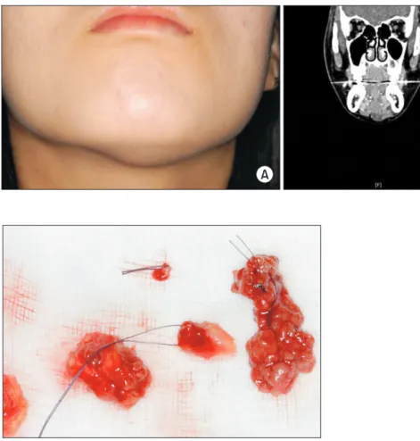
A female patient aged 16 years complained of left sub- mental swelling in 2008.(Fig. 1. A) Intraorally, there was no swelling on the mouth floor. Computed tomography (CT) revealed a mutiloculated cystic lesion at the left sublingual and submental space.(Fig. 1. B) The tentative diagnosis was a plunging ranula.
Under general anesthesia, the left sublingual gland was removed transorally.(Fig. 2) Because the cystic wall was so delicate and fragile, the cystic component could not be com- pletely removed, and gauze packing was placed. The gauze was changed regularly for two weeks, and secondary wound healing was good. The histologic diagnosis was plunging ranula.(Fig. 3) There has been no recurrence, and submental swelling disappeared.
Moon-Gi Choi
Department of Oral and Maxillofacial Surgery, College of Dentistry, Wonkwang University, 460 Iksan-daero, Iksan 54538, Korea
TEL: +82-63-859-2921 FAX: +82-63-859-2926 E-mail: omschoi@wonkwang.ac.kr
ORCID: https://orcid.org/0000-0003-3502-7652
2. Case 2
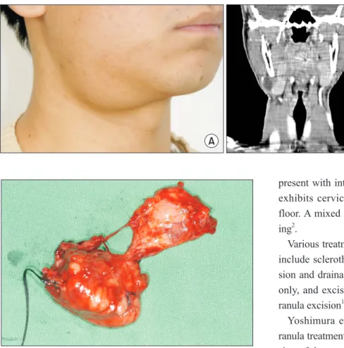
A male patient aged 22 years visited our facility. He had complained of right submandibular swelling in 2005. On CT, a cystic mass was observed around the right submandibular gland.(Fig. 4) The tentative diagnosis was a ranula originat- ing from the submandibular gland.
Because the submandibular gland was difficult to remove transorally, a submandibular approach was taken to remove the affected gland. Intraoperatively, a cystic mass was at- tached to both the sublingual and submandibular glands, so both glands were removed.(Fig. 5) The histologic diagnosis was a plunging ranula. There has been no recurrence since then.
3. Case 3
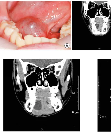
A male patient aged 12 years complained of a foreign body sensation due to swelling on the right of the mouth floor in
2015. On CT, a cystic mass, sized 3×7 cm2 was observed.(Fig.
6)
The tentative diagnosis was a simple ranula. Our depart- ment recommended removing the sublingual gland, but his parents opted for a more conservative treatment. Therefore, marsupialization was performed. Unlooping was carried out and the cystic wall and mucosa of the mouth floor were su- tured together. The histologic diagnosis was a ranula.
The healing process was uneventful. However, four months later, the patient complained of slight submandibular swell- ing. There was no intraoral swelling. On CT, the ranula was observed to have increased in size (Fig. 7) and was deter- mined to have recurred.
Our department recommended sublingual gland removal.
Under general anesthesia, the affected sublingual gland was removed.(Fig. 8) The patient experienced no further recur-
10 cm
A B
Fig. 1. A. The patient showed left sub- mental swelling. B. On computed to- mography, a mutilocuated cystic lesion at the left sublingual and submental space was observed.
Moon-Gi Choi: Case report of the management of the ranula. J Korean Assoc Oral Maxillofac Surg 2019
Fig. 2. Because the cystic wall was fragile, the cyst ruptured. Cys- tic fluid was suctioned. Only the affected left sublingual gland and attached ruptured cystic component were removed transorally.
Moon-Gi Choi: Case report of the management of the ranula. J Korean Assoc Oral
Maxillofac Surg 2019 Fig. 3. A mucus-containing space lined fibrous connective tissue
or granulation tissue with various sizes of vascular lumen (H&E staining, ×33).
Moon-Gi Choi: Case report of the management of the ranula. J Korean Assoc Oral Maxillofac Surg 2019
rence.
4. Case 4
A female patient aged 22 years complained of swelling of the right mouth floor in 2018. The amount of swelling was small. However, CT revealed a cystic mass that was attached to the sublingual gland.(Fig. 9) The tentative diagnosis was a simple ranula.
Because the cystic mass was attached to the sublingual gland, the sublingual gland was removed to prevent recur- rence. Under general anesthesia, the ranula and sublingual glands were removed together. The patient has had no further recurrence.
Table 1 details the cases treated by our department.
III. Discussion
Ranulas can be classified into three groups. Oral ranulas
present with intraoral swelling only, while a pluning ranula exhibits cervical swelling without swelling of the mouth floor. A mixed ranula has both intraoral and cervical swell- ing2.
Various treatments for ranulas have been suggested. These include sclerotherapy with OK-432, marsupialization, inci- sion and drainage, aspiration of cystic fluid, ranula excision only, and excision of the sublingual gland with or without ranula excision1,3,5-7.
Yoshimura et al.8 compared three different methods of ranula treatment. The recurrence rate was 25.0% for the exci- sion of the ranula only, 36.4% for marsupialization, and 0%
for excision of sublingual gland along with the ranula. Their study concluded that removal of the sublingual gland with the ranula was the most effective treatment modality.
Zhao et al.2 compared the recurrence rates of 580 ranulas treated using different surgical methods. They showed that recurrence was not associated with the type of ranula, but was correlated with the surgical method; the recurrence rate was 66.7% for marsupialization, 57.69% for excision of the ranula, and 1.2% for excision of the sublingual gland either with or without the ranula. Their study stressed that transoral sublingual gland removal was a basic and essential measure required to reduce the recurrence of any type of ranula.
Harrison3 surveyed the literature about the success rate of ranula treatments. In oral ranulas, the success rate was 100%
for the removal of the sublingual gland, 99% for removal of both the sublingual gland and the ranula, 63% for the re- moval of the ranula only, 55% for marsupialization only, 82%
for marsupialization with packing, 73% for injection of OK- 432, and 0% for incision and drainage. In plunging ranulas, the success rate was 96% for the removal of the sublingual gland, 95% for removal of both the sublingual gland and the ranula, 38% for the removal of the ranula only, 38% for mar- supialization only, 100% for marsupialization with packing,
A B
9 cm
Fig. 4. A. The patient showed sub- mandibular swelling. B. On computed tomography, cystic mass was observed around the right submandibular gland.
Moon-Gi Choi: Case report of the management of the ranula. J Korean Assoc Oral Maxillofac Surg 2019
Fig. 5. Intraoperatively, cystic mass attached to both sublingual and submandibular gland, so both glands were removed.
Moon-Gi Choi: Case report of the management of the ranula. J Korean Assoc Oral Maxillofac Surg 2019
59% for the injection of OK-432, and 4% for incision and drainage. For plunging ranulas, excision of the sublingual gland had an almost 100% success rate, and the addition of
marsupialization with packing could further reduce the failure rate. In the case of excision of the sublingual gland, the suc- cess rate was not 100% in all cases, which meant a sublingual gland that was source of extravasion had not been removed completely.
Shelley et al.9 reported on patients with extensive plung- ing ranulas. The initial lesion was a simple ranula managed by marsupialization only. However, the lesion recurred as a massive plunging ranula. Although extensive plunging ranula may penetrate into peripheral areas, this type of ranula can still be managed by the transoral removal of the sublingual gland and drainage of the cystic fluid.
Several authors have reported that marsupialization has resulted in a high rate of recurrence: 66.76% by Zhao et al.2, 61% by Crysdale et al.7, 52.6% by Parekh et al.4, and 20% by Patel et al.10.
Marsupialization performed with unroofing drains the cys-
8 cm
Fig. 7. On computed tomography, more increased sized ranula was observed.
Moon-Gi Choi: Case report of the management of the ranula. J Korean Assoc Oral Maxillofac Surg 2019
Fig. 8. Ranula and sublingual gland were removed together.
Moon-Gi Choi: Case report of the management of the ranula. J Korean Assoc Oral Maxillofac Surg 2019
12 cm
Fig. 9. On computed tomography, a small cystic mass attached to sublingual gland.
Moon-Gi Choi: Case report of the management of the ranula. J Korean Assoc Oral Maxillofac Surg 2019
9 cm
A B
Fig. 6. A. A right mouth floor swelled. B.
On computed tomography, cystic mass, sized 3×7 cm2 was observed.
Moon-Gi Choi: Case report of the management of the ranula. J Korean Assoc Oral Maxillofac Surg 2019
tic fluid and causes the collapse of the cavity, which quickly heals the oral surface or roof of the ranula and isolates the ranula from the mucosa of the floor of the mouth. The caus- ative extravasation is usually located in the deeper portion of the sublingual gland, so the source of leakage is not elimi- nated, and the ranula therefore has a tendency to recur within several weeks. To reduce the incidence of recurrence, a modi- fied marsupialization including packing of the cavity with gauze has been introduced instead of leaving the unroofed ranula cavity open with subsequent collapse of the cystic cav- ity. Gauze packing can bring about the fibrosis of ruptured acini of the sublingual gland and seal the leaking area11.
Morita et al.12 reported that marsupialization can be a useful treatment method for small-sized oral ranulas. In a report of nine patients treated with marsupialization, the ranulas only recurred in three patients. Baurmash13 was against sublingual gland removal for the treatment of oral ranula regardless of size and stressed that if the cyst is shallow and superficial, marsupialization with unroofing can be used to achieve low recurrence rates.
Marsupialization and simple cyst removal have varying success rates, but their recurrence rates are thought to depend on complete removal of the cyst along with the involved sub- lingual gland2. Removal of the sublingual gland is the recom- mended primary treatment for plunging ranulas.
According to Crysdale et al.7, when the ranula is excised along with the involved sublingual gland, the success rate is almost 100%; it is recommended that oral ranulas greater than 1 cm in diameter and plunging ranulas should be treated by excision of the ranula along with the sublingual gland as a primary therapy. If the ranula recurs after marsupialization, the involved sublingual gland must be removed1.
Instead of total removal of the sublingual gland, Chung et al.14 reported that partial sublingual glandectomy with ranula
excision, which removes the feeding portion and degenerative acinar cells, yielded good outcomes. As a new conservative method, Chung et al.14 recommended a partial removal of the affected sublingual gland. However, that study only included 10 patients, and 1 of these experienced a recurrence after par- tial removal. Therefore, further research will be needed.
Complications may occur due to damage to the lingual nerve and submandibular duct during excision of the sublin- gual gland. Careless manipulation of the distal lingual nerve may lead to paresthesia of the tongue and stenosis of the sub- mandibular duct due to extensive scarring, which may further lead to obstructive submandibular sialadenitis. Baurmash11,13 insisted that to reduce the incidence of this complication, complete removal of the ranular component may not be nec- essary. In addition, ductal laceration may induce additional salivary leakage11.
There is a dispute over the need for a transcervical incision when treating a plunging ranula. The most common cause of plunging ranula is a herniated portion of extravasated mu- cin that protrudes through the mylohyoid muscle from the sublingual gland. Plunging ranulas are also considered to be peusdocysts, which means there is no need for removal of the epithelial lining. So, if a problematic sublingual gland is to be excised transorally, there is no need for a transcervical ap- proach except in the case of plunging from the submandibu- lar gland6.
In ranulas that originate from the submandibular gland, it is essential to excise the ranula along with the submandibu- lar gland through a cervical approach. When the sublingual gland sticks to the ranula, it is wise to remove this sublingual gland along with the submandibular gland at the same time6. Kim and Simental15 also recommended that transoral removal of the sublingual gland with placement of a drain to the left of the residual ranula cavity for two weeks can avoid the
2 M 22 Removal of the ranula along with the sublingual and submandibular glands
No - Ranula originated from
the submandibular gland 13 yr
3 M 12 Marsupialization Yes Removal of ranula with
sublingual gland, no recurrence
Oral ranula 3 yr
4 F 22 Removal of both the ranula and the
sublingual gland No - Oral ranula 5 mo
(F: female, M: male)
Moon-Gi Choi: Case report of the management of the ranula. J Korean Assoc Oral Maxillofac Surg 2019
morbidity associated with invasive therapy without requiring a transcervical approach.
Injection of a sclerosing agent such as OK-432 into the cavity of the ranula has been shown to be a highly effective treatment method for cystic lesions, including ranulas1,10,16. The cytokines released after an injection of OK-432 induce inflammation in the cystic wall, fibrosis, and eventual shrink- age of the cyst16. According to Kono et al.16, the result was a complete regression in 78.2% of patients and a partial re- gression in 13%. However, a 100% rate of recovery was not possible10. Although they were not severe, fever and painful swelling after injection with OK-432 have been reported as side effects. Because no patients reported damage to the facial nerve and there were few complications compared to surgery, OK-432 may be the method of choice for treatment of ranulas, especially in pediatric populations1. For complete regression, however, multiple injections are sometimes re- quired; if there is no response to OK-432, surgery should be carried out10.
After reviewing several cases and the literature2,3,6,7,9,11,14, our department established a strategy for the treatment of ranulas. Because the source of leakage of mucus into the sur- rounding tissue is the sublingual gland itself, ranulas should be excised together with the involved sublingual salivary gland except in the case of a superficial, small oral one. In very small, superficial oral ranulas, marsupialization can be considered. Transoral excision must be carried out along with preservation of the Wharton’s duct and the lingual nerve.
The ranula’s cystic wall is fragile and difficult to remove completely; sometimes a portion remains following surgery.
Because ranulas have no true epithelial lining, removal of the cystic component is not mandatory; the residual cystic wall does not cause any problem. In contrast, removal of the sublingual gland is of the utmost importance. In plunging ranulas, transoral excision of the sublingual gland is required, but it is not necessary to remove cystic wall completely. The transcervical approach might leave a noticeable scar without any prominent effects. In questionable cases, drain or gauze packing may also be helpful. Recurrent oral ranulas must be excised along with the sublingual gland. Ranulas that arise from the submandibular gland are difficult to remove tran- sorally, so a transcervical approach should be used in those patients.
Author’s Contributions
The data collection and manuscript was performed by
M.G.C.
Acknowledgements
This paper was supported by Wonkwang University in 2017.
Consent for Publishing Photographs
Written informed consent was obtained from the patients for publication of this article and accompanying images.
Conflict of Interest
No potential conflict of interest relevant to this article was reported.
References
1. Zhi K, Gao L, Ren W. What is new in management of pediatric ranula? Curr Opin Otolaryngol Head Neck Surg 2014;22:525-9.
2. Zhao YF, Jia Y, Chen XM, Zhang WF. Clinical review of 580 ranulas. Oral Surg Oral Med Oral Pathol Oral Radiol Endod 2004;98:281-7.
3. Harrison JD. Modern management and pathophysiology of ranula:
literature review. Head Neck 2010;32:1310-20.
4. Parekh D, Stewart M, Joseph C, Lawson HH. Plunging ranula:
a report of three cases and review of the literature. Br J Surg 1987;74:307-9.
5. Zhi K, Wen Y, Ren W, Zhang Y. Management of infant ranula. Int J Pediatr Otorhinolaryngol 2008;72:823-6.
6. Anastassov GE, Haiavy J, Solodnik P, Lee H, Lumerman H. Sub- mandibular gland mucocele: diagnosis and management. Oral Surg Oral Med Oral Pathol Oral Radiol Endod 2000;89:159-63.
7. Crysdale WS, Mendelsohn JD, Conley S. Ranulas--mucoceles of the oral cavity: experience in 26 children. Laryngoscope 1988;98:296-8.
8. Yoshimura Y, Obara S, Kondoh T, Naitoh S. A comparison of three methods used for treatment of ranula. J Oral Maxillofac Surg 1995;53:280-2; discussion 283.
9. Shelley MJ, Yeung KH, Bowley NB, Sneddon KJ. A rare case of an extensive plunging ranula: discussion of imaging, diagnosis, and management. Oral Surg Oral Med Oral Pathol Oral Radiol Endod 2002;93:743-6.
10. Patel MR, Deal AM, Shockley WW. Oral and plunging ranulas:
what is the most effective treatment? Laryngoscope 2009;119:1501- 11. Baurmash HD. Marsupialization for treatment of oral ranula: a sec-9.
ond look at the procedure. J Oral Maxillofac Surg 1992;50:1274-9.
12. Morita Y, Sato K, Kawana M, Takahasi S, Ikarashi F. Treatment of ranula--excision of the sublingual gland versus marsupialization.
Auris Nasus Larynx 2003;30:311-4.
13. Baurmash HD. A case against sublingual gland removal as primary treatment of ranulas. J Oral Maxillofac Surg 2007;65:117-21.
14. Chung IK, Lee HJ, Hwang DS, Kim YD, Park HR, Shin SH, et al.
Partial sublingual glandectomy with ranula excision: a new conser- vative method for treatment. J Korean Assoc Oral Maxillofac Surg 2012;38:160-5.
15. Kim PD, Simental A Jr. Treatment of ranulas. Oper Tech Otolaryn-